Introduction
Acetylcholine (ACh) is widely distributed in the nervous system, and has been implicated in a variety of behavioral and physiological functions, such as learning and short-term memory. There is evidence that cortical ACh is involved in attentional effort and the detection of behavioral significant stimuli (Klinkenberg et al., Reference Klinkenberg, Sambeth and Blokland2011). As shown in the visual and barrel cortices, ACh is also likely to play a critical role in neuronal plasticity (Bruel-Jungerman et al., Reference Bruel-Jungerman, Lucassen and Francis2011). Neurotransmission of ACh in the visual cortex is considered as one of the nonvisual inputs and is thought to serve as a neurochemical basis of attention, arousal, and motivation (Gu, Reference Gu2003). Evidence suggest that ACh takes part in visual cortex plasticity and that therefore adaptive changes in the visual cortex not only depend on visual input but also on the functional state of this cortical area (Gu, Reference Gu2003). Indeed, visual perception can be enhanced by boosting plasticity in the visual cortex with ACh (Kang et al., Reference Kang, Huppe-Gourgues and Vaucher2014); and on the electrophysiological level (Brocher et al., Reference Brocher, Artola and Singer1992; Kirkwood et al., Reference Kirkwood, Rozas, Kirkwood, Perez and Bear1999; Origlia et al., Reference Origlia, Kuczewski, Aztiria, Gautam, Wess and Domenici2006), it has been shown that the visual cortex expresses ACh-dependent long-term depression (LTD) and long-term potentiation (LTP).
The cholinergic input to the cerebral cortex originates mostly from cholinergic neurons of the basal forebrain (Henderson, Reference Henderson1987), whereas only a small fraction comes from intrinsic cortical neurons (as reviewed in Gu, Reference Gu2003). The basal cholinergic neurons highly depend on the expression of neurotrophins and their receptors not only during development, but also during adulthood (Niewiadomska et al., Reference Niewiadomska, Mietelska-Porowska and Mazurkiewicz2011). Mice with a reduced expression of brain-derived neurotrophic factor (BDNF) expression show decreased choline acetyltransferase activity within the brain (Chourbaji et al., Reference Chourbaji, Hellweg, Brandis, Zorner, Zacher, Lang, Henn, Hortnagl and Gass2004). Nerve-growth factor (NGF), another neurotrophin, is known to represent a neurotrophic factor for forebrain cholinergic neurons (Hefti & Will, Reference Hefti and Will1987). NFG can act through the high-affinity receptor trkA as well as through the low-affinity receptor p75NTR. BDNF on the other hand acts through the high-affinity receptor trkB as well as through the low-affinity receptor p75NTR. Concerning the cholinergic basal forebrain neurons, it is known that cholinergic neurons in the septum and in the nucleus basalis of Meynert express trkA (Sobreviela et al., Reference Sobreviela, Clary, Reichardt, Brandabur, Kordower and Mufson1994) as well as p75NTR (Heckers et al., Reference Heckers, Ohtake, Wiley, Lappi, Geula and Mesulam1994). Moreover, co-expression of p75NTR and trkA has been shown in these brain regions (Sobreviela et al., Reference Sobreviela, Clary, Reichardt, Brandabur, Kordower and Mufson1994). In addition, cholinergic neurons within the nucleus basalis of Meynert, at least in humans, express trkB receptors (Boissiere et al., Reference Boissiere, Faucheux, Agid and Hirsch1997). Aside from the septum and nucleus basalis of Meynert, cholinergic neurons can be found in the striatum. These neurons seem not to express p75NTR, but trkA (Sobreviela et al., Reference Sobreviela, Clary, Reichardt, Brandabur, Kordower and Mufson1994; Fagan et al., Reference Fagan, Garber, Barbacid, Silos-Santiago and Holtzman1997).
Concerning the role of the trkA receptor in the cholinergic system, the analysis of trkA deficient mice has revealed that NGF/trkA interactions are critical for the survival and maintenance of cholinergic neurons. Thus, heterozygous trkA deficient mice display a reduction in the size and number of cholinergic neurons in the septum and a prominent reduction in cholinergic fiber densities of the hippocampus and cortex (Fagan et al., Reference Fagan, Garber, Barbacid, Silos-Santiago and Holtzman1997). The analysis of conditional forebrain specific (homozygous) trkA deficient mice revealed no change in the number or size of basal forebrain cholinergic neurons or striatal cholinergic neurons as compared to controls (Sanchez-Ortiz et al., Reference Sanchez-Ortiz, Yui, Song, Li, Rubenstein, Reichardt and Parada2012). However, loss of trkA does not affect the number of the basal forebrain cholinergic neurons, but the loss of trkA caused a marked decrease in ChAT (choline acetyltransferase, the transferase enzyme responsible for the synthesis of acetylcholine) levels (Sanchez-Ortiz et al., Reference Sanchez-Ortiz, Yui, Song, Li, Rubenstein, Reichardt and Parada2012). In contrast, mice deficient for the low-affinity neurotrophin receptor p75NTR displayed increased numbers (Naumann et al., Reference Naumann, Casademunt, Hollerbach, Hofmann, Dechant, Frotscher and Barde2002) and size of basal forebrain cholinergic neurons (Yeo et al., Reference Yeo, Chua-Couzens, Butcher, Bredesen, Cooper, Valletta, Mobley and Longo1997; Greferath et al., Reference Greferath, Bennie, Kourakis, Bartlett, Murphy and Barrett2000), as well as increased ChAT activity (Yeo et al., Reference Yeo, Chua-Couzens, Butcher, Bredesen, Cooper, Valletta, Mobley and Longo1997) and target innervation (Yeo et al., Reference Yeo, Chua-Couzens, Butcher, Bredesen, Cooper, Valletta, Mobley and Longo1997; Dokter et al., Reference Dokter, Busch, Poser, Vogt, von Bohlen und Halbach, Gass, Unsicker and von Bohlen und Halbach2015).
The cholinergic neurons in the cortex seem to represent local interneurons, which do not express p75NTR (Heckers et al., Reference Heckers, Ohtake, Wiley, Lappi, Geula and Mesulam1994). The cholinergic innervation of the visual cortex arises from the cholinergic neurons of the basal forebrain that co-express p75NTR. The available data indicate that the p75NTR system and the cholinergic system are closely linked together. Since reductions in p75NTR levels in the basal forebrain increase the number of cholinergic neurons, we hypothesized that deletion of p75NTR also increases the cholinergic innervation of the cortex. Therefore, we analyzed whether deficiency for p75NTR might affect both, the densities of cholinergic neurons within the visual cortex and the cholinergic innervation pattern of the visual cortex. Since these cholinergic neurons within the cortex are negative for p75NTR, cell counts were also made in order to control the integrity of the acetylcholine machinery. We next determined densities of cholinergic fibers (originating from the basal forebrain) within the visual cortex of adult p75NTR deficient (p75NTRExIII−/−) mice and age-matched controls. For this analysis, we used an immunofluorescent approach for labeling choline acetyltransferase.
Several p75NTR knockout mouse models are available. In 1992, the so-called p75NTR Exon III knockout mice have been created (Lee et al., Reference Lee, Li, Huber, Landis, Sharpe, Chao and Jaenisch1992). These mice lack the full-length p75NTR, whereas a short isoform (without a neurotrophin binding site) is still expressed. Several years later, in 2001, p75NTR Exon IV knockout mice have been created. These mice neither express the full-length nor the truncated isoform of the receptor (von Schack et al., Reference von Schack, Casademunt, Schweigreiter, Meyer, Bibel and Dechant2001). However, these p75NTR Exon IV knockout mice may still express an intracellular fragment of the p75NTR with proapoptotic properties (Paul et al., Reference Paul, Vereker, Dickson and Barker2004). Thus, in 2011, a new mouse line was created that neither expresses the full-length receptor nor the truncated isoform nor the intracellular fragment (Bogenmann et al., Reference Bogenmann, Thomas, Li, Kim, Yang, Pierchala and Kaartinen2011).
Since we were interested to analyze the direct effects of a deletion of the low-affinity neurotrophin receptor p75NTR (only interacting with the neurotrophins), we took advantage of the p75NTR Exon III deficient mice. In addition, the p75NTR Exon III knockout mice, in contrast to the other mouse models (von Schack et al., Reference von Schack, Casademunt, Schweigreiter, Meyer, Bibel and Dechant2001; Bogenmann et al., Reference Bogenmann, Thomas, Li, Kim, Yang, Pierchala and Kaartinen2011), did not display severe disturbances of the hind limb system. Thus, concerning the interest of altered cholinergic innervation in the visual cortex and possible related behavioral alterations, the p75NTR Exon III mice may represent a suitable model for future behavioral studies.
Material and methods
Animals
Given the conflicting results reported for p75NTRExIII−/− mice concerning the number and size of cholinergic neurons in the septum (for details concerning this issue see: Naumann et al., Reference Naumann, Casademunt, Hollerbach, Hofmann, Dechant, Frotscher and Barde2002; Dokter et al., Reference Dokter, Busch, Poser, Vogt, von Bohlen und Halbach, Gass, Unsicker and von Bohlen und Halbach2015) it is mandatory to compare mutant animals with wild-type mice of the same line (Naumann et al., Reference Naumann, Casademunt, Hollerbach, Hofmann, Dechant, Frotscher and Barde2002). For our analysis we obtained p75NTRExIII mutant mice (Lee et al., Reference Lee, Li, Huber, Landis, Sharpe, Chao and Jaenisch1992) in their original background from Charles River Laboratories (homozygous and wildtype B6.129S4-Ngfr tm1Jae /J; stock number: 002213). Adult homozygous knockout mice (p75NTRExIII−/−) were analyzed in comparison to age-matched controls (p75NTRExIII+/+) with an age of 4–6 months. All animal experiments were performed in accordance with German animal rights regulations and with permission of the Landesamt für Landwirtschaft, Lebensmittelsicherheit und Fischerei (LALLF) Mecklenburg–Vorpommern, Germany.
Animals were euthanized with an overdose of ether and transcardially perfused with phosphate-buffered saline (PBS: 2.0 g NaH2PO4, 10.73 g Na2HPO4 and 9.0 g NaCl in 1000 ml distilled water, pH 7.2) followed by perfusion with 4% paraformaldehyde (PFA; dissolved in PBS). Brains were removed and immersed in the same fixative for about four days. Thereafter the brains were transferred into a new solution containing 4% PFA, dissolved in PBS and stored at 4 °C. Data were obtained from n = 4 mice per group.
30 µm thick coronal sections were made using a vibratome (VT 1000; Leica, Germany) and collected in 20% ethanol. Free-floating sections were incubated for 72 h with polyclonal goat anti choline-acetyltransferase (ChAT) antiserum (1:200; Millipore, USA) in the presence of 0.1% Triton-X100 (Serva, Germany) at 4°C. After rinsing, the sections were transferred to biotinylated antigoat IgG (1:200; Vector, Burlingame, USA) for 2 h at room temperature. After washing, sections were incubated in Cy3-conjugated streptavidin (1:2000; Jackson Immunoresearch, USA) for 2 h at room temperature. Sections were counterstained with DAPI (1:10,000; Molecular Probes, Netherlands) washed, and coverslipped in fluorescent mounting medium (Moviol; Merck, Germany).
The number of cholinergic neuronal somata within the visual cortex were estimated within a region of interest (ROI, frame-size: 1562500 µm2) using an Olympus BX 63 (Olympus, Hamburg, Germany) with a 20× objective and a DP80 camera (Olympus, Germany), connected to a personal computer. The number of visible cells within the ROI was counted within one focal plane. Thus, not cell numbers, but cell densities were determined (von Bohlen und Halbach, Reference von Bohlen und Halbach2013). Since cell densities were determined in one focal plane and not in a volume probe, no corrections for section thickness and height of the stained cells were made, as e.g., used for the Abercrombie’s method (Hedreen, Reference Hedreen1998). Since in vibratome section z-axis distortion can bias the counting, especially near the surface of the section (Baryshnikova et al., Reference Baryshnikova, von Bohlen und Halbach, Kaplan and von Bartheld2006), the focal plane was set near the middle of the section. Analysis was performed at different positions of the primary visual cortex [layers II–III, between Bregma −2.3 (rostral); and −3.16 (caudal)] with the help of a mouse brain atlas (Franklin & Paxinos, Reference Franklin and Paxinos2007). Since serial sections were collected between Bregma −2.3 and −3.16, no distinction between the binocular (V1b) or monocular (V1m) can be made. Per mouse, eight different ROIs were sampled. Images were stored on a computer and analyzed off-line using the software Neurolucida version 10 (MBF, USA).
Densities of ChAT-positive fibers were quantified using an Axioplan 2 imaging microscope (Zeiss, Germany). The randomly chosen microscope field of interest (50 × 50 µm) either located within layer IV or V of the primary visual cortex was captured by an AxioCam video camera (Zeiss, Germany), connected to a personal computer. Images were stored on a computer and analyzed off-line. A grid consisting of single pixels spaced 2.5 × 2.5 µm apart (in the x-by y-plane) was overlaid on the image. Fibers intercepting grid points were counted. Relative fiber densities were expressed as Q = G i/G o. G i is the number of fibers intercepting the grid points and G o is the total number of grid points within the region of interest (von Bohlen und Halbach & Unsicker, Reference von Bohlen und Halbach and Unsicker2003; von Bohlen und Halbach et al., Reference von Bohlen und Halbach, Minichiello and Unsicker2005). Between six and eight different ROIs were analyzed per zone and animal at different rostro-caudal positions of the primary visual cortex, spaced 120 µm (in the z-axis) apart. Data were collected and calculated using Excel (Version 2003; Microsoft, USA).
Statistics
Nonparametric Mann–Whitney-test was used for statistical evaluation of cholinergic cell densities and cholinergic fiber densities. Data were presented as means ±SEM. Significance for all tests was set at P < 0.05. Statistical analyses were performed using Prism 6.01 (GraphPad Software, USA).
Results
No alteration in the densities of ChAT-positive neurons in the visual cortex of p75NTRExIII−/− mice
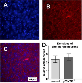
The visual cortex displays a low number of cholinergic neurons. Low densities of these cells are located in layer II–III of the adult murine visual cortex and seem to represent interneurons (Consonni et al., Reference Consonni, Leone, Becchetti and Amadeo2009). In the visual cortex of both, adult control as well as p75NTR deficient mice, sparsely distributed ChAT-positive neuronal somata can be detected. No significant difference in the densities of those cells was found by comparing both group of mice (Fig. 1). In addition, some scattered ChAT-positive neurons were found to be located in layer IV of the visual cortex (Fig. 2B and 2C).
Cholinergic neurons within the visual cortex. (A–C) Sparsely distributed cholinergic (ChAT-positive) neurons can be found in layer III of the visual cortex [(A) cell nuclei in the visual cortex visualized with DAPI (in blue); (B) ChAT-positive fibers and cells (in red); (C) overlay A–B]. (D) Analysis of the densities of ChAT-positive neurons in the visual cortex (layers II and III) revealed no significant difference between p75NTR deficient mice and their age-matched controls.

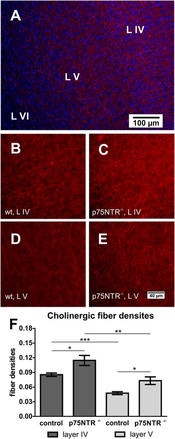
Cholinergic innervation of the visual cortex. (A) Overview of the cholinergic innervation of the visual cortex (L IV = layer IV; L V = layer V; L VI = layer VI). The cell nuclei were visualized with DAPI (in blue). Cholinergic structures were stained with an antibody directed against ChAT (in red). (B) ChAT-positive fibers in the visual cortex (layer IV) of a control mouse. (C) ChAT-positive fibers in the visual cortex (layer IV) of a p75NTR deficient mouse. (D) ChAT-positive fibers in the visual cortex (layer V) of a control mouse. (E) ChAT-positive fibers in the visual cortex (layer V) of a p75NTR deficient mouse. (F) In the murine brain, the cholinergic fiber density is higher in layer IV than in layer V. Moreover, p75NTR deficient mice display significant higher cholinergic fiber densities, as compared to age-matched controls, in both, layer IV and V.

Increased densities of cholinergic fibers in the visual cortex of p75NTRExIII−/− mice
Within the visual cortex the laminar distribution pattern of cholinergic fibers are different among species (Gu, Reference Gu2003). Since the density of the cholinergic innervation of the mouse visual cortex (Fig. 2A) differs between the layers (Gu, Reference Gu2003), we examined in detail layer IV and layer V. We could confirm that the density of the cholinergic innervation of layer IV was significantly higher as compared to layer V (+56%; P = 0.0079) in adult control mice (Fig. 2B, 2D, and 2F).
We could demonstrate for the first time that deletion of p75NTR has a strong impact upon the cholinergic innervation pattern of the primary visual cortex. Thus, in p75NTR deficient mice, the cholinergic innervation of layer IV (in comparison to layer V) was strongly increased (+64%; Fig. 2C, 2E, and 2F).
Concerning both, layer V and layer IV, deletion of p75NTR has an effect upon the cholinergic innervation. In detail, layer V of p75NTR−/− mice displays a significant higher cholinergic innervation as compared to controls (+34.3%; P = 0.036). Moreover, in case of layer IV, the difference was even more pronounced, since p75NTR deficient mice displayed a significant increase in the cholinergic density of about 53.7% (P = 0.036), as compared to age-matched controls (Fig. 2F).
Discussion
Acetylcholine plays an important role within the visual system. There is evidence that cholinergic deficits in the human primary visual cortex may underlie some of the abnormalities in visual processing and global cognitive performance seen in Alzheimer’s disease (Ikonomovic et al., Reference Ikonomovic, Mufson, Wuu, Bennett and DeKosky2005).
The visual cortex receives a substantial cholinergic innervation from the basal forebrain cholinergic system. The innervation of the primary visual and the prefrontal cortices by this system is involved in cognitive processing of sensory stimuli (Laplante et al., Reference Laplante, Morin, Quirion and Vaucher2005). Since these cholinergic neurons are involved in the processing of information, an animal model that allows investigating the consequences of increased cholinergic innervation of the visual cortex would be helpful. We thought that mice with a deletion in neurotrophins or their receptors might be interesting candidates for such a model. For this we focused on mice which carry a deletion of the full-length, low-affinity pan-neurotrophin receptor p75NTR since:
-
(1) The low-affinity pan-neurotrophin receptor p75NTR is expressed within the rodent visual cortex (Rossi et al., Reference Rossi, Sala and Maffei2002). The p75NTR immunoreactive signal in the cortex coincides with the laminar distribution of cholinergic afferents (Pioro & Cuello, Reference Pioro and Cuello1990), which hinted for a localization of p75NTR on cholinergic fibers but not on neurons of the cortex.
-
(2) Mice deficient for the low-affinity neurotrophin receptor p75NTR displayed increased numbers of basal forebrain cholinergic neurons along with an increased cholinergic target innervation of the hippocampus (Yeo et al., Reference Yeo, Chua-Couzens, Butcher, Bredesen, Cooper, Valletta, Mobley and Longo1997; Naumann et al., Reference Naumann, Casademunt, Hollerbach, Hofmann, Dechant, Frotscher and Barde2002; Dokter et al., Reference Dokter, Busch, Poser, Vogt, von Bohlen und Halbach, Gass, Unsicker and von Bohlen und Halbach2015).
Based on this we analyzed mice deficient for the full-length p75NTR concerning the cholinergic innervation of the visual cortex. We could show that the cholinergic innervation of the visual cortex in these mice was increased in both layers analyzed (layer IV and V), but that the densities of cholinergic neurons in the visual cortex was unaffected.
Densities of cholinergic neurons
Immunotoxic lesions using an antibody directed against p75NTR (192 IgG), coupled to the cytotoxin saporin, (192 IgG-saporin), results in a selective immunolesion of the forebrain cholinergic system (Nilsson et al., Reference Nilsson, Leanza, Rosenblad, Lappi, Wiley and Bjorklund1992), including a nearly complete loss of cholinergic fibers in the hippocampus and neocortex (Heckers et al., Reference Heckers, Ohtake, Wiley, Lappi, Geula and Mesulam1994). Concerning the cortex, it is known that a subpopulation of cortical cholinergic interneurons exists. These nonpyramidal neurons can be found frequently in the somatosensory cortex and in low densities in the visual cortex (Consonni et al., Reference Consonni, Leone, Becchetti and Amadeo2009). Treatment with 192 IgG-saporin, which results in a loss of different populations of cholinergic neurons, did not result in a loss of cholinergic neurons in the cortex (Heckers et al., Reference Heckers, Ohtake, Wiley, Lappi, Geula and Mesulam1994), indicating that these neurons did not express p75NTR. Therefore, we speculated that loss of p75NTR in mice would have no effect upon the densities of ChAT-positive neurons located in the visual cortex. The analysis of mice deficient for the full-length isoform of p75NTR revealed indeed that this cholinergic population was not affected. This for instance is in line with results concerning the striatal cholinergic neurons. These neurons, also do not express p75NTR (Yeo et al., Reference Yeo, Chua-Couzens, Butcher, Bredesen, Cooper, Valletta, Mobley and Longo1997). In a study, analyzing wild-type and p75NTR null mutant mice, no difference in the number or in the ChAT activity of these striatal cholinergic neurons was found (Yeo et al., Reference Yeo, Chua-Couzens, Butcher, Bredesen, Cooper, Valletta, Mobley and Longo1997).
Densities of cholinergic fibers
In contrast to unaltered densities of cholinergic neurons in the visual cortex, we found that lack of full-length p75NTR leads to an increase of cholinergic fiber densities in the visual cortex. This is in line with data demonstrating a crucial role of p75NTR for cholinergic neurons of the basal forebrain. For example, monkeys treated with immunotoxic 192 IgG-saporin loss the cholinergic innervation of the neocortex and, in addition, they were unable to learn a simple visual discrimination task (Ridley et al., Reference Ridley, Pugh, Maclean and Baker1999).
In the process of learning to discriminate temporally modulated visual stimuli acetylcholine seems to play a decisive role in the visual cortex (Minces et al., Reference Minces, Alexander, Datlow, Alfonso and Chiba2013). This can be tested, for example, by using a visual Go/No-Go task to assess the ability to learn to discriminate fine differences in the temporal structure of visual stimuli (Minces et al., Reference Minces, Alexander, Datlow, Alfonso and Chiba2013).
Acetylcholine is important for boosting visual cortex functions and plasticity in order to enhance visual perception (Kang et al., Reference Kang, Huppe-Gourgues and Vaucher2014). In that context, an interesting behavioral test for mice might be the recently introduced visually guided task. In this test head-fixed mice learned to discriminate two grating patterns in a virtual reality environment in which the animals' were running in a controlled position in a corridor (Poort et al., Reference Poort, Khan, Pachitariu, Nemri, Orsolic, Krupic, Bauza, Sahani, Keller, Mrsic-Flogel and Hofer2015). This test has been used to examine learning related changes in area V1 in mice (Poort et al., Reference Poort, Khan, Pachitariu, Nemri, Orsolic, Krupic, Bauza, Sahani, Keller, Mrsic-Flogel and Hofer2015) and might be of interest for getting insight into the role of acetylcholine in these processes. In addition, electrophysiological recordings have shown that altered cholinergic innervation is able to modulate the responsiveness of visual cortical cells (Siciliano et al., Reference Siciliano, Fornai, Bonaccorsi, Domenici and Bagnoli1999). Moreover, electrophysiological recordings in visual cortex slices further demonstrated that acetylcholine could act on membrane potential and modulate synaptic transmission (see for review Gu, Reference Gu2003).
In summary, we could show that deficiency for p75NTR induces an increase in the cholinergic innervation of the visual cortex but does not alter the density of cholinergic cortical interneurons. Based on the critical role of the cholinergic system in neuronal plasticity mice with altered cholinergic innervation of the visual cortex, like p75NTR deficient mice, may represent an interesting model to study the possible roles of acetylcholine in the visual cortex.
Acknowledgments
We wish to thank Mrs S. Hanisch for excellent technical assistance and Mrs S. Seidel for performing some of the immunohistochemical stainings. This study was supported by grants from the German Research Foundation (DFG; BO 1971/5-1) and from the EU (EFRE).