Introduction
Schistosomatidae actually contain 14 genera of medical and veterinary importance (Littlewood & Johnston, Reference Littlewood and Johnston1995; Snyder & Loker, Reference Snyder and Loker2000; Lockyer et al., Reference Lockyer, Olson, Ostergaard, Rollinson, Johnston, Attwood, Southgate, Horák, Snyder, Le, Agatsuma, McManus, Carmichael, Naem and Littlewood2003; Olson et al., Reference Olson, Cribb, Tkach, Bray and Lttlewood2003; Snyder, Reference Snyder2004; Brant et al., Reference Brant, Morgan, Mkoji, Snyder, Rajapakse and Loker2006; Loker & Brant, Reference Loker and Brant2006). Nine are known to parasitize birds and some of them are currently recognized as agents of human cercarial dermatitis (swimmer's itch). Historically, cases of cercarial dermatitis have been described in Europe (Szidat, Reference Szidat1930; Brumpt, Reference Brumpt1931; Desportes, Reference Desportes1945; Dönges, Reference Dönges1965; van den Broek, Reference van den Broek1965). Recently, the occurrence of bird schistosomes and swimmer's itch has become more of a problem in recreational areas, having important direct (health complications) and indirect (impact on travel industry) consequences (Kolářová et al., Reference Kolářová, Gottwaldova, Cechova and Sevcova1989, Reference Kolářová, Horák and Fajfrlik1992, Reference Kolářová, Horák and Sitko1997, Reference Kolářová, Skírnisson and Horák1999; Zbikowska et al., Reference Zbikowska, Franckiewicz Grygon and Wojcik2001). The pathogenicity of bird schistosome cercariae has been studied in experimental and natural hosts – birds, mammals and humans (Lévine et al., Reference Lévine, Clark and Hanon1956; Haas & Pietsch, Reference Haas and Pietsch1991; Gay et al., Reference Gay, Bayssade-Dufour, Grenouillet, Bourezane and Dubois1999; Horák & Kolářová, Reference Horák and Kolářová2000, Reference Horák and Kolářová2001; Bayssade-Dufour et al., Reference Bayssade-Dufour, Martins and Vuong2001, Reference Bayssade-Dufour, Vuong, René, Martin-Loehr and Martins2002; Zbikowska, Reference Zbikowska2003).
For a long time, ocellate furcocercariae were mostly identified as Trichobilharzia ocellata (LaValette, 1855) Brumpt, Reference Brumpt1931 (Eklu-Natey et al., Reference Eklu-Natey, Al-Khudri, Gauthey, Dubois, Wüest, Vaucher and Huggel1985). The present status and non-validity of this species has been reviewed by Rudolfová et al. (Reference Rudolfová, Hampl, Bayssade-Dufour, Lockyer, Littlewood and Horák2005) who conclude that Trichobilharzia szidati Neuhaus, Reference Neuhaus1952 should be the correct name for cercariae usually released from Lymnaea stagnalis snails in Europe (Neuhaus, Reference Neuhaus1952). Recently, systematic investigations after experimental infection of ducks led to the description of three new species of Trichobilharzia in Europe: Trichobilharzia franki Müller et Kimmig, 1994, Trichobilharzia regenti Horák Kolářová et Dvořák, 1998 and Trichobilharzia salmanticencis Simon-Martin et Simon-Vicente, 1999 (Müller & Kimmig, Reference Müller and Kimmig1994; Horák et al., Reference Horák, Kolářová and Dvořák1998; Simon-Martin & Simon-Vicente, Reference Simon-Martin and Simon-Vicente1999). Additionally, adults of bird schistosomes have been repeatedly identified in naturally infected hosts in Europe (Rudolfová et al., Reference Rudolfová, Sitko and Horák2002, Reference Rudolfová, Littlewood, Sitko and Horák2007; Kolářová et al., Reference Kolářová, Rudolfová, Hampl and Skírnisson2006; Skírnisson & Kolářová, Reference Skírnisson and Kolářová2008).
In France, several authors have reported cases of cercarial dermatitis in different freshwater areas (de Gentile et al., Reference de Gentile, Picot, Bourdeau, Bardet, Kerjan, Piriou, Guennic, Bayssade-Dufour, Chabasse, Mott and Le Guennic1996; Gay et al., Reference Gay, Bayssade-Dufour, Grenouillet, Bourezane and Dubois1999; Léger & Martin-Loehr, Reference Léger and Martin-Loehr1999; Caumes et al., Reference Caumes, Felder-Moinet, Couzigou, Darras-Joly, Latour and Léger2003). However, all these studies were mainly looking for cercariae and snails as agents of the disease. Ferté et al. (Reference Ferté, Depaquit, Carré, Villena and Léger2005) and Jouet et al. (Reference Jouet, Ferté, Depaquit, Rudolfová, Latour, Zaniella, Kaltenbach and Léger2008) used molecular systematics to study ocellate furcocercariae (agents of human dermatitis) from naturally infected molluscs. These authors identified the cercariae emitted by the snail hosts L. stagnalis and Radix auricularia collected in the artificial Der-Chantecoq Lake as T. szidati and T. franki, respectively. Interestingly, they found four unique haplotypes of schistosome cercariae emitted by one species of snail (Radix peregra) in Annecy Lake. The only investigation in aquatic birds in France was performed by Bayssade-Dufour et al. (Reference Bayssade-Dufour, Jouet, Rudolfová, Horák and Ferté2006) and focused on Bilharziella polonica (Kowalewski, 1895) and Dendritobilharzia pulverulenta (Braun, 1901).
The present work continues the investigation of bird schistosomes in France, but with a focus on the wildfowl from the Champagne-Ardenne and Annecy Lake areas. Our aim was to identify adult schistosome stages and their corresponding final hosts under natural conditions.
Materials and methods
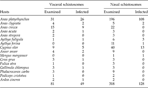
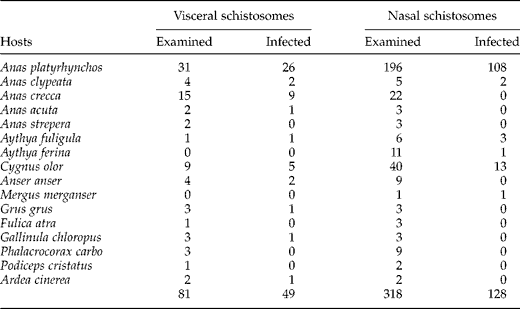
From 2005 to 2008, a total of 318 heads and 81 viscera of aquatic birds were dissected. They originated mainly from the Champagne-Ardenne and Annecy lakes and from one recreational lake (in Beauvais, Oise). The birds belonged to 16 species from five orders (table 1): Anseriformes – Anas platyrhynchos (mallard), Anas crecca (common teal), Anas clypeata (northern shoveller), Anas strepera (gadwall), Anas acuta (northern pintail), Aythya ferina (common pochard), Aythya fuligula (tufted duck), Cygnus olor (mute swan), Anser anser (greylag goose) and Mergus merganser (goosander); Gruiformes – Fulica atra (common coot), Gallinula chloropus (common moorhen), Grus grus (common crane); Pelecaniformes – Phalacrocorax carbo (great cormorant); Podicipediformes – Podiceps cristatus (great crested grebe); Ciconiiformes – Ardea cinerea (grey heron). Birds were either shot during the hunting season or found dead by members of the SAGIR Network (National Network for surveillance of wildlife diseases in France) and ONCFS (Office National de la Chasse et de la Faune Sauvage).
Results of wildfowl examination for nasal and visceral bird schistosomes in France.
All birds were kept frozen until autopsy. The examination of birds for nasal schistosomes was performed by perfusion of nostrils with water (eggs) and dissection of nasal tissue (adults). Intestinal wall, liver and mesenteric veins were examined for visceral species according to Skírnisson & Kolářová (Reference Skírnisson and Kolářová2008). When parasites were found, some specimens (piece of worms) were conserved for staining in chlorhydric carmine. Eggs and fragments of the adult forms of schistosomes were fixed in 95% ethanol and frozen for molecular investigations. After removing all ethanol from samples, DNA extraction was done using the Qiamp DNA mini kit (Qiagen, Germany) following the manufacturer's instructions. During the first step (tissue lysis), eggs and fragments of worms were crushed one by one using a piston pellet (Treff, Switzerland). DNA was eluted in 50 μl of elution buffer as provided by the manufacturer.
Sequencing of the D2 domain and the second internal transcribed spacer (ITS-2) of the 28S subunit of ribosomal DNA were used for the identification of avian schistosomes obtained from naturally infected birds. Polymerase chain reaction (PCR) was performed in a 50 μl volume using 5 μl of DNA, and 50 pmol of each of the primers. The PCR mix contained (final concentrations) 10 mm Tris HCl (pH 8.3), 1.5 mm MgCl2, 50 mm KCl, 0.01% Triton X100, 200 μm dNTP (each base), and 1.25 units of Taq polymerase (Eppendorf, Germany). The D2 domain was amplified using the primers used by Mollaret et al. (Reference Mollaret, Jamieson, Adlard, Hugall, Lecointre, Chombard and Justine1997): C2′B and D2. ITS-2 was amplified using ITS3Trem and ITS4Trem primers as previously described by Dvořák et al. (Reference Dvořák, Vancova, Hampl, Flegr and Horák2002). The initial denaturation at 94°C for 3 min was followed by 40 cycles of denaturation at 94°C for 45 s, annealing at 50°C for 45 s and extension at 68°C for 2 min, with a final elongation time of 10 min at 68°C. PCR products were directly sequenced in both directions with the primers used for DNA amplification (Qiagen). The sequences are deposited in GenBank under accession numbers FJ793813 to FJ793922.
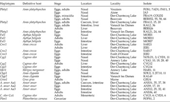
Sequence alignment was performed using the ClustalW routine included in the MEGA version 3.1 software (Kumar et al., Reference Kumar, Tamura and Neï2004) and checked by eye. The D2 domain (554 bp) and the ITS-2 region (373 bp) were used for tree construction and rooted with the outgroup taxon. Homologous sequences in D2 and ITS-2 were pooled under the same haplotype (table 2). Previous samples reported by us were included in this work for complementary analyses (Bayssade-Dufour et al., Reference Bayssade-Dufour, Jouet, Rudolfová, Horák and Ferté2006; Jouet et al., Reference Jouet, Ferté, Depaquit, Rudolfová, Latour, Zaniella, Kaltenbach and Léger2008).
Isolates of bird schistosomes from France used for phylogenetic analyses.

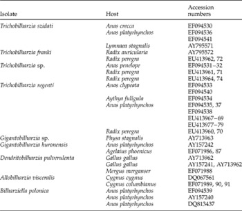
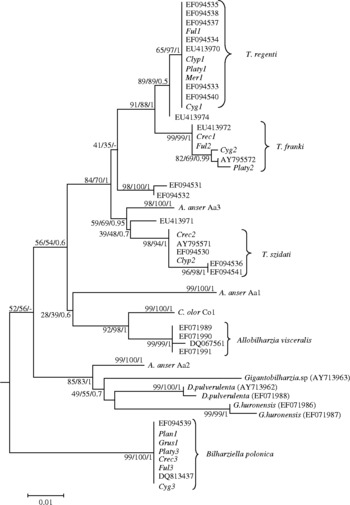
Phylogenetic analyses based on the D2 and ITS-2 domains were performed using haplotypes obtained from this study (table 2) and from those of Trichobilharzia regenti, T. szidati, Trichobilharzia sp., Allobilharzia visceralis, Dendritobilharzia pulverulenta, Gigantobilharzia sp., G. huronensis and Bilharziella polonica contained in the GenBank database (table 3). Sequences of Schistosoma mansoni (GenBank accession numbers Z46503 and SMU22168) served as outgroups. Phylogenetic reconstruction using neighbour-joining (NJ) with the Tajima-Nei model and uniform rates among sites was performed using the MEGA version 3.1 software (Kumar et al., Reference Kumar, Tamura and Neï2004). A maximum likelihood (ML) analysis was performed in PHYML Online (Guindon et al., Reference Guindon, Lethiec, Duroux and Gascuel2005). Model and model parameters were chosen using the hierarchical likelihood ratio test implemented in Modeltest 3.7 (Posada & Crandall, Reference Posada and Crandall2001). For both NJ and ML analysis, gaps were treated as missing data and internal node support assessed by bootstrapping over 500 replicates. Bayesian inference (BI) was performed using MrBayes 3.1.1 (Huelsenbeck & Ronquist, Reference Huelsenbeck and Ronquist2001); four simultaneous Markov Monte Carlo chains (temperature 0.2) were run for 2,000,000 generations under a HKY+Γ substitution model and sampled every 100 generations. The first 5000 trees were discarded as ‘burn-in’. Since all methods rooted with Schistosoma mansoni yielded results with a similar topology, the tree based on ML for the ITS-2 is the only one presented here (see fig. 1).
GenBank sequences of bird schistosomes used for phylogenetic analyses.

Phylogenetic tree of bird schistosome isolates based on the ITS-2 region of rDNA, constructed using the maximum likelihood (ML) method in PHYML (HKY+Γ model of substitution). Schistosoma mansoni was used as outgroup. To save space, this taxon is not shown. The node support is given in neighbour-joining (NJ) and ML bootstraps, and Bayesian posterior probability. The scale shows the number of nucleotide substitutions per site between DNA sequences.

Results
Eight lineages of nasal and visceral schistosomes (four previously identified species and four unknown unique lineages) were recovered from three French lakes. Identifications were based on molecular data and confirmed with morphological characters when possible. A count of all schistosomes recovered from aquatic birds is shown in table 1.
Nasal schistosomes (eggs and/or adults) were found in 128 out of 318 (40%) heads of aquatic birds from only six anseriform species, Anas platyrhynchos, Anas clypeata, Aythya ferina, Aythya fuligula, Cygnus olor and Mergus merganser. Birds from other orders were not positive for nasal schistosomes. All eggs of nasal schistosomes had the same morphological features (spindle-shaped eggs), except for those from Anas clypeata which were crescent-shaped. Species determination was not possible as only fragments of adults were isolated, which lacked distinguishing morphological characters. A high prevalence of nasal schistosomes was observed in A. platyrhynchos and C. olor from Annecy Lake. In infected C. olor, the number of lesions was very high in the nasal mucosa and contained many eggs relative to the infected ducks.
Visceral schistosomes were found in 49 out of 81 (60%) viscera of aquatic birds. Eggs and/or adults were found from ten species of birds from three orders. Of the schistosomes recovered, only eggs and adults from the genus Bilharziella were described based on specific morphological features (oval egg with spine and long tail, digestive and genital apparatus of adults). Bilharziella polonica was the single schistosome in 17 birds (from species A. platyrhynchos, A. crecca, Aythya fuligula, C. olor, Grus grus, Gallinula chloropus and Ardea cinerea) and was a co-parasite with Trichobilharzia, Allobilharzia or Dendritobilharzia in 15 other birds (from species A. platyrhynchos, A. crecca, A. clypeata, C. olor and Aythya fuligula). The adults of Bilharziella were located in the hepatic or mesenteric vessels and the eggs were located in the caecal mucosa.
As described previously, crescent-shaped eggs resembling T. szidati were observed in four samples from A. platyrhynchos and A. crecca, and a new host record A. clypeata was found.
Anser anser were infected by three visceral schistosomes. These were characterized by the morphology of the eggs or adults and determined as: (Haplotype ‘A. anser Aa1’) oval eggs with spine and large adults; (Haplotype ‘A. anser Aa2’) small, spindle-shaped eggs without adults; (Haplotype ‘A. anser Aa3’) large, spindle-shaped eggs and small adults. Fragments of schistosome adults were also found in mesenteric vessels of four C. olor and displayed morphological features similar to those of the genus Allobilharzia. They were determined as Haplotype ‘C. olor Co1’.
The phylogenetic analysis was based on the D2 domain and the internal transcribed spacer ITS-2. The phylogenetic tree based on the sequences of our isolates and the sequences deposited in GenBank is shown in fig. 1. Based on our haplotype data, all nasal schistosomes isolated from A. platyrhynchos (Haplotype ‘Platy1’), C. olor (Haplotype ‘Cyg1’), Aythya fuligula (Haplotype ‘Ful1’) and Mergus merganser (Haplotype ‘Mer1’) were identical with haplotypes of Trichobilharzia regenti deposited in GenBank for D2 and ITS-2. Only sequences from A. clypeata (Haplotype ‘Clyp1’) showed intraspecific variations (two nucleotides) in D2.
We recovered Bilharziella polonica, Trichobilharzia franki, T. szidati and four other unique genetic lineages of visceral schistosomes. For the visceral schistosomes belonging to the genus Trichobilharzia, 11 isolates had sequences that grouped with the T. franki branch. Sequences of the eggs and adults from A. platyrhynchos (Haplotype ‘Platy2’) and C. olor (Haplotype ‘Cyg2’) are identical in D2 but differ by only one site within the AY795572 haplotype (cercariae from Radix auricularia). Other isolates from Aythya fuligula (Haplotype ‘Ful2’) and A. crecca (Haplotype ‘Crec1’) were homologous in D2 and ITS-2 with the haplotype of cercariae isolated from Radix peregra (accession numbers EU413962/72). These two populations differ by four and three intraspecific variations in D2 and ITS-2, respectively. All isolates (Haplotypes ‘Platy3’, ‘Crec3’, ‘Grus1’, ‘Ful3’ and ‘Cyg3’) morphologically determined as Bilharziella were found to be identical to the haplotypes of Bilharziella polonica deposited in GenBank, based on their D2 and ITS-2 sequences and phylogenetic positions. Ocellate furcocercariae with short furca were also isolated from two Planorbarius corneus (Haplotype ‘Plan1’) in France. Sequences of these cercariae were found to be identical with all other sequences of B. polonica described previously.
DNA extraction of the crescentic eggs from A. platyrhynchos failed. Consequently, only sequences isolated from A. crecca and A. clypeata were included in the phylogenetic analysis. All of them were identical to haplotypes of Trichobilharzia szidati isolated from A. crecca and Lymnaea stagnalis (accession numbers EF094530 and AY795571). Yet, they differ by three nucleotides in ITS-2 from those of T. szidati isolated from A. platyrhynchos in Poland and Czech Republic (accession numbers EF094536 and EF094541).
In addition to the above species, we also identified four unique haplotype lineages that could not be assigned to any species group. Phylogenetic analyses of the D2 and ITS-2 domains of the three visceral schistosomes from Anser anser confirmed the exclusive position of these three new haplotypes. From C. olor, sequences of adults differ by two and six variations in D2 and ITS-2, respectively, from sequences of Allobilharzia visceralis available in GenBank. This new haplotype from C. olor forms a sister-group of those isolated from Cygnus cygnus in Iceland and Cygnus columbianus in North America (Kolářová et al., Reference Kolářová, Rudolfová, Hampl and Skírnisson2006; Brant, Reference Brant2007).
Discussion
In a recent study, we showed that Trichobilharzia spp. could infest several different species of snails and were not host specific ( Jouet et al., Reference Jouet, Ferté, Depaquit, Rudolfová, Latour, Zaniella, Kaltenbach and Léger2008). Moreover, the presence of larval stages of T. regenti, T. franki, T. szidati and two new haplotypes of Trichobilharzia sp. in different areas in France was also confirmed. Our new results indicate that adults of bird schistosomes (similar to their larvae) are also able to parasitize different species of final hosts under natural conditions. The high prevalence of infested aquatic birds observed here is in agreement with previous studies in Europe (Rudolfová et al., Reference Rudolfová, Sitko and Horák2002, Reference Rudolfová, Littlewood, Sitko and Horák2007; Bayssade-Dufour et al., Reference Bayssade-Dufour, Jouet, Rudolfová, Horák and Ferté2006). Eight lineages of avian schistosomes were found in our study, based on their morphological features and molecular sequence data; four species T. regenti, T. franki, T. szidati and Bilharziella polonica are well-known causative agents of cercarial dermatitis in Europe. The three new haplotypes of visceral avian schistosomes from Anser anser and the new visceral haplotype from Cygnus olor plus the unique larval haplotypes from our previous studies, clearly demonstrate the high diversity of avian schistosomes found in France (Jouet et al., Reference Jouet, Ferté, Depaquit, Rudolfová, Latour, Zaniella, Kaltenbach and Léger2008).
Based on our results, the host spectrum of B. polonica observed by Bayssade-Dufour et al. (Reference Bayssade-Dufour, Jouet, Rudolfová, Horák and Ferté2006) is to be expanded to include four new final hosts in France: Anas crecca, Aythya fuligula, Gallinula chloropus and Grus grus. The only study that reported a visceral examination of G. grus was performed by Gottschalk & Prange (Reference Gottschalk and Prange2002), but they did not find any schistosomes. Therefore, our finding of B. polonica represents the first report of a bird schistosome from this species and other Gruiformes. Cercariae of B. polonica were also found in planorbid snails of the species Planorbarius corneus, known to host B. polonica (Zbikowska, Reference Zbikowska2004). Phylogenetic analyses, based on their D2 and ITS-2 sequences proved that they belong to one species and confirmed the monospecificity of the genus Bilharziella.
For T. regenti, T. franki and T. szidati, all life-cycle stages are well established in different sites in France. All our findings of nasal schistosomes belonged to the same species, T. regenti. However, morphological and molecular variation observed for the eggs isolated from A. clypeata needs to be confirmed, notably by a comparison with all the different nasal species described by Fain (Reference Fain1955a, Reference Fainb, Reference Fain1956a, Reference Fainb, Reference Fain1959) from African aquatic birds. Except for eggs previously described using morphological features by Skírnisson & Kolářová (Reference Skírnisson and Kolářová2008), T. franki was isolated for the first time in its natural conditions in final hosts (A. platyrhynchos, A. crecca, Aythya fuligula and C. olor), and confirmed by molecular phylogenetic analyses. Previous studies describing A. platyrhynchos as the definitive host of T. franki were based on experimental infections (Müller & Kimmig, Reference Müller and Kimmig1994). Based on molecular analyses, two populations have been found for this species: one from A. platyrhynchos, C. olor and cercariae emitted by Radix auricularia, and another from Aythya fuligula, A. crecca and cercariae isolated from R. peregra. This pattern will be studied to determine if an adaptive phenomenon of differential host use could be responsible for the observed molecular clustering of these parasites. Finally, several samples corresponding to crescentic eggs were found to belong to the T. szidati branch: eggs were isolated from A. crecca and A. platyrhynchos (based only on shape of the eggs), as described in Europe by Rudolfová et al. (Reference Rudolfová, Littlewood, Sitko and Horák2007), and from a new species, A. clypeata.
Only two morphological studies have detected eggs and adults of avian schistosomes from Anser anser (Wojcinski et al., Reference Wojcinski, Barker, Hunter and Lumsden1987; Skírnisson & Kolářová, Reference Skírnisson and Kolářová2008). Our results showed three new haplotypes of bird schistosomes as found by examination of viscera of Anser anser. All of them differ by the morphological features of the eggs and adults (when present) and their phylogenetic position. Identification of the species will be confirmed according to the recommendations of Rudolfová et al. (Reference Rudolfová, Hampl, Bayssade-Dufour, Lockyer, Littlewood and Horák2005).
An interesting point is the relationship between C. olor and bird schistosomes. A high prevalence of infected C. olor was observed, and five species of schistosomes were isolated from this host (T. regenti, T. franki, Dendritobilharzia pulverulenta (unpublished), B. polonica and a new haplotype named ‘C. olor Co1’) alone or co-parasitized. Our finding of T. regenti in C. olor represents the first report of this species from this host. Previous studies described parasites of the genus Dendritobilharzia as the agent of encephalitis in swans (Lévine et al., Reference Lévine, Clark and Hanon1956; Wilson et al., Reference Wilson, New and Scholtens1982) but only one nasal schistosome was described in the past from C. olor, without species determination (Palmer & Ossent, Reference Palmer and Ossent1984). In addition, many lesions were found in the nasal mucosa of infected birds and each lesion contained many eggs, and these high levels of parasitism are a health risk for birds (see Lévine et al., Reference Lévine, Clark and Hanon1956; Wilson et al., Reference Wilson, New and Scholtens1982; Palmer & Ossent, Reference Palmer and Ossent1984; van Bolhuis et al., Reference van Bolhuis, Rijks, Dorrestein, Rudolfová, van Dijk and Kuiken2004). Adults of ‘C. olor Co1’ were found in mesenteric vessels. Based on phylogenetic analyses, ‘C. olor Co1’ appears more closely related to Allobilharzia visceralis isolated from Cygnus cygnus and Cygnus columbianus (Kolářová et al., Reference Kolářová, Rudolfová, Hampl and Skírnisson2006; Brant, Reference Brant2007). However, its unique features suggest that it might be a new species. Morphological investigations should thus be undertaken to confirm this hypothesis.
In conclusion, France is particularly concerned with avian schistosomes because of the position of this country in the migratory flyways of birds. Complete life cycles (cercariae/snails and adults–eggs/birds) of T. regenti, T. franki (first description), T. szidati and B. polonica were found in different ponds, particularly in Annecy Lake. This distribution of avian schistosomes in association with the increasing populations of protected waterbirds (e.g. C. olor populations in French lakes) could explain the increased risk of cercarial dermatitis in these tourist/recreational areas. The putative new species (lineages) found in these final hosts need to be compared with species occurring in other continents, especially Africa, North America and Iceland, as these are the wintering and breeding-ground destinations for birds that fly through France. The phylogenetic position of these haplotypes should now be verified by experimental infections to test viability of eggs. Further investigations concerning all these new haplotypes as causal agents of human cercarial dermatitis should also be performed in the future.
Acknowledgements
The authors thank Sara Brant for proofreading the manuscript, Karl Skírnisson, Libuse Kolářová, Nicole Léger and Emmanuel Liénard for their help and fruitful comments. They also thank Y. Maupois and the ONCFS technical staff (Office National de la Chasse et de la Faune Sauvage) of Der-Chantecoq Lake, the SAGIR (LVD10 and LVD74) network and Bruno Hornoy for their help in providing samples, and Monique Boutry, Chantal Grimplet, Mélanie Vittier for their technical assistance. Financial support for this study was provided by ONCFS, SILA (Syndicat Mixte du Lac d'Annecy) and the town council of Beauvais.