Mitral valve prolapse is a common valvular disease with an incidence of 2–6%.Reference Bonow, Carabello and Leon 1 Mitral valve prolapse is caused by progressive myxomatous degeneration of mitral valve leaflets and chordae tendinea with superior displacement of one or two mitral leaflets by more than 2 mm above the plane of mitral annulus into the left atrium. Mitral valve prolapse can be sporadic, familial, or coexist with the connective tissue disorders – for example, Marfan syndrome, Ehler Danlos syndrome.Reference Glesby and Pyeritz 2 , Reference Nesta, Leyne and Yosefy 3 Abnormal collagen composition, accumulation of proteoglycans, and increased matrix metalloproteinase activity are the components in pathogenesis of mitral valve prolapse.Reference Rabkin, Aikawa, Stone, Fukumoto, Libby and Schoen 4 , Reference Grande-Allen, Griffin, Calabro, Ratliff, Cosgrove and Vesely 5 Polymorphisms found in genotypes of collagen, fibrillin-1, and Urokinase plasminogen activator, which is a metalloproteinase activator in sporadic mitral valve prolapse, contribute to the underlying mechanism.Reference Chou, Shi, Hsu and Tsai 6 – Reference Chou, Hung, Chen, Wu and Tsai 8 Mitral valve prolapse is more common (30%) in patients with benign joint hypermobility syndrome, which has overlapping features with genetic disorders such as Marfan syndrome and Ehler Danlos syndrome.Reference Pitcher and Grahame 9 Mitral valve prolapse is considered as a systemic and progressive disease since it is not found in newborns and its prevalence is low in early childhood despite genetic influences.Reference Nascimento, Freitas and Teixeria 10 , Reference Hickey and Wilcken 11 From this point of view, every structure of the heart, myocardium, valves, and great vessels can be affected from attenuation of supportive structure of extracellular matrix because of ongoing metalloproteinase activity, elastin degradation, and abnormal collagen composition. Heart cavities and great vessels may be widened. Great vessels may show different elastic properties and left ventricle systolic and diastolic functions may be altered.
There are animal studies concerning endothelial dysfunction in the early course of valve disease.Reference Moesgaard, Olsen and Viuff 12 , Reference Mow and Pedersen 13 These findings lead us to question the condition of vascular health in patients with mitral valve prolapse.
We investigated systolic and diastolic diameters and functions of the left ventricle and left atrium; systolic and diastolic diameters of the aorta and its elastic properties; carotid intima-media thickness, which is an early indicator for atherosclerosis; and flow-mediated dilation of brachial artery for endothelial function in children with mitral valve prolapse.
Material and methods
Patients
The study group consisted of 43 patients with mitral valve prolapse, their ages between 6 and20 years (mean±SD=13.4±3.9; median: 14, male/female=12/31), and the control group, which consisted of 42 healthy children between 6 and 19 years of age (mean ± SD=12.8±3.4; median: 13, male/female=12/30). Patients with more than mild mitral regurgitation or other valvular disease, systemic disease, heritable disorders of the connective tissue, genetic disease, associated CHD, or rheumatic heart disease were excluded. None of the patients had inherited disorders of the connective tissue such as Marfan syndrome, Ehler Danlos syndrome, or Loeys Dietz syndrome. The control group included healthy children referred to paediatric cardiology clinics for evaluation of an innocent murmur or atypical chest pain and found to have no cardiac abnormality by clinical evaluation or echocardiography. Physical examination findings, weight and height measurements, and blood pressures of all patients were recorded. Patients were divided into two groups according to the leaflet thickness (classical and non-classical mitral valve prolapse) because a previous adult study has shown increased aortic distensibility and decreased stiffness in classical form of mitral valve prolapse.Reference Unlu, Demirkol and Aparci 14 Patients were also divided into groups (younger versus older) according to age (6–12 years versus 13–20 years) because aortic stiffness increases with age in the normal population.Reference Fritze, Romero and Scleicher 15 The control group was also divided into two groups according to age and comparisons between the study and control groups were carried out between corresponding age groups. Patients gave written consent forms and the study was approved by the local ethics committee.
Echocardiography
Echocardiography was performed using Philips IE33 Echocardiography machine (Philips Medical Systems, Bothell, WA, United States of America) equipped with 5 MHz and L11 Mhz linear transducers. All patients underwent M-Mod, two-dimensional, and Doppler echocardiographic examinations. Left ventricular end-diastolic diameter, left ventricular end-systolic diameter, left ventricular posterior wall thickness, and left atrial diameter were measured; ejection fraction and fractional shortening were calculated from M-mode echocardiographic measurements in a standard fashion using parasternal long axis view of the left ventricle. Left ventricular and left atrial dimensions were adjusted to body surface area of the children. In the parasternal long axis view, the mitral annulus diameter was measured at mid-diastole and adjusted to body surface area of the children. Mitral inflow parameters, mitral peak early velocity, mitral peak late velocity, isovolumetric relaxation time, and deceleration time were measured by Pulse Wave Doppler echocardiography for assessment of left ventricular diastolic functions. The measurements were obtained with standard techniques according to the recommendations of the American Society of Echocardiography.Reference Lopez, Colan and Frommelt 16
Mitral valve prolapse was assessed at parasternal long axis and defined as superior displacement of one or two mitral leaflets by more than 2 mm above the plane of mitral annulus into the left atrium during systole. Mitral valve prolapse was defined as classical mitral valve prolapse if leaflet thickness at mid-diastole was more than 5 mm and non-classical mitral valve prolapse if leaflet thickness was lower than 5 mm.Reference Heyek, Gring and Griffin 17 – Reference Levine, Stathogiannis and Newell 19 Mitral regurgitation was assessed according to the European Association of Echocardiography Recommendations of 2010.Reference Lancellotti, Moura and Pierard 20
Systolic and diastolic diameters of the aortic annulus, sinus valsalva, sinotubular junction, ascending, descending, and abdominal aorta were measured and z-scores obtained by using Halifax data.Reference Warren, Boyd, O’Connell and Dodds 21 Aortic strain, aortic distensibility, and aortic stiffness index were calculated using the formulas below:
 $$\eqalignno{ {\rm Aortic}\,{\rm strain}\,\left( \,\%\, \right){\equals}100{\times}\cr\left( {{\rm Systolic}\,{\rm diameter}{\minus}{\rm Diastolic}\,{\rm diameter}} \right)\,/\,{\rm Diastolic}\,{\rm diameter}$$
 $$\eqalignno{ {\rm Aortic\,stiffness\,index}\,{\equals}\,{\rm logarithm\,(Systolic\,blood}\cr\left( {{\rm pressure/Diastolic\,blood\,pressure}} \right)[{\rm (Systolic\,diameter}\cr{\minus}\,{\rm Diastolic\,diameter)}/{\rm Diastolic\,diameter]}$$
 $$\eqalignno{ {\rm Aortic\,distensibility\,(cm^2/dyn^2/10^6)}\,{\equals}\,{\rm 2{\times}}\cr\left( {{\rm Systolic\,diameter{\minus}Diastolic\,diameter}} \right)[{\rm (Systolic}\cr{\rm blood\,pressure/Diastolic\,blood\,pressure)}{\times}{\rm Diastolic\,diameter]}$$
Carotid intima-media thickness
Carotid intima-media thickness was measured from the right carotid artery using a 11 MHz linear array probe when the patient is in supine position, with the neck turned 45° towards the opposite side. Images were acquired at the end-diastolic phase simultaneous with the tip of R-wave on electrocardiogram. Neck vessel was shown in a cross-sectional plane first and then the transducer was rotated clockwise to a longitudinal plane. Measurements were obtained when the longitudinal distance of the carotid artery walls were visible at least 10 mm long on both sides. Measurement of carotid intima-media thickness was performed at the far wall of the carotid artery. Carotid intima-media thickness was the distance between two bright lines measured edge to edge. An average carotid intima-media thickness value was obtained from three separate video-loop measurements.Reference Dalla Pozza, Ehringer-Schetitska and Fritsch 22
Flow-mediated dilation
Flow-mediated dilation was performed by using a L11 MHz linear array transducer after 10 minute resting in supine position from the right brachial artery. Above antecubital fossa, a non-branching, straight segment of the brachial artery was identified and images were recorded. Baseline brachial artery diameter was measured offline at the tip of the R-wave of simultaneous electrocardiogram. A pneumatic cuff on the forearm was then inflated up to 50 mmHg above systolic blood pressure for 5 minutes and then released. After cuff-deflation, brachial artery diameter was measured at every 30 seconds of 3 minutes at end-diastolic phase. Flow-mediated dilatation was calculated as the percentage change in diameter from baseline value to the biggest value after cuff-deflation.Reference Celermajer, Sorensen and Gooch 23
Statistical analysis
Statistical analysis was performed using SPSS for Windows (Version 22.0; IBM Corp., Armonk, NY, United States of America) Data are presented as mean ± SD for continuous variables and median (range) for non-contiguous variables. For comparisons between groups, t-test, χ2test, and Mann–Whitney U were used, whichever is appropriate. Correlation between variables was expressed by using the Spearman rank correlation coefficient. The p-values <0.05 were considered statistically significant.
Results
Patient characteristics
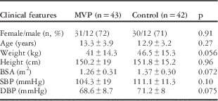
There was no statistically significant difference on comparing age, weight, height, body mass index, systolic and diastolic blood pressures of patients with mitral valve prolapse with the control groups (Table 1). There were more female patients than the male patients in each group but there was no statistically significant difference in terms of female/male ratio between the two groups (p=0.91). The patients with classical mitral valve prolapse were taller than the patients with non-classic mitral valve prolapse (p=0.034). Of 43 mitral valve prolapse patients, 13 had chest pain, four had palpitation, five had syncope, and two had arthralgia as the symptom of presentation. In total, 28 of the mitral valve prolapse patients had late systolic murmur with or without mid-systolic click, and 15 of them had no murmur. In 43 patients with mitral valve prolapse, 23 had prolapse of anterior leaflet, five had prolapse of posterior leaflet, and 15 had prolapse of both leaflets. In total, 20 patients in the study group had trivial or mild mitral regurgitation.
Patient characteristics of mitral valve prolapse (MVP) and control groups.

BSA=body surface area; DBP= diastolic blood pressure; SPB=systolic blood pressure.
Data are expressed as number (n) and percentage(%) or mean±SD as appropriate
Left ventricle and left atrium
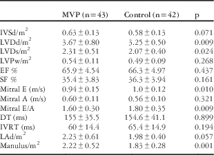
In the assessment of left ventricular dimensions, left ventricular diastolic and systolic diameters, and mitral annulus diameter adjusted to the body surface area were larger in the mitral valve prolapse group than the control group (p=0.009, p=0.024, p=0.001) (Table 2). Left ventricular dimensions, systolic, and diastolic functions are shown in Table 2. There was no significant difference between classical and non-classical groups in terms of left ventricle measurements. Leaflet thickness had no correlation to left ventricular dimensions (p>0.05). In older patients (13–20 years of age), left atrial diameter adjusted to the body surface area was significantly larger than that in the control group (p=0.039). In the assessment of left ventricular diastolic functions, mitral peak early velocity and peak early-to-late velocity ratio were lower in patients with mitral valve prolapse compared with the control group (p=0.010, p=0.009).
Echocardiographic features of mitral valve prolapse (MVP) and control groups.

DT=deceleration time; EF=ejection fraction; IVRT=isovolumic relaxation time; IVSd=interventricular septum systolic diameter; LAd=left atrial diameter; LVDd=left ventricular diastolic diameter; LVDs=left ventricular systolic diameter; LVPw=left ventricular posterior wall diameter; m2=body surface area; Manulus=mitral anulus; Mitral A=mitral peak late wave velocity; Mitral E=mitral peak early wave velocity; SF=shortening fraction
z-Scores of aorta
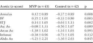
z-Scores of patients with mitral valve prolapse at the aortic annulus, sinus valsalva, and sinotubular junction were higher than the control group (p=0.008, p=0.003, p=0.002, respectively) (Table 3). z-Scores of three patients in the mitral valve prolapse group were over +2. When we excluded these three patients, z-scores of mitral valve prolapse at the aortic annulus and sinotubular junction were still higher compared with the control group (p=0.043, p=0.011). Patients with mitral valve prolapse in younger age had higher z-scores at the arcus aorta, isthmus, and abdominal aorta compared with control patients (p=0.047, p=0.006, p=0.041). Patients with mitral valve prolapse in older age had higher z-scores at the aortic root (p=0.025, p=0.035, p=0.033). z-Scores did not differ significantly between classical and non-classical patients with mitral valve prolapse.
z-Scores of aorta.

Abdo Ao=abdominal aorta; ; Arcus Ao=arcus aorta; Asc Ao=ascending aorta; MVP=mitral valve prolapse; STJ=sinotubular junction; SV=sinus valsalva
Elasticity parameters of the aorta
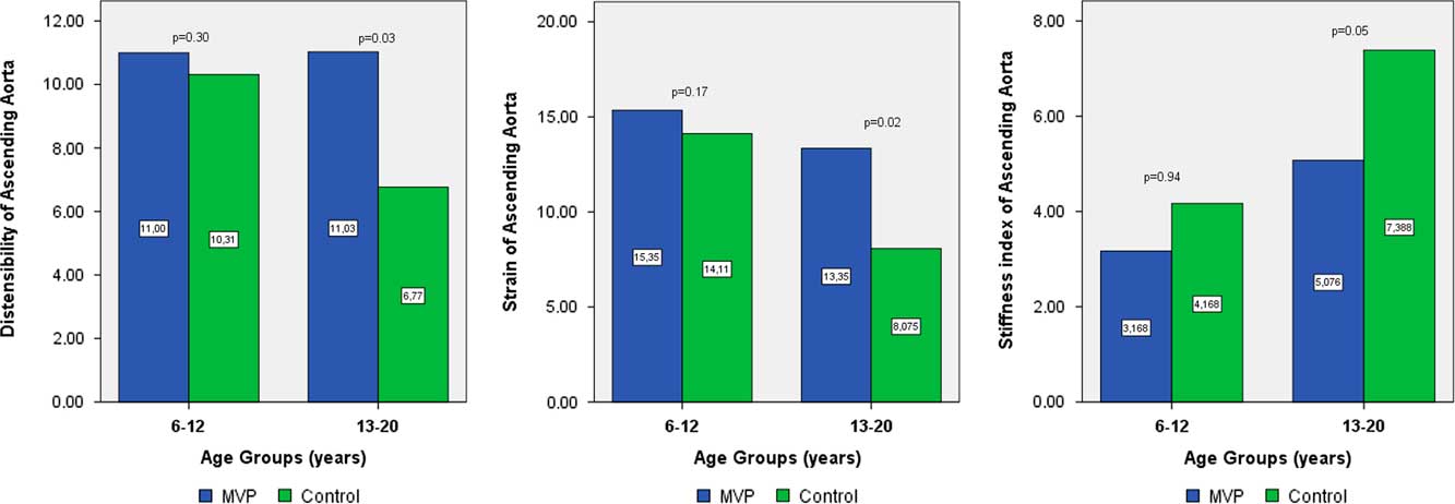
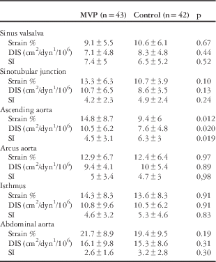
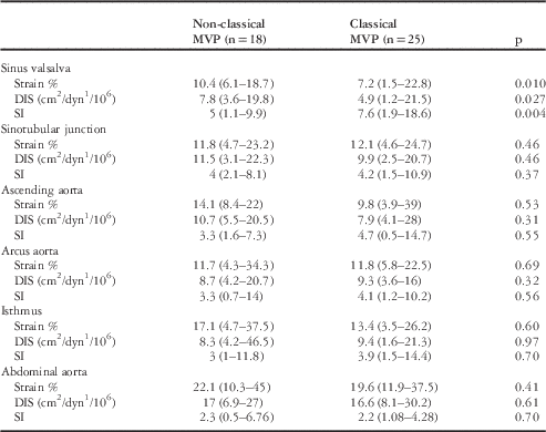
In the mitral valve prolapse group, aortic strain and distensibility increased and stiffness decreased at the ascending aortic level compared with controls (p=0.012, p=0.020, p=0.019; respectively). There was no difference in terms of elasticity parameters at other aortic levels. Elasticity parameters are shown in Table 4. When we excluded patients with mitral regurgitation from the mitral valve prolapse group, the difference did not change (p=0.006, p=0.010, p=0.013 for strain, distensibility, stiffness index, respectively). Exclusion of the patients with a dilated aorta over +2 SD did not change the difference between the study and control groups in terms of strain, distensibility, and stiffness at the level of the ascending aorta (p=0.007, p=0.005, p=0.009). Classical mitral valve prolapse had lower strain and distensibility, and higher stiffness of the aorta at sinus valsalva level (p=0.010, 0.027, 0.004, respectively). Elasticity parameters of classical and non-classical mitral valve prolapse are shown in Table 5. Patients with mitral valve prolapse at older age had higher strain and distensibility and lower stiffness at the level of the ascending aorta compared with the control group (p=0.021, 0.034, 0.05, respectively) (Fig 1). In the younger age group we found the same difference which did not reach statistical significance. Distensibility was lower and stiffness was higher in older age than in younger age in both groups but reached statistical significance only in the control group (p=0.008, p=0.094, p=0.003, for strain, distensibility, stiffness index, respectively, in the control group).
Comparison of distensibility (left), strain (middle), stiffness index (right) of ascending aorta (Asc Ao) between younger and older age groups. Patients with mitral valve prolapse (MVP) at older age had higher strain and distensibility and lower stiffness at the level of Asc Ao than the control group.

Aortic elasticity parameters of patients in groups of mitral valve prolapse (MVP) and control groups.

DIS=distensibility; SI=stiffness index
Aortic elasticity parameters of patients with classical and non-classical mitral valve prolapse (MVP).

DIS=distensibility; SI=stiffness index
Carotid intima-media thickness
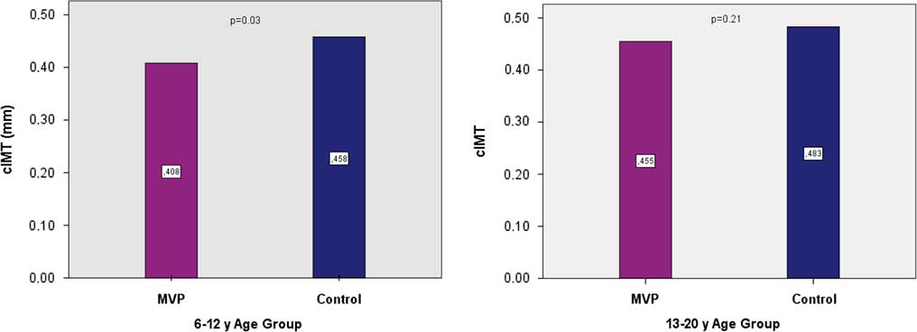
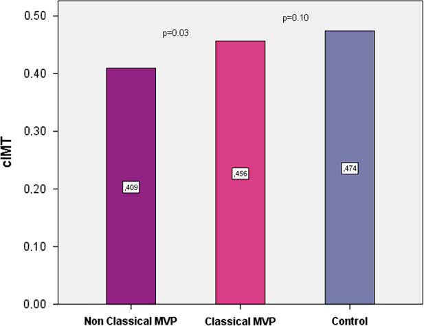
Patients with mitral valve prolapse had thinner carotid intima-media thickness (median=0.43 mm; range: 0.33–0.54) than the control patients (median= 0.46 mm; range: 0.40–0.55) (p=0.020). Carotid intima-media thickness was thinner in younger patients with mitral valve prolapse than in the control group (p=0.03, Fig 2). Carotid intima-media thickness was thicker in the classical mitral valve prolapse group (0.45±0.07 mm) than in the non-classical mitral valve prolapse group (0.40±0.06 mm), but there was no statistically significant difference between the classical mitral valve prolapse group and the control patients (p=0.037, Fig 3).
Mitral valve prolapse (MVP) group had thinner carotid intima-media thickness (cIMT) than control group in group 6–12 years.

Carotid intima-media thickness (cIMT) was thicker in patients with classical mitral valve prolapse (MVP) than non-classical MVP but there was no statistically significant difference between classical MVP and control patients.

In both mitral valve prolapse and control groups, carotid intima-media thickness correlated to stiffness index of sinus valsalva (p=0.001, r=0.54; p=0.007, r=0.47, respectively) and inversely correlated to strain and distensibility of sinus valsalva (p=0.005, r=−0.47; p=0.001, r=−0.49, respectively).
Flow-mediated dilation
There was no statistically significant difference in comparison of flow-mediated dilatation among groups.
Discussion
Mitral valve prolapse was more prevalent in female gender in our study group, as is the case in the literature.Reference Avierinos, Gersh and Melton 24 Although there was no difference between the ages of classical and non-classical mitral valve prolapse groups, classical mitral valve prolapse patients were taller than non-classical mitral valve prolapse patients. Symptoms like syncope, presyncope, orthosthatic hypotension, palpitation, and chest pain are common in mitral valve prolapse patients. Chest pain is the most common symptom in our patients, followed by symptoms like palpitation and syncope. Activation of neuroendocrine and autonomic nervous systems are the main causes for symptoms in patients with mitral valve prolapse.Reference Boudoulas and Wooley 25
Our study demonstrated left ventricular and left atrial enlargement in patients with mitral valve prolapse. Community-based studies in adult patients showed the enlargement of left ventricle in patients with mitral valve prolapse but the effect of mitral regurgitation, age, hypertension, diabetes mellitus, and coronary artery diseases could not be excluded in these studies.Reference Avierinos, Gersh and Melton 24 , Reference Freed, Benjamin and Levy 26 Yiginer et al excluded all these factors and demonstrated increased left ventricle dimensions in adults with classical mitral valve prolapse.Reference Yiginer, Keser and Ozmen 27 To our knowledge this is the first study showing left ventricular and left atrial enlargement in children with mitral valve prolapse. None of our patients had more than mild regurgitation, and even when we excluded the patients with mitral regurgitation, the difference between study and control groups in terms of left atrial and left ventricular dimensions remained. Therefore, we can conclude that left ventricular enlargement is not a consequence of valvular regurgitation. Since there was no patient with systemic connective tissue disease or systemic hypertension in both groups, the enlargement of the cardiac chambers is not related to these factors. Rather it may be due to endogenous factors within the myocardial tissue in patients with mitral valve prolapse. In the literature, there are patients with Marfan syndrome having dilated ventricles or dilated cardiomyopathy in infancy.Reference Meijboom, Timmermans and van Tintelen 28 , Reference Booms, Cisler and Mathews 29 In fibrillin-1-deficient mice models, it was shown that abnormal mechanosignalling in the myocardial tissue results in dilated cardiomyopathy. Mechanotransducer apparatus provides the connection between sarcomer and extracellular matrix and transmit myocardial stretch to intracellular level. Altered fibrillin-1 and extracellular matrix proteins in Marfan syndrome disrupts the mechanosignalling and the left ventricle becomes more vulnerable to mechanical stress.Reference Cook, Carta and Bénard 30 Furthermore, changes in matrix metalloproteinases are not restricted to the mitral valve only; altered matrix metalloproteinase expressions are also found in right and left atrial appendages of Marfan patients undergoing mitral valve surgery.Reference Anné, Willems and Roskams 31
In our study, we found that mitral peak early velocity and mitral early-to-late velocity ratio of patients with mitral valve prolapse were lower than that of the control group. Altered extracellular matrix may cause impairment in relaxation. This finding suggests that the patients with mitral valve prolapse have a more restrictive diastolic physiology compared to healthy individuals. This difference is not thought to be related to increased afterload of the left ventricle as the systolic and diastolic blood pressures were similar in both groups.
Aortic diameters were also enlarged in patients with mitral valve prolapse in our study. Even when we excluded three patients with a z-score of the aortic root above +2, the difference between study and control groups remained statistically significant. Aortic root dilatation is expected in heritable connective tissue disorders such as Marfan, Loeys Dietz, Ehler Danlos syndromes, in which mitral valve prolapse is a common finding.Reference Wheeler, Ikonomidis and Jones 32 One previous study in adults found mitral valve prolapse as an independent predictor of a larger aortic root.Reference Matos-Souza, Fernandes-Santos, Hoehne, Franchini and Nadruz 33 We found a similar association in non-syndromic children with mitral valve prolapse. Higher aortic root z-scores were found in the older age group in our study. It is well known that aortic dimensions are increased in patients with Marfan syndrome and patients with isolated bicuspid aortic valve. Loss of vascular smooth muscle cells due to apoptosis with matrix metalloproteinase activity and cystic medial necrosis have been found to be causes for aortic wall weakness and aortic dilation in these patients.Reference Pisano, Maresi and Balistreri 34 In Marfan syndrome this pathogenesis is likely to be related fibrillin-1 mutation.Reference Hollister, Godfrey and Sakai 35 Abnormal extracellular matrix might cause increased aortic dimensions in isolated patients with mitral valve prolapse. We did not carry out genetic or histologic investigation in our study. However, none of the patients had dysmorphic features and the Ghent score for assessment of Marfan syndrome was within the normal range in all patients. Further histologic and molecular studies are needed in order to exclude the effect of genetic factors controlling the function and structure of the connective tissue in this patient group.
We found that ascending aorta elasticity was increased in the mitral valve prolapse patients compared with the control patients. There is no similar study conducted in the paediatric age group with non-syndromic mitral valve prolapse patients. On the other hand, in young adults with mitral valve prolapse, aortic elasticity was found to be decreased and stiffness increased in a few studies.Reference Yazici, Ataoglu and Makarc 36 , Reference Kardesoglu, Ozmen, Aparci, Cebeci, Uz and Dincturk 37 Our results do not seem to correlate with these studies. However, vascular functions may not be the same in all age groups. It is more sensible to suggest increased elasticity in presence of a weak connective tissue. Long-term increased distensibility and strain in the vascular wall may cause injury and scarring, which may increase stiffness progressively. This may be the cause of discrepancy between our study and previous studies in young adults. Adult mitral valve prolapse patients with benign joint hypermobility syndrome were shown to have increased distensibility and reduced stiffness compared to mitral valve prolapse patients without benign joint hypermobility syndrome.Reference Yazici, Ataoglu and Makarc 36 The pathophysiology of aortic distensibility in non-syndromic mitral valve prolapse seems to be in accordance with benign joint hypermobility syndrome rather than Marfan syndrome. Persistence of the increased aortic elasticity in mitral valve prolapse even after exclusion of patients with dilated aorta over +2 SD may suggest an unidentified connective tissue disorder in this patient group.
Distensibility decreased in the older age group both in the mitral valve prolapse and control groups in our study. Decreasing aortic distensibility and increasing stiffness with age has already been known in normal population.Reference Fritze, Romero and Scleicher 15 Ageing causes both collagen accumulation and increased elastin degradation in the normal human aorta. In Marfan syndrome, fibrillin-1 deficiency causes augmented elastinolysis and loss of distensibility in the aorta earlier even in childhood.Reference Akazawa, Motoki and Tada 38 In adults, aortic stiffness was found to be an independent predictor of progressive dilatation of the aorta in patients with Marfan syndrome.Reference Nollen, Groenink, Tijssen, Van Der Wall and Mulder 39 Studies investigating collagen composition of myxomatous mitral valve found increased type III collagen and increased amount of proteoglycans.Reference Cole, Chan, Hickey and Wilcken 40 Further investigation is needed in order to clarify whether the aortic wall and mitral valve leaflets of the patients with mitral valve prolapse have similar collagen components which differ from the normal population. The effect of abnormal collagen composition and non-intense matrix metalloproteinase activity in the aortic wall may play role in elasticity of mitral valve prolapse patients and cause reduced stiffness and increased distensibility in early age. Ageing is an important factor affecting connective tissue functions. Mitral valve prolapse is not found in infancy or very young children; it becomes detectable later in childhood. Linear growth during adolescence causes progression in disease severity. Long-term follow-up of aortic elasticity parameters in this group may clarify pathophysiology of the disease.
Classical mitral valve prolapse had increased stiffness at sinus valsalva level in our study. There is only one adult study comparing elasticity parameters of aorta between classical and non-classical mitral valve prolapse; it found reduced aortic stiffness in the ascending aorta in classical mitral valve prolapse patients than in non-classical mitral valve prolapse patients.Reference Unlu, Demirkol and Aparci 41
Autonomic functions may affect aortic distensibility and stiffness.Reference Nemes, Takács and Gavallér 42 Excessive vagal tone and alfa-adrenergic hyperactivity was found in mitral valve prolapse patients. Symptoms like orthosthatic hypotension, palpitation, fatigue, and exercise intolerance in patients with mitral valve prolapse syndrome were referred to increased vagal tone and abnormal adrenergic activity.Reference Gaffney, Karlsson and Campbell 43 Decrease in heart rate variability was demonstrated in patients with mitral valve prolapse.Reference Strano, De Castro and Ferruci 44 Decreased aortic stiffness compared with control patients may be related to high parasympathetic autonomic tone. Dominant sympathetic autonomic tone during prepubertal age may also affect aortic wall functions.
There are no studies investigating endothelial function in patients with mitral valve prolapse. Endothelial function may be studied using carotid intima-media thickness and flow-mediated dilatation. Increased carotid intima-media thickness and decreased flow-mediated dilatation indicates high risk for atherosclerotic coronary artery disease. Increased carotid intima-media thickness was found in various disease states such as obesity, systemic hypertension, diabetes, Kawasaki disease, and late after coarctation repair, which are known risk factors for atherosclerotic heart disease.Reference Dietz, Tacke and Gort 45 – Reference Vriend, de Groot, de Wall, Zijta, Kastelein and Mulder 49 We found thinner carotid intima-media thickness in patients with mitral valve prolapse. This difference was more prominent in non-classical mitral valve prolapse. We may consider that mitral valve prolapse is protective for atherosclerosis. However, this suggestion needs more detailed analysis focussing especially on this field. Previous studies found that carotid intima-media thickness is correlated to arterial stiffnessReference Gullu, Erdogan and Calıskan 50 , which also supports our findings. Our study showed this correlation both in the mitral valve prolapse and control groups.
Conclusion
We found that patients with mitral valve prolapse had larger left cardiac chambers and aortic root and more distensible ascending aorta in childhood, which may lead to increased risk for dilatation, dissection, or rupture of the aorta. As thinner carotid intima-media thickness was detected in the patients with mitral valve prolapse, we suggested that there is no increased risk for atherosclerosis in these patients. New genetic and histopathologic studies and similar studies with long-term follow-up may enlighten us about the pathogenesis and prognosis of mitral valve prolapse in childhood.
Acknowledgement
This study was presented in the Annual Congress of European Association of Pediatric Cardiology (AEPC) 2017, Lyon and awarded with the second prize in oral presentations of Imaging abstracts.
Financial Support
This research received no specific grant from any funding agency, commercial, or not-for-profit sectors.
Conflicts of Interest
None.
Ethical Standards
The authors assert that all procedures contributing to this work comply with the ethical standards of the relevant national guidelines on human experimentation at Marmara University and with the Helsinki Declaration of 1975, as revised in 2008, and has been approved by the institutional committees Marmara University.