Pulmonary sequestration is a congenital bronchopulmonary malformation that receives a systemic arterial blood supply. Pulmonary atresia with intact ventricular septum is frequently associated with several coronary artery abnormalities including ventriculo-coronary arterial communication and a right ventricle-dependent coronary circulation. Patients with pulmonary atresia with intact ventricular septum need to undergo Blalock–Taussig shunt operation to maintain pulmonary artery blood flow and coronary arterial circulation. We present a rare case of pulmonary atresia with intact ventricular septum combined with pulmonary sequestration, in which the patient experienced repetitive myocardial ischaemic attacks after modified Blalock–Taussig shunt operation. Coil occlusion of the aberrant arteries to the pulmonary sequestration proved to be successful in controlling attacks.
Case report
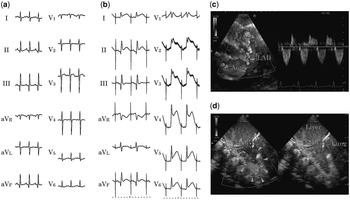
The patient had not been identified as having any congenital cardiac abnormalities on foetal ultrasonography, and was born at 38 weeks of gestation, weighing 2826 g, with no sign of neonatal asphyxia. Severe cyanosis was identified and he was referred to our hospital for further examination and treatment 10 hours after birth. Pulmonary atresia with intact ventricular septum, a hypoplastic right ventricle, ventriculo-coronary arterial communication, and patent ductus arteriosus were diagnosed by ultrasonography and by contrast-enhanced multi-detector row computed tomography; however, pulmonary sequestration was not detected in this examination. Prostaglandin E1 was administered to maintain the ductus arteriosus. A modified Blalock–Taussig shunt operation was performed at 13 days of age. Although the operation was successful, he occasionally experienced attacks of bradycardia, low blood pressure, and low oxygen saturation from the 9th postoperative day. Cardiopulmonary resuscitation was necessary on several occasions. Echocardiography showed left ventricular asynergy, and electrocardiography revealed ST-segment elevation in V2-6 – the anterolateral chest lead (Fig 1a and b).
Electrocardiogram showing myocardial ischaemia, and ultrasonography showing the pulmonary sequestration. ( a ) Echocardiography recording before modified Blalock–Taussig shunt operation. ( b ) Echocardiography showing ST-segment elevation in V2-6 at the time of a myocardial ischaemic attack. Atrial pacing was performed to maintain the heart rate for treatment. ( c ) Echocardiography demonstrates ventriculo-coronary arterial communication. Doppler flow imaging was recorded with the sample volume located at the left anterior descending artery. The flow pattern reveals bidirectional flow, indicating the presence of ventriculo-coronary arterial communication. ( d ) Ultrasonography shows a wedge-shaped echogenic feature. Blood supply is marked (arrowheads). The mass was diagnosed as pulmonary sequestration. Ao=aorta; LAD=left anterior descending coronary artery.

He underwent a bidirectional Glenn operation and removal of the Blalock–Taussig shunt at 59 days of age; however, his circulatory dynamics continued to be unstable. Pulmonary sequestration of the right inferior lobe was diagnosed 42 days after the bidirectional Glenn operation. Ultrasonography showed a wedge-shaped, echogenic mass supplied by several arteries (Fig 1c). Contrast-enhanced multi-detector row computed tomography demonstrated multiple aberrant arteries to the sequestrated lung tissue. The diagnosis was finally confirmed by selective angiography.
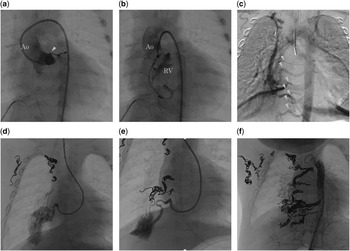
Furthermore, the angiography demonstrated a ventriculo-coronary arterial communication and stenosis of the left main trunk of the left coronary artery with hypoplastic left coronary arteries (Fig 2a and b). These findings indicated that myocardial ischaemia had resulted from coronary arterial steal induced by the modified Blalock–Taussig shunt and the aberrant systemic arteries feeding the pulmonary sequestration.
Angiography and coil occlusion of pulmonary sequestration. ( a ) Left coronary angiography shows stenosis (arrowhead) and hypoplastic appearance. ( b ) Right coronary angiography reveals the presence of the ventriculo-coronary arterial communication. ( c ) Aortography demonstrates aberrant arteries to the pulmonary sequestration. ( d and e ) Selective angiography reveals the arterial supply to the sequestered lung. Collateral arteries other than vessels to the pulmonary sequestration were already occluded just before this angiography. ( f ) Aortography after treatment using a total of 53 coils. Ao=aorta; RV=right ventricle.

In addition, he underwent coil occlusion of the aberrant arteries to the pulmonary sequestration. Aortography showed multiple feeding arteries to the right lower lung provided by the descending aorta. Selective-feeding angiography detected the sequestrated lung tissue and return of flow to the pulmonary vein. Coil occlusion was performed twice for nine feeding arteries (Fig 2c and d). After coil occlusion, his myocardial ischaemic attacks improved. He was successfully extubated 6 days after coil occlusion.
Discussion
Pulmonary sequestration is a rare congenital pulmonary abnormality that accounts for 0.15–6.4% of all pulmonary abnormalities. Sequestered lung tissue has a systemic arterial supply and no bronchial communication, and thus has no ventilatory function. This disease was first reported by Pryce in 1946 and is currently classified into two types.Reference Chien, Huang, Lin, Lee, Hsieh and Weng 1 Extralobar sequestration is separated from the normal lung by visceral pleura, whereas intralobar sequestration shares a common visceral pleura with the normal lung. More than 90% of patients with pulmonary sequestration receive an aberrant systemic arterial supply, not from the pulmonary artery.Reference Marine, Valdes, Mertens, Bergoeing and Kramer 2 The patient we encountered showed ten aberrant arteries from the thoracic aorta. In most cases, patients with pulmonary sequestration are asymptomatic and are diagnosed after experiencing recurrent infections of the sequestered lung. This was a rare case in which the feeding artery caused problems in terms of systemic and coronary circulatory failure. A few reports have described pulmonary sequestration complicated by CHD.
We present a case of pulmonary atresia with intact ventricular septum associated with ventriculo-coronary arterial connections.Reference Ahmed, Snodgrass and Kaine 3 Furthermore, the left coronary artery was stenotic and hypoplastic.
The presence of a modified Blalock–Taussig shunt flow together with pulmonary sequestration presumably induced the coronary arterial ischaemia. Surgery is the conventional method of treatment for pulmonary sequestration, but the merits of this approach are debatable in asymptomatic patients. Endovascular treatment of pulmonary sequestration, with selective embolisation of the inflow arteries, offers an attractive, minimally invasive therapy compared with conventional surgery.Reference Greil, Schoebinger and Kuettner 4 He therefore underwent a bidirectional Glenn operation, which was effective in preventing coronary arterial ischaemia. Furthermore, he underwent coil occlusion of the feeding arteries to the sequestered lung. These treatments were quite effective in improving symptoms.
Although pulmonary sequestration complicated by CHD is rare, evaluations must be conducted for other cardiovascular diseases, including pulmonary sequestration, which can lead to decreases in coronary blood flow.