Clinical case
A male patient was followed by hepatology and haematology services from age 16 due to abdominal discomfort and splenomegaly. Blood tests showed thrombocytopenia (66–143 × 109/L), anaemia (0.258–0.376 L/L), elevated lactate dehydrogenase (507–605 IU/L), and intermittently increased bilirubin (20–66 umol/L). Haemolytic anaemia and myeloproliferative disorders were excluded (negative coombs and JAK mutation test), and no evidence of myeloma multiple (negative Bence jones proteins). Infectious diseases (negative hepatitis screening, negative human immunodeficiency virus serology, negative TB screening) and autoimmune screening (autoimmune hepatitis, primary biliary cirrhosis, lupus) were also negative. Liver MRI showed signs of chronic liver disease with remodelling and heterogeneous signal return from the parenchyma and significant splenomegaly, but no aetiology was found despite these extensive investigations. He remained under outpatient surveillance.
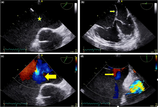
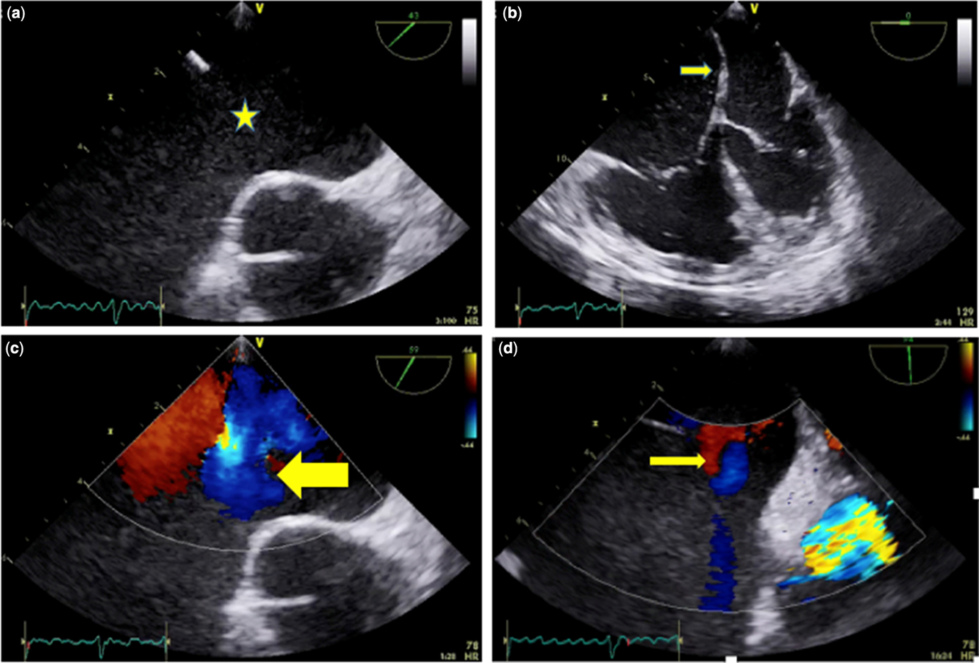
At age 21, the patient was admitted with a urinary tract infection and atrial fibrillation. He described mild breathlessness on effort (NYHA FC II) and had resting oxygen saturations of 95%. Mild obesity was noted (body mass index of 32), as well as hepatosplenomegaly, mildly elevated jugular venous pressure with normal atrial waves, no Kussmaul’s sign, a left parasternal heave, and wide fixed splitting of the second heart sound with an ejection systolic murmur at the upper left sternal edge. A transthoracic echocardiogram showed a large secundum atrial septal defect with a left-to-right shunt and severe dilation of the right heart chambers. A cardiac MRI showed increased right ventricle volumes (end-diastolic 205 ml/m2, end-systolic 95 ml/m2) with normal right ventricle systolic function (RVEF 56%) without late gadolinium enhancement and normal pericardium. A transoesophageal echocardiogram confirmed an isolated secundum atrial septal defect not suitable for percutaneous closure because of deficient retroaortic and superior caval vein rims (Fig 1). Cardiac catheterisation showed a significant pulmonary to systemic flow ratio (Qp:Qs) of 2:1, mild pulmonary hypertension (pulmonary pressure 40/20/30 mmHg, pulmonary wedge pressure 20 mmHg), secondary to a left-to-right shunt and elevated left end-diastolic pressure (left ventricle 116/19 mmHg, right ventricle 40/19 mmHg), with normal pulmonary vascular resistance 2.2 WU. No signs of pericardial constriction, restriction, or effusion were found during the preoperative workout. A full blood count showed a persistent thrombocytopenia (114 × 109/L), mild anaemia (haematocrit 406 L/L; haemoglobin 129 g/L; mean corpuscular volume 105 fl; haptoglobin <0.05 g/L), and mildly abnormal liver function (alanine transferase 77 IU/L; bilirubin 24 umol/L; albumin 41 g/L; normal APPT; slightly prolonged PT, 12.2 seconds).
Transoesophageal echocardiogram at first presentation showing a single secundum atrial septal defect (23 mm, stair) with absence of aortic rim. (a) Four-chamber view showing dilated right atrium (RA) and right ventricle (RV). Leftward bowing of the interatrium septum, suggestive of increased RA pressures. (b) Colour flow mapping showing significant left-to-right shunt (arrow) (c) and small right-to-left shunt (arrow) (d).
In the absence of a significant pulmonary vascular disease, the patient was advised surgical closure of the atrial septal defect. He underwent surgical repair with a porcine pericardial patch. Four months later, he was seen in clinic complaining of shortness of breath on exertion and had low saturations (sO2 90%). A transthoracic echocardiogram and transoesophageal echocardiogram showed a significant residual atrial septal defect with bidirectional shunting. Repeat cardiac catheterisation showed raised right atrial pressure (30/25/25 mmHg), pulmonary pressure (60/31/41 mmHg) with elevation in both left and right ventricular end-diastolic pressures (right ventricle 60/28 mmHg, left ventricle 131/24 mmHg, wedge 25/28/24 mmHg). The calculated Qp:Qs was 2:1, and pulmonary vascular resistance was still low at 2.2 WU, and a further surgery was recommended.
The patient underwent a further cardiac surgery for atrial secundum defect closure with a bovine pericardial patch. Over the course of a year following the second surgical repair, he became progressively more limited and breathless (NYHA FC III, IV) and additionally developed significant lower limb oedema and abdominal distension, necessitating hospital admission for intravenous diuretic therapy. Due to persistent anaemia, upper endoscopy was performed, which showed oesophageal varices, for which he underwent banding. He had multiple percutaneous attempts at arrhythmia ablation (flutter ablation) with recurrence and adoption of a rate control strategy with beta-blocker.
A transthoracic echocardiogram showed an intact atrial septum with significant bi-atrial enlargement and a prominent “septal bounce” (early diastolic movement of the interventricular septum to the left side on inspiration and to the right side during expiration). These findings were not present on his prior echocardiogram. The tissue Doppler in the basal interventricular septum showed an increased E′ (0.17 m/second), with a normal E/E′ ratio. Cardiac MRI reported thickening of the pericardium without signs of fibrosis or inflammation, and again a septal bounce (Video 1, Supplementary material), with persistent right heart dilatation, signs of portal hypertension with progression of his liver disease to a cirrhotic pattern. Cardiac CT showed thickening of the pericardium as well (5 mm).
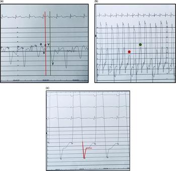
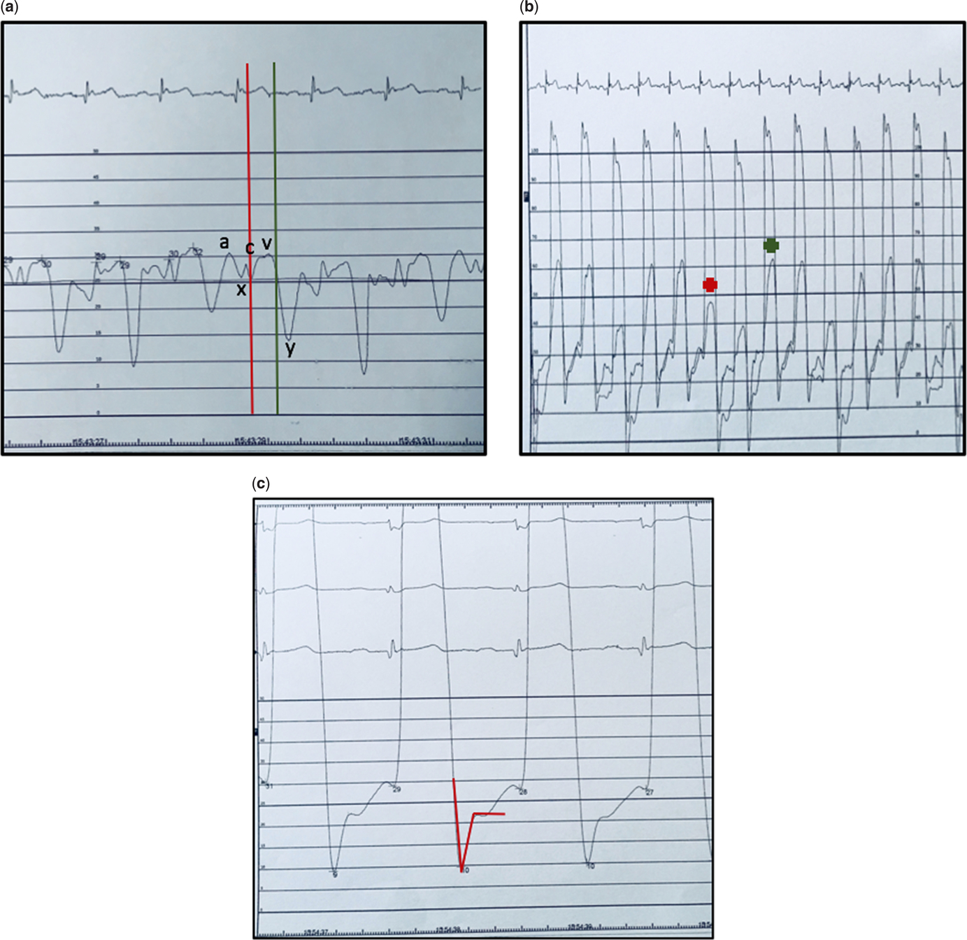
Repeat cardiac catheterisation showed findings consistent with the haemodynamic features of pericardial constriction, including “square root sign” and “discordant respiratory variation between LV/RV” (Fig 2). A diagnosis of constrictive pericarditis was made, and the patient underwent pericardiectomy at 3 years after the initial presentation to cardiac services. The surgery was uneventful, and at 1 year post-pericardiectomy, he showed a significant improvement of his symptoms with no evidence of systemic venous congestion on a small dose of oral diuretic. He continues to suffer from chronic anaemia secondary to chronic liver disease with regular intravenous iron therapy.
Right heart catheterisation: Equalisation of LV and RV diastolic pressure variations of the peak systolic pressure in both ventricles during respiration especially in the right ventricle (red asterisk: inspiration; green asterisk: expiration). (a) Right atrium tracing with red line indicating the end of QRS, i.e. end-systole, and green line indicating the end of the T-wave, i.e., end-diastole, with raised right atrium pressure (mean 25 mmHg) and prominent Y descent (indicative of prominent early diastolic filling). (b) Respiratory variation of peak systolic pressure mainly in the right ventricle. (c) Left ventricle tracings showing a “square root sign” (red line) as an expression of a sudden increase in end-diastolic pressure at early diastole secondary to a pericardial constriction/restraint.
Discussion
We presented a case of constrictive pericarditis and atrial septal defect that posed significant diagnostic challenges. To our knowledge, this is the first case of likely combined idiopathic pericardial constriction and atrial septal defect. Constrictive pericarditis is notoriously a difficult diagnosis to make, and in this case, the pathognomonic features were blunted by the co-existing atrial secundum defect.Reference Gatzoulis, Webb and Daubeney1–Reference Garcia3
At initial diagnosis and following the first attempt at surgical repair, the patient showed clinical features that were unusual for a “simple atrial septal defect”, including hepatosplenomegaly, atrial fibrillation, and resting desaturation. Co-existing pulmonary vascular disease was considered at this stage, and when excluded, a decision to proceed to surgical closure was made. Atrial secundum defects associated with high pulmonary vascular resistance occur in 8% of patients with atrial secundum defects.Reference Garcia3–Reference Engelfriet, Duffels and Møller5 The symptomatic deterioration with systemic venous congestion was precipitated by a complete closure of the atrial septal defect, where the transmission of high diastolic intracardiac pressures could not be equalised at atrial level, causing worsening of systemic venous congestion. Despite causing clinical deterioration, the presence of an intact septum facilitated the diagnosis by unmasking the pathognomonic findings such as septal bounce, and made the haemodynamic features of constriction more evident on both cross-sectional imaging and diagnostic catheterisation.Reference Welch and Oh6
We postulate that this patient had both conditions at first presentation, because of the presence of hepatosplenomegaly at diagnosis with a rapid progression to portal hypertension and varices, suggesting chronicity of constrictive pericarditis, as well as some atypical cardiac features at initial cardiac presentation with early atrial fibrillation and mild desaturation.
The first physiological clue, in the presence of an intact atrial septum following the second atrial septal defect closure, was the septal bounce seen on echocardiography and CMR. This prompted an in-depth analysis of the state of cardiac filling on doppler and CMR, as well as an evaluation of the pericardial thickness on CMR and CT.
The third right and left cardiac catheterisation was the conclusive test in this case, showing hallmark features in atrial and ventricular waveforms.
Conclusion
Worsening of a clinical status after a secundum atrial septal defect repair in the absence of pulmonary hypertension demands further investigation of an underlying pathology. In our case, constrictive pericarditis was the co-existing lesion and the main culprit of clinical deterioration.
Learning points
1. Simple congenital heart diseases can co-exist with another lesion. Their co-existence can worsen the clinical status and survival of the patient.
2. Therefore, a thorough investigation using transthoracic and transoesophageal echo or cardiac MRI is highly warranted while seeking for the underlying pathology.
3. Recognising the co-existence of cardiac pathologies will confer a better understanding and a prompt treatment of future complications.
Supplementary material
To view supplementary material for this article, please visit https://doi.org/10.1017/S1047951119001677
Acknowledgements
None.
Financial Support
This research received no specific grant from any funding agency, or from commercial or not-for-profit sectors.
Conflicts of Interest
None.