INTRODUCTION
Vinpocetine is extracted from the periwinkle plant, Vinca minor, originally discovered and marketed in 1978 under the trade name Cavinton. Since then, Vinpocetine has been widely used in the treatment of cerebrovascular disorders and the improvement of cognitive impairment including stroke, Alzheimer's disease and memory disturbances (Bagoly et al., Reference Bagoly, Fehér and Szapáry2007). Its beneficial clinical effect is based on a wide range of action including anti-oxidant, anti-inflammatory, vasodilating and neuroprotective effects. However, the precise mechanism underlying these effects is not yet well understood.
Previous studies have demonstrated that Vinpocetine is a cerebral vasodilator that improves brain-blood flow (Tamaki and Matsumoto, Reference Tamaki and Matsumoto1985), and acts as a cerebral metabolic enhancer by enhancing oxygen and glucose uptake and increasing neuronal adenosine triphosphate (ATP) production (Szobor and Klein, Reference Szobor and Klein1976). Vinpocetine appears to affect several cellular targets, such as Ca2+/calmodulin-stimulated cyclical nucleotide phosphodiesterase-1 and voltage-dependent Na+ channels and Ca2+ channels (Bönöczk et al., Reference Bönöczk, Gulyás, Adam-Vizi, Nemes, Kárpáti and Kiss2000). In recent studies, Vinpocetine inhibits inflammatory responses in endothelia/macrophages and in a mouse model of lung inflammation (Jeon et al., Reference Jeon, Xu, Aizawa, Lim, Jono and Kwon2010; Medina, Reference Medina2010).
The inflammation of the central nervous system (CNS) is now recognized to be a major feature in several neurological disorders (Amor et al., Reference Amor, Puentes, Baker and van der Valk2010; Lue et al., Reference Lue, Kuo, Beach and Walker2010; Galimberti and Scarpini, Reference Galimberti and Scarpini2011; Rossi et al., Reference Rossi, Angiari, Zenaro, Budui and Constantin2011). Microglia are resident immune cells of the CNS, while microglial activation plays an important role in chronic inflammatory and degenerative diseases of the CNS (Perry et al., Reference Perry, Nicoll and Holmes2010). In the brain, translocator protein (TSPO), previously known as the peripheral-type benzodiazepine receptor (PBR) (Papadopoulos et al., Reference Papadopoulos, Baraldi, Guilarte, Knudsen, Lacapère and Lindemann2006), is expressed in activated microglia (Myers et al., Reference Myers, Manjil, Cullen, Price, Frackowiak and Cremer1991; Stephenson et al., Reference Stephenson, Schober, Smalstig, Mincy, Gehlert and Clemens1995; Banati, Reference Banati2002; Chen and Guilarte, Reference Chen and Guilarte2008; Papadopoulos and Lecanu, Reference Papadopoulos and Lecanu2009) and used as a biomarker of microglial activation and as a therapeutic target for neurological and psychiatric disorders (Rupprecht et al., Reference Rupprecht, Papadopoulos, Rammes, Baghai, Fan and Akula2010).
Early autoradiographic studies on whole hemisphere human brain slices with [11C] Vinpocetine have defined that the radiolabelled compound shows specific binding to TSPO with moderate affinity (Hall et al., Reference Hall, Varnäs, Sandell, Halldin, Farde and Vas2002; Gulyas et al., Reference Gulyas, Halldin, Vas, Banati, Shchukin and Finnema2005). Further investigations with positron emission tomography (PET) in non-human primates and human volunteers have supported the hypothesis that radiolabelled Vinpocetine may serve as a TSPO biomarker (Gulyas et al., Reference Gulyas, Toth, Vas, Shchukin, Kostulas and Hillert2012). Although Vinpocetine binds with a moderate affinity to TSPO (IC50: 200 nM), it can be a usable PET radioligand for in vivo exploration of the TSPO system in the human brain (Gulyas et al., 2005).
As Vinpocetine has a binding affinity to TSPO induced in activated microglia, thereby affecting the function of activated microglia, the microenvironment of inflammation and the outcome of neurons. In the present study, we used the Vinpocetine, as TSPO-specific ligand, to explore the role of Vinpocetine in BV-2 microglia and hypoxic mice. Our results demonstrate that Vinpocetine exhibits anti-inflammatory and neuroprotective effects in vitro and in vivo experiments.
MATERIALS AND METHODS
Vinpocetine
Vinpocetine injection (brand name: Cavinton, 10 mg per 2 ml) was obtained from Gedeon Richter Ltd, Hungary, which is a semisynthetic derivative alkaloid of vincamine (sometimes described as ‘a synthetic ethyl ester of apovincamine’), an extract from the periwinkle plant. Most clinical application used 10 mg Vinpocetine 2–3 times per day i.v., or 30, 45 and 60 mg day−1 orally.
Microglial culture
The BV-2 immortalized microglial cell line (Bocchini et al., Reference Bocchini, Mazzolla, Barluzzi, Blasi, Sick and Kettenmann1992) was purchased from ShenKe Biological Technology Co., LTD, Shanghai, China, and cultured in Dulbecco's modified Eagle's medium (DMEM, Gibco), supplemented with 10% fetal bovine serum (Gibco), 100 U ml−1 penicillin and 100 µg ml−1 streptomycin (Gibco) at 37°C in a humidified cell incubator under 95%/5% (v/v) mixture of air and CO2. Cells were plated on a 96-well plate (Corning, USA) at a density of 1.0 × 10⁶ ml−1, 200 µl per well for proliferation assay, on a 24-well plate at 1.0 × 10⁶ ml−1, 500 µl per well for cytokine assay and at 0.2 × 10⁶ ml−1, 500 µl per well for immunocytochemistry staining.
Primary neuron culture
Embryonic day 18 mouse cortex (Shanghai SLAC Laboratory Animal Co. Ltd) were dissected under a microscope, and then dissociated in Neurobasal-A-medium (Gibco) supplemented with 2% B27 (Gibco), 100 U ml−1 penicillin and 100 µg ml−1 streptomycin (Gibco), by trituration. The cells were then plated on cover slips coated with poly-d-lysine (0.1 mg ml−1, Sigma) at a density of 1 × 10⁶ cells cm−2. After 7 days, neurons were incubated with different conditioned medium.
Treatment of LPS
In preliminary experiments, different concentrations (0.1, 1 and 10 µg ml−1) and exposure times (6, 12, 24 and 48 h) of LPS (Endotoxin is ≤0.1 ng ml−1, Sigma, USA) were carried out to determine the optimal concentration and exposure time of LPS in BV-2 microglia (supplementary fig. 1 online). In the present experiment, BV-2 microglia were incubated in a medium containing 1 µg ml−1 of LPS (Sigma, St. Louis, MO) for 12 h. In order to study the effect of Vinpocetine on cell proliferation and death, BV-2 microglia were exposed to 10–80 µM of Vinpocetine in the presence or absence of LPS (1 µg ml−1). For the production of nitric oxide (NO) and cytokines, Vinpocetine (20 and 50 µM per ml) was added at 12 h after LPS treatment or immediately after LPS treatment according to previous study (Jeon et al., Reference Jeon, Xu, Aizawa, Lim, Jono and Kwon2010). For the measurement of nuclear factor-κB (NF-κB) and activator protein-1 (AP-1) as well as the preparation of Vinpocetine-conditioned medium, Vinpocetine was used at a dose of 50 µM ml−1. Vinpocetine treatment was maintained for 24 h.
Treatment of oxygen–glucose deprivation (OGD)
In BV-2 microglia, normal media were replaced with no glucose media and culture plates were placed in a Forma Anaerobic System at 37°C incubator (Thermo, USA). The chamber was flushed with 85% N2/10% H2/5% CO2. After OGD, no glucose media were replaced with normal DMEM and cultures were returned to a 37°C normoxic incubator (95% air and 5% CO2) for reoxygenation. In preliminary experiments, hypoxia (1.5, 3 and 6 h) and reoxygenation (6, 12, 24 and 48 h) were carried out to determine the optimal hypoxia/reoxygenation in BV-2 microglia. Subsequent experiments focused on 6 h of OGD combined with 24 h of reoxygenation, unless stated otherwise. To explore the effect of Vinpocetine in BV-2 microglia treated with OGD, Vinpocetine (20 and 50 µM) was added at 6 h after OGD treatment or immediately after OGD treatment according to previous study (Jeon et al., Reference Jeon, Xu, Aizawa, Lim, Jono and Kwon2010). For the measurement of NF-κB and AP-1 as well as the preparation of Vinpocetine-conditioned medium, Vinpocetine was used at dose of 50 µM ml−1. Vinpocetine treatment was maintained for 24 h.
Preparation of Vinpocetine-conditioned medium in BV-2 microglia
First, BV-2 microglia were treated with LPS (1 µg ml−1) for 12 h (phosphate-buffered saline (PBS) as control), or treated with OGD for 6 h (Normoxia as control). Next, Vinpocetine (50 µM) was added to LPS- or OGD-treated BV-2 microglia for 24 h (PBS- or Normoxia-treated BV-2 microglia as control). Subsequently, BV-2 microglia were washed three times with PBS to remove Vinpocetine and LPS, and then cultured for a further 24 h. The supernatants of Vinpocetine-conditioned medium were collected, and then added to primary cultured neurons.
Mouse model of hypoxia/reoxygenation and Vinpocetine treatment
Male C57/B6 mice approximately 10–12 w, weighing 25–30 g (Shanghai SLAC Laboratory Animal Co. Ltd), were randomly divided into three groups (six mice per group): normoxia = mice was kept under normoxic conditions (21% oxygen), hypoxia control = mice were exposed to hypoxia (8% oxygen) and injected with normal saline, and Vinpocetine treatment = mice were exposed to hypoxia, and injected with Vinpocetine.
Hypoxia was achieved through the use of a custom-designed plexiglass chamber (35 cm × 25 cm × 30 cm), capable of holding 12 mice. Air flow through the chamber was provided by compressed mixed gases (8% O2 and 92% N2) and holes drilled into the chamber. After 10 min, the oxygen meter displayed the oxygen concentration at 8%, and hypoxia then performed for 6 h. Animals in the normoxic group were housed in similar cages as hypoxic animals with oxygen levels maintained at 21%. Subsequently, hypoxic mice were injected i.p. with 5 mg kg−1 of Vinpocetine everyday for 7 consecutive days (start at 6 h after hypoxia to 7 days after hypoxia). The control group was injected with saline in the same manner.
After 7 days, mice were anesthetized with 10% chloral hydrate and perfused via the left ventricle with 50 ml of 0.9% saline. The whole brains were obtained and immediately stored in −80°C for western blot and cytokine measurement.
MTT assay
Cell proliferation of BV-2 microglia was detected by MTT assay. In brief, 100 µl of MTT solution (0.5 mg ml−1 Duchefa) was added to cultured cells and incubated for an additional 4 h at 37°C, until the medium turned purple. Absorbance at 570 nm was measured by a microplate reader after addition of 100 µl dimethyl sulfoxide. Each experiment was performed in triplicate and repeated three times with separate cell preparations. Results were expressed as a percentage of the control value at 570 nm.
LDH assay
The death of BV-2 microglia and primary neurons was measured by lactate dehydrogenase (LDH) release in the culture medium. Levels of LDH release in supernatants of cultured cells were measured using the cytotoxicity detection kit (Promega) according to the manufacturer's protocol. Maximum LDH release (OD 570 nm) was determined following the treatment of cells with lysis buffer, and considered as control value (100% LDH release). Data were expressed as a percentage of the control value.
Nitrite assay
NO of BV-2 microglia was assayed by Griess reaction, measuring the end product nitrite. Supernatants (100 µl) of cultured cells were mixed with 100 µl of Griess reagent for 10 min at room temperature. Absorbance was measured at 510 nm in an automated plate reader. Concentrations of nitrite were determined by reference to a standard curve of sodium nitrite (Sigma). Determinations were performed in duplicate and repeated in three independent experiments. The results were expressed as μM.
Cytokine enzyme-linked immunosorbent assay (ELISA) assay
Supernatants from BV-2 microglia and primary neurons were harvested and measured for cytokine concentrations of IL-1β, IL-6 and TNF-α (Pepro Tech Inc., USA) by sandwich ELISA kits following the manufacturer's instructions. The concentrations of cytokines were quantified by reference to the standard curve. Determinations were performed in duplicate and repeated in three independent experiments. The results were expressed as pg ml−1.
Immunocytochemistry staining
BV-2 microglia were plated onto poly-lysine-coated cover slips in a 24-well plate. For immunofluorescence, the cells subjected to different treatments were fixed with 4% paraformaldehyde for 30 min, and blocked with 1% bovine serum albumin (BSA) for 30 min. The cells were then incubated overnight with rabbit anti-TSPO (1:1000; R&D Systems, Inc.), rabbit anti-NF-κB (1:200; Chemicon, Temecula, CA) or rabbit anti-AP-1 (1:300; Chemicon). Subsequently, the cells were incubated with Alexa Fluor 555-conjugated anti-rabbit secondary antibodies (1:1000, Invitrogen), and counter-stained with Hochest. Following these procedures, cells were air dried, cleared in xylene and coverslipped. All cells were independently and blindly examined and quantified by two investigators under fluorescent microscopy (Olympus BX60) equipped for fluorescence and an automated quantitative system.
Western blot analysis
Cells were homogenized with a glass homogenizer using ice-cold lysis buffer supplemented with protease inhibitors. Lysates were centrifuged at 10,000 g for 15 min at 4°C, and supernatants were collected. In all cases, protein concentrations were determined by a Bradford protein assay with BSA used as standard (Sigma, CA). Cell extracts (30 µg) were loaded on SDS–polyacrylamide gels and transferred onto nitrocellulose membrane (Millipore). The membranes were then incubated at 4°C overnight with antibodies against TSPO (1:500), NF-κB (1:200; Chemicon) and AP-1 (1:300; Chemicon). The next day, the membranes were incubated with the horseradish peroxidase-conjugated secondary antibody (Cell Signaling Technology) for 1 h. The immunoblots were developed with an enhanced chemiluminescence (ECL) system (GE Healthcare Life Sciences) and measured with Quantity Software (Bio-Rad, Hercules, CA). To compare protein loading, an antibody directed against β-actin (Sigma) was used.
Flow cytometry analysis
For the mitochondrial impairment of oxidative stress, BV-2 microglia were exposed to 1 mM H2O2 solution in culture medium with or without Vinpocetine for 12 h. Afterwards, the cells were washed twice with Hank's Buffered Salt Solution (HBSS) and used to monitor the mitochondrial parameters as described below.
Mitochondria were assessed using the flurometric probe, tetramethyl rhodamine methyl ester (TMRM, Molecular Probes Inc., Leiden, The Netherlands), a non-toxic, monovalent cation, that is a membrane potential-dependent probe of a functioning mitochondrion. For each experiment a mitochondrial aliquot of 50 µl (1 mM TMRM stock, 1:1000 diluted to 1 µM in HBSS) was added in 1 ml of cell suspension for 10 min at 37°C. To demonstrate normal mitochondrial depolarization, we used the mitochondrial uncoupler, carbonylcyanide p-(trifluoromethoxy) phenylhydrazone (FCCP, 10 µM) as a positive control. Cytofluorimetric analysis was performed using a Beckman Coulter Epics XL flow cytometer (Beckman Coulter) equipped with a 488 nm argon ion laser. The TMRM signal was analyzed in the FL2 channel. Data were acquired on a logarithmic scale. Mitochondrial depolarization is represented as percentage TMRM shifts in FL2 channel. At least 10,000 gated events were acquired for each sample. Data were analyzed by means of WinMDI 2.8 software (The Scripps Research Institute, La Jolla, CA).
Statistical analyses
Multiple comparisons among more experimental groups were performed by ANOVA followed by post hoc tests. Comparisons between two experimental groups were made by Student's t-test. P value less than 0.05 was considered significant.
RESULTS
TSPO is unregulated in BV-2 microglia treated with LPS and OGD
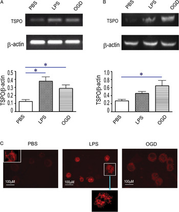
To determine the pharmacological characteristics of Vinpocetine targeting TSPO, we required BV-2 microglia that expresses relatively high levels of TSPO. Using RT-PCR, western blot and immunocytochemistry, we found that TSPO was unregulated in BV-2 microglia treated with LPS or OGD, as compared with untreated cells (Fig. 1A,B). In Fig. 1C, the intensity of fluorescence TSPO staining was enhanced in BV-2 microglia treated with LPS or OGD, as compared to untreated cells. These results show that LPS-stimulated or OGD-treated BV-2 microglia express relatively high levels of TSPO and therefore constitute a reliable cell model to determine the pharmacological effect of Vinpocetine targeting TSPO with specific affinity.
The expression of TSPO in BV-2 microglia treated with LPS and OGD. (A) The level of mRNA by RT-PCR, (B) the level of protein by western blot and (C) immunocytochemistry staining. Small photo inserted in PBS was positive mitochondrial staining with MitoRed (left). Image amplification shows diffuse cytoplasmic mitochondrial staining (middle). BV-2 microglia were exposed to LPS (1 µg ml−1) for 12 h, or treated with OGD for 6 h. Representative images derived from at least three independent experiments are shown in (C), middle and right. Values indicate means ± SD from three independent experiments. *P < 0.05 versus PBS.

Vinpocetine inhibits proliferation of BV-2 microglia treated with LPS
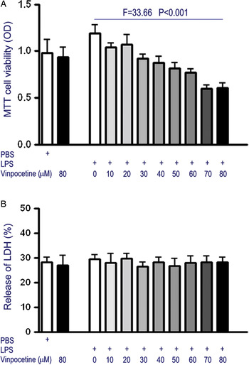
The effect of TSPO-specific ligand on proliferation of BV-2 microglia was assessed using MTT assay. BV-2 microglia were exposed to 10–80 µM of Vinpocetine in the presence or absence of LPS. The results show that Vinpocetine inhibited the proliferation of BV-2 microglia activated with LPS in a dose-dependent manner (Fig. 2A, P < 0.001), but did not influence the proliferation of BV-2 microglia in the absence of LPS (Vinpocetine = 80 µM).
The proliferation and death of BV-2 microglia treated with LPS and Vinpocetine. (A) Cell proliferation by MTT assay (F = 33.66, P < 0.001), and (B) cell death by LDH assay. BV-2 microglia were exposed to 10–80 µM of Vinpocetine in the presence or absence of LPS (1 µg ml−1) for 24 h. Values indicate means ± SD from three independent experiments.

To determine whether Vinpocetine was cytotoxic against BV-2 microglia, we exposed BV-2 microglia to 10–80 µM of Vinpocetine in the presence or absence of LPS, and measured the amount of LDH release. Vinpocetine did not have a significant effect on LDH release relative to vehicle-treated microglia, indicating that Vinpocetine-TSPO binding is not cytotoxic to BV-2 microglia at the concentrations tested (Fig. 2B).
Vinpocetine reduces inflammatory factors from BV-2 treated with LPS or OGD
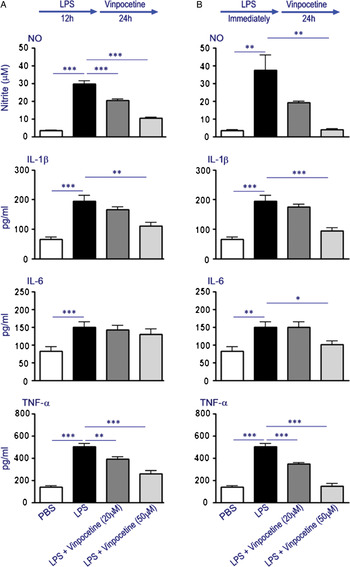
We next examined the effect of Vinpocetine on the release of NO and inflammatory cytokine IL-1β, IL-6 and TNF-α in BV-2 microglia treated with LPS and OGD. LPS stimulated BV-2 microglia to produce NO and inflammatory cytokine IL-1β, IL-6 and TNF-α relative to vehicle control (Fig. 3A,B). After 12 h of LPS stimulation, the addition of Vinpocetine at 20 and 50 µM reduced the release of NO ranging 31.5–64.9% decrease, IL-1β ranging 14.8–43.5% decrease, IL-16 ranging 4.7–13.4% decrease and TNF-α ranging 21.9–49.0% decrease relative to LPS treatment without Vinpocetine (Fig. 3A).
Inhibition of NO, IL-1β, IL-6 and TNF-α from BV-2 microglia stimulated with LPS and Vinpocetine. (A) After 12 h of LPS (1 µg ml−1) stimulation, BV-2 microglia were treated with Vinpocetine at 20 and 50 µM for 24 h. (B) BV-2 microglia were treated with Vinpocetine at 20 and 50 µM for 24 h, immediately after LPS (1 µg ml−1) stimulation. Values are means ± SD from three independent experiments, *P < 0.05, **P < 0.01 and ***P < 0.001.

Immediately after LPS stimulation, the addition of Vinpocetine at 20 and 50 µM also reduced the release of NO ranging 48.5–89.7% decrease, IL-1β ranging 10.7–52.3% decrease, IL-16 ranging 0–32.8% decrease and TNF-α ranging 31.4–70.7% decrease relative to LPS treatment without Vinpocetine (Fig. 3B).
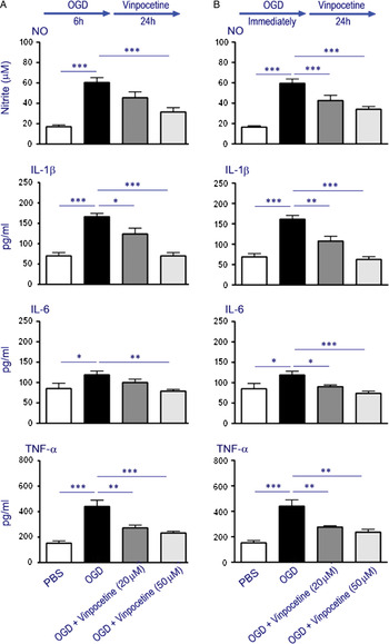
Simultaneously, OGD triggered BV-2 microglia to produce NO and inflammatory IL-1β, IL-6 and TNF-α relative to PBS-control (Fig. 4A,B). After 6 h of OGD treatment, the addition of Vinpocetine at 20 and 50 µM reduced the release of NO ranging 25.0–48.3% decrease, IL-1β ranging 25.9–58.4% decrease, IL-16 ranging 16.1–36.7% decrease and TNF-α ranging 38.1–47.7% decrease relative to OGD treatment without Vinpocetine (Fig. 4A).
Inhibition of NO, IL-1β, IL-6 and TNF-α from BV-2 microglia treated with OGD and Vinpocetine. (A) After 12 h of OGD treatment, BV-2 microglia were treated with Vinpocetine at 20 and 50 µM for 24 h. (B) BV-2 microglia were treated with Vinpocetine at 20 and 50 µM for 24 h, immediately after OGD treatment. Values are means ± SD from three independent experiments, *P < 0.05, **P < 0.01 and ***P < 0.001.

Immediately after OGD treatment, the addition of Vinpocetine at 20 and 50 µM also reduced the release of NO ranging 37.7–56.3% decrease, IL-1β ranging 33.1–61.8% decrease, IL-16 ranging 24.5–38.1% decrease and TNF-α ranging 37.4–46.1% decrease relative to OGD treatment without Vinpocetine (Fig. 4B).
Vinpocetine suppresses NF-κB and AP-1 expression in BV-2 treated with LPS
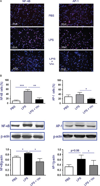
As NF-κB and AP-1 play a critical role in regulating inflammatory response, we explored whether Vinpocetine acts as an anti-inflammatory agent by inhibiting NF-κB and/or AP-1. It was reported that Vinpocetine inhibited the TNF-α-induced activation of NF-κB in vascular smooth muscle cells, umbilical vein endothelial cells and lung epithelial cells (Jeon et al., Reference Jeon, Xu, Aizawa, Lim, Jono and Kwon2010). In this study, western blot analysis and immunocytochemistry staining show that Vinpocetine treatment did not influence the phosphorylation of JNK, p38 and AKT (supplementary fig. 3 online). However, the levels of both NF-κB and AP-1 expression were elevated at 12 h after stimulation with LPS. The up-regulation of both NF-κB and AP-1 expression in LPS-stimulated BV-2 microglia was suppressed by the treatment of Vinpocetine (Fig. 5).
Inhibition of NF-κB and AP-1 in BV-2 microglia stimulated with LPS and Vinpocetine. (A) Representative images and quantitative analysis of immunocytochemistry staining, immunostaining of NF-κB or AP-1 (red) was restained with Hoechst (blue). (B) Representative bands and quantitative analysis of western blot. BV-2 microglia were treated with Vinpocetine at 50 µM for 24 h, immediately after LPS (1 µg ml−1) stimulation. Values are means ± SD from three independent experiments, *P < 0.05.

Vinpocetine-conditioned medium exhibits neuroprotective effect
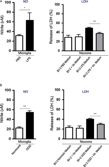
To confirm the reactivity of the cells to LPS and OGD, BV-2 microglia were treated with LPS (1 µg ml−1, 12 h) and OGD (6 h), then NO was measured. As shown in Fig. 6A,B, the production of NO was obviously triggered by LPS (P < 0.05) or OGD (P < 0.001), as compared to PBS and normoxia. Subsequently, Vinpocetine (50 µM) was added to LPS- or OGD-treated BV-2 microglia for 24 h. Different conditioned medium was collected to detect their neuroprotective effect when exposing primary cultured neurons. We observed that the Vinpocetine-conditioned medium significantly reduced the death of primary neurons, as compared with the control-conditioned medium. The results show that levels of LDH release were reduced in primary neurons incubated with the Vinpocetine-conditioned medium in LPS-stimulated BV-2 microglia compared with the control-conditioned medium (from 46.0 ± 3.7 to 35.3 ± 4.23%; P < 0.001, Fig. 6A). Simultaneously, levels of LDH release were also reduced in primary neurons incubated with the Vinpocetine-conditioned medium in OGD-treated BV-2 microglia compared with the control-conditioned medium (from 45.7 ± 3.6 to 34.7 ± 4.5%; P < 0.001, Fig. 6B). These results further demonstrate that Vinpocetine has a neuroprotective effect.
Vinpocetine-conditioned medium prevents neuron damage by LDH release. First, BV-2 microglia were treated with LPS for 12 h (A) or treated with OGD for 6 h (B), and production of NO was measured to define the activation of BV-2 microglia. Next, Vinpocetine (50 µM) was added to LPS- or OGD-treated BV-2 microglia for 24 h. Subsequently, BV-2 microglia were washed three times with PBS to remove Vinpocetine and LPS, and then cultured for a further 24 h. The supernatants of Vinpocetine-conditioned medium were collected, and then added to primary cultured neurons. Death of neurons was determined by LDH release. Values are means ± SD from three independent experiments, ***P < 0.001 versus control medium.

Vinpocetine exhibits anti-inflammatory effect in mouse model of hypoxia
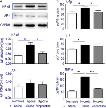
Based on anti-inflammatory effect of Vinpocetine in vitro experiment, we further investigated whether they can exhibit similar effect in vivo system. Hypoxic mice were injected i.p. with 5 mg kg−1 of Vinpocetine and saline everyday for 7 consecutive days. The levels of NF-κB and AP-1 expression in hypoxic brain were elevated than in normoxic mice (Fig. 7A, P < 0.01 for NF-κB and P > 0.05 for AP-1). The administration of Vinpocetine significantly inhibited the expression of NF-κB (Fig. 7A, P < 0.05), but only slightly decreased the expression of AP-1 (Fig. 7A, P > 0.05). The levels of inflammatory cytokine TNF-α, IL-1β and IL-6 were increased in hypoxic brain, Vinpocetine significantly inhibited TNF-α and IL-1β production (Fig. 7B, P < 0.001 and P < 0.05, respectively). These results further indicate that Vinpocetine has an anti-inflammatory effect in vitro and in vivo experiment.
Inhibition of inflammatory molecules in hypoxic mice treated with Vinpocetine.(A) The expression of NF-κB and PA-1 by western blot. (B) The levels of IL-1β, IL-6 and TNF-α production by ELISA. Values are means ± SD from six mice, *P < 0.05, **P < 0.01 and ***P < 0.001.

DISCUSSION
Vinpocetine, a specific TSPO ligand, has long been used for the treatment of cerebrovascular disorders and cognitive impairment (Bagoly et al., Reference Bagoly, Fehér and Szapáry2007; Rupprecht et al., Reference Rupprecht, Papadopoulos, Rammes, Baghai, Fan and Akula2010). Vinpocetine dilates blood vessels and enhances circulation in the brain, improving oxygen utilization and glucose uptake from blood, which activate cerebral metabolism and neuronal ATP bioenergy production (Szobor and Klein, Reference Szobor and Klein1976; Tamaki and Matsumoto, Reference Tamaki and Matsumoto1985). In addition, vinpocetine elicits neuronal protective effect that increases the resistance of the brain to hypoxia and ischemic injury (Adám-Vzi, Reference Adám-Vizi2000).
In the present study, we found: (1) LPS and OGD induced the up-regulation of TSPO expression on BV-2 microglia; (2) Vinpocetine inhibited inflammatory response in BV-2 microglia treated with LPS or exposed to OGD; and (3) Vinpocetine-conditioned medium in BV-2 microglia is neuroprotective.
Microglia have an important neuroprotective function in the brain by removal of damaged cells, promoting neurogenesis, inducing the reestablishment of a functional neuronal environment by restoration of the myelin sheath, and by releasing neurotrophic factors and anti-inflammatory molecules (Ziv et al., Reference Ziv, Ron, Butovsky, Landa, Sudai and Greenberg2006; Walter and Neumann, Reference Walter and Neumann2009). We detected the levels of neurotrophic factor nerve growth factor (NGF), brain-derived neurotrophic factor (BDNF) and glial cell line-derived neurotrophic factor (GDNF) in Vinpocetine-treated BV-2 microglia, but did not find any significant change (supplementary fig. 3 online). For the beneficial effect of Vinpocetine, the fine regulation of neuroinflammatory response plays a key role in neuroprotection (Emmanouil et al., Reference Emmanouil, Taoufik, Tseveleki, Vamvakas and Probert2011), either directly or indirectly, via inhibition of inflammatory factors (Ceulemans et al., Reference Ceulemans, Zgavc, Kooijman, Hachimi-Idrissi, Sarre and Michotte2010). In the present study, our results show that Vinpocetine is an anti-inflammatory agent in LPS-activated or OGD-treated BV-2 microglia as well as hypoxic mice. Both LPS and OGD stimulate BV-2 microglia to produce NO, IL-1β, IL-6 and TNF-α which contribute to neuronal damage. However, Vinpocetine effectively inhibits the production of these inflammatory factors in a dose-dependent manner, especially NO and TNF-α, regardless of LPS-stimulated or OGD-treated BV-2 microglia. Thus, our results, together with the fact that the beneficial effect is observed in stroke and cognitive impairment (Feigin et al., Reference Feigin, Doronin, Popova, Gribatcheva and Tchervov2001), identify Vinpocetine as a highly promising and attractive anti-inflammatory candidate for the treatment of CNS inflammatory diseases. Moreover, the potential cytotoxicity of Vinpocetine at higher concentrations needs to be investigated.
The Vinpocetine is a phosphodiesterase-1(PDE-1) inhibitor (Shimizu et al., Reference Shimizu, Murata, Watanabe, Sato, Morita and Tagawa2009; Giachini et al., Reference Giachini, Lima, Carneiro, Tostes and Webb2011). PDEs are involved in the regulation of intracellular levels of the second messengers cyclic adenosine monophosphate (cAMP) and cyclic guanosine monophosphate (cGMP), and play an important role in regulating the proliferation of cells (Shimizu et al., Reference Shimizu, Murata, Watanabe, Sato, Morita and Tagawa2009), thereby linking PDE activity not only in the regulation of vascular tone but also in the control of proliferation. It was reported that cell growth was inhibited by the PDE-1 inhibitor Vinpocetine (Shimizu et al., Reference Shimizu, Murata, Watanabe, Sato, Morita and Tagawa2009). Thus, Vinpocetine can prevent the proliferation of BV-2 possible through inhibiting PDE-1.
The particular interest in this study is the identification of BV-2 microglia-conditioned medium treated with Vinpocetine in primary neurons. It remains controversial as to whether the inflammatory response plays a beneficial or detrimental role for cerebral tissue and cells. There is a substantial evidence that molecules of the innate immune reaction is harmful to neurons and oligodendrocytes, whereas other observations indicate that inflammation is actually beneficial to recovery after brain injury (Rivest, Reference Rivest2011). To explore the relationship between anti-inflammation and neuroprotection in BV-2 microglia treated with Vinpocetine, we collected Vinpocetine-conditioned medium in BV-2 microglia, and observed its effect on primary cultured neurons. Upon exposing primary cultured neurons with conditioned medium, Vinpocetine-conditioned medium in both LPS- or OGD-treated BV-2 microglia markedly declined the death of neurons, suggesting a neuroprotective role in vitro. In the last decade, the involvement of PBR/TSPO in mitochondrial function was extensively investigated using ligands or over-expressing cells. Several studies indicated that PBR/TSPO-mediated changes in mitochondrial function can play a key role during cell death. In this study, we did not observe that Vinpocetine influences mitochondrial function in both resting or H2O2-treated BV-2 microglia (supplementary fig. 4 online), indicating that the neuroprotective effect of Vinpocetine could be mediated by inhibiting microglial inflammatory response.
NF-κB and AP-1 play pivotal roles in cell proliferation and cell survival. Interestingly, our results further show that Vinpocetine down-regulated the levels of Nuclear Factor-KappaB (NF-κB) and AP-1 in LPS-BV-2 microglia and hypoxic mice. In the present study, the anti-proliferative and anti-inflammatory effects of Vinpocetine were correlated with a reduced expression of NF-κB and AP-1. We therefore propose that the inhibitory effect of Vinpocetine on NF-κB and AP-1 results in the reduction of proliferation and inflammatory factors in BV-2 microglia treated with LPS or OGD. These findings may lead to the development of previously undescribed therapeutical strategies for various human inflammatory diseases, including pulmonary inflammatory disease, arthritis and atherosclerosis (Okamoto, Reference Okamoto2006).
In conclusion, it is evident that Vinpocetine acts as an anti-inflammatory agent in BV-2 microglia and hypoxic mice. Microglia-conditioned medium treated with Vinpocetine exhibits neuroprotective effect on primary cultured neurons injured by OGD. Vinpocetine inhibits the expression of NF-κB and AP-1, thus becoming an attractive therapeutical candidate for treating inflammatory diseases. Further studies should address the mechanisms underlying the neurotrophic support of Vinpocetine, which may involve the stimulation of neuroprotection and neurorepair by glial cells.
ACKNOWLEDGEMENTS
This work was supported by project grants from the National Natural Science Foundation of China (Code 30572164).
Statement of interests
The authors declare that they have no conflict of interest.
Supplementary material
The supplementary material referred to in this article can be found online at journals.cambridge.org/ngb.