Introduction
Recent confocal microscopy studies with fluorescent phallotoxins as actin-specific probes have greatly improved our understanding of the digenean muscle system, which was previously very limited. A great deal of this research was devoted to exploring the musculature of cercariae and adult worms (Czubaj & Niewiadomska, Reference Czubaj and Niewiadomska1997; Mair et al., Reference Mair, Maule, Shaw, Johnston and Halton1998, Reference Mair, Maule, Day and Halton2000, Reference Mair, Maule, Fried, Day and Halton2003; Kumar et al., Reference Kumar, McGeown, Reynoso-Ducoing, Ambrosio and Fairweather2003; Stewart et al., Reference Stewart, Marks and Halton2003a, Reference Stewart, Mousley, Koubková, Šebelová, Marks and Haltonb; Bulantová et al., Reference Bulantová, Chanová, Houžvičková and Horák2011; Krupenko, Reference Krupenko2011, Reference Krupenko2014b; Tolstenkov et al., Reference Tolstenkov, Prokofiev, Terenina and Gustafsson2011, Reference Tolstenkov, Akimova, Chrisanfova, Terenina and Gustafsson2012a, Reference Tolstenkov, Akimova, Terenina and Gustafssonb, Reference Tolstenkov, Akimova, Terenina and Gustafssonc, Reference Tolstenkov, Akimova, Terenina and Gustafssond), while the architecture of metacercarial muscles has been studied in only a few species (Apatemon cobitidis proterorhini, Bucephaloides gracilescens, Echinostoma caproni and Microphallus pygmaeus) (Stewart et al., Reference Stewart, Marks and Halton2003a, Reference Stewart, Mousley, Koubková, Šebelová, Marks and Haltonc; Šebelová et al., Reference Šebelová, Stewart, Mousley, Fried, Marks and Halton2004; Krupenko, Reference Krupenko2010). In the digenean life cycle, metacercariae are an intermediate larval stage between free-swimming cercariae and adult worms. Over the course of development, metacercariae resorb provisional cercarial structures, often growing in size and undergoing morphogenetic changes. The magnitude of these processes can vary widely in different taxonomic groups.
The most complex form of development occurs in metacercariae of the subfamily Diplostomoidea (see, for example, Szidat, Reference Szidat1924, Reference Szidat1929; Erasmus, Reference Erasmus1958; Wiśniewski, Reference Wiśniewski1958; Nasir, Reference Nasir1960; Shigin, Reference Shigin1986; Ginetsinskaya, Reference Ginetsinskaya1988; Galaktionov & Dobrovolskij, Reference Galaktionov and Dobrovolskij2003). There is evidence (see, for example, Szidat, Reference Szidat1924, Reference Szidat1929; Nasir, Reference Nasir1960; Shigin, Reference Shigin1986; Ginetsinskaya, Reference Ginetsinskaya1988) that the development of these metacercariae proceeds with a profound metamorphosis, involving a massive transformation of the muscular system. These observations were partially corroborated by electron microscopy studies of body-wall muscles and anterior organ/oral sucker in developing and infective metacercariae of Diplostomum pseudospathaceum (syn. D. chromatophorum) (Podvyaznaya & Dobrovolsky, Reference Podvyaznaya and Dobrovolsky2001; Podvyaznaya, Reference Podvyaznaya2013). The purpose of the present study was to use phalloidin staining and confocal laser scanning microscopy to provide additional information on changes in muscle architecture in this diplostomoid species during development from free-swimming cercariae to infective metacercariae.
Metacercariae of D. pseudospathaceum are very common pathogenic parasites of many species of freshwater Palaearctic fish (Shigin, Reference Shigin1986; Niewiadomska, Reference Niewiadomska1996). Cercariae of this species are produced by daughter sporocysts parasitizing the lymnaeid snails Stagnicola palustris and Lymnaea stagnalis. Once the free-living larvae encounter their prospective host fish, they attach to it with ventral suckers, cast away their tails and penetrate the host tissue (Shigin, Reference Shigin1986; Höglund, Reference Höglund1991). Post-penetration larvae (diplostomula) migrate by various routes to the eye lens of the fish (Shigin, Reference Shigin1986; Höglund, Reference Höglund1991; Niewiadomska, Reference Niewiadomska1996). Penetration and migration are actively assisted by a specialized penetration apparatus comprising a number of various structures: cercarial spines, anterior organ, penetration glands and the larval intestine, which functions as a glandular organ (Shigin, Reference Shigin1986; Höglund, Reference Höglund1991; Podvyaznaya, Reference Podvyaznaya2013). Larvae usually reach the lens within 24 h and establish there as metacercariae (Shigin, Reference Shigin1986; Höglund, Reference Höglund1991). Metacercarial development is accompanied by a series of morphogenetic transformations. Penetration glands and larval spines undergo degeneration (Shigin, Reference Shigin1986; Podvyaznaya, Reference Podvyaznaya1999). The anterior organ is transformed into an oral sucker and the cercarial intestine into typical digenean caeca; these transformations cause radical changes in the functions of these organs (Podvyaznaya, Reference Podvyaznaya2013). Extensive morphogenesis occurs in the excretory and nervous systems (Niewiadomska & Moczoń, Reference Niewiadomska and Moczoń1984; Shigin, Reference Shigin1986). Metacercariae also form lappets and a holdfast, specialized diplostomoid musculo-glandular organs that become fully functional only in adult worms: the holdfast plays an active role in feeding, while the lappets are used primarily for attachment to the host tissues (see, for example, Erasmus, Reference Erasmus1969, Reference Erasmus1970). In the definitive host (larid birds), the worms develop a hindbody containing the reproductive organs; in metacercariae it is present only as a small primordium. In this study we provide a detailed description of the musculature of the body wall, anterior organ/oral sucker, acetabulum, pharynx and intestine in cercariae and metacercariae of various ages, and we also examine the development of the lappets and holdfast. These data may offer some additional insight into a possible functional role of these organs at different ontogenetic stages.
Materials and methods
Metacercariae of D. pseudospathaceum of various ages were obtained from experimentally infected female guppies (Poecilia reticulata). Guppies were infected with cercariae released by naturally infected molluscs, S. palustris. Snails were collected from Lake Pertozero in the outskirts of Petrozavodsk (62°10′N, 33°58′E). Each guppy was placed for 1.5–2 h in a Petri dish (80 mm × 35 mm) filled with water containing 25–30 cercariae. All fish were then maintained in a common tank at 22–23°C and dissected 3, 6, 7, 8, 10 and 20 days after infection. Infection intensity was 3–6 parasites per fish. Twenty-day-old larvae were found to have all features of fully developed metacercariae and are referred to in the following text as infective metacercariae.
In addition, metacercariae of later stages of development and infective metacercariae were extracted from eye lenses of Rutilus rutilus caught in Lake Pertozero near the habitat of infected S. palustris. In the following, larvae of later stages of development are referred to as pre-infective metacercariae. They were of nearly the same size as infective metacercariae, had clearly identifiable lappets, holdfast organ and hindbody, and contained excretory granules in their paranephridial excretory system. These metacercariae differed from infective metacercariae by being more translucent and mobile. By their state of maturity, pre-infective metacercariae occupied an intermediate position between 10- and 20-day-old metacercariae collected from experimentally infected hosts. Preliminary species identification of metacercariae obtained from naturally infected fish was made by examining them under a light microscope. Further comparative analysis of phalloidin-stained whole mounts confirmed that metacercariae from experimentally and naturally infected hosts belong to the same species. Species identification and age evaluation of live cercariae and metacercariae were performed on a Leica DMLS light microscope (Leica Microsystems, Wetzlar, Germany).
For confocal microscopy, metacercariae extracted from the eye lenses of fish, and cercariae released by molluscs, were fixed for 15–17 h with 4% paraformaldehyde in 0.01 m phosphate-buffered saline (PBS), and were kept in 0.01 m PBS containing 0.1% sodium azide. Animals were permeabilized for 2 h with 0.5% Triton X-100 in PBS, stained for 2–3 h with tetramethylrhodamine B isothiocyanate (TRITC)-conjugated phalloidin (1:150, Sigma-Aldrich, P1951; Sigma-Aldrich, St. Louis, Missouri, USA), rinsed in PBS and then mounted with Vectashield (Vector Laboratories Inc., Burlingame, California, USA) or 80% glycerol/PBS. Confocal images were collected on a Leica TCS SP5 confocal laser scanning microscope. All confocal images of phalloidin-stained whole mounts were presented as maximum intensity projections of the Z-stacks.
Results
Body-wall musculature, parenchymal muscles and hindbody
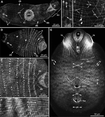
The body-wall musculature of cercariae is composed of outer circular (fig. 1A,C, cb), intermediate longitudinal (fig. 1A–C, lb) and inner diagonal muscles (fig. 1A–C, db). Circular muscle fibres have a diameter of 0.3–0.5 μm and are typically grouped into bands of two strands. This double-stranded arrangement is especially evident dorsally in the postacetabular region. Longitudinal muscles are broad and ribbon-shaped (fig. 1B), especially ventrally in the midbody region, and are generally wider (up to 2.5 μm) than other muscles in the body wall. Many of the longitudinal muscles show more or less distinct separation into several (usually 2 or 4) branches (fig. 1B) and, like circular muscles, might actually constitute closely packed bundles of muscle fibres. At the posterior end of the body, the longitudinal muscles bend inwards and sink beneath the level of the body wall to terminate at the constriction between the body and the tail. The body wall above these muscles is doubled into a fold that forms a caudal pocket around the base of the tail. Distal portions of individual muscle fibres, both in the longitudinal and circular musculature, often diverge from their own muscle bands and merge with adjacent bands (fig. 1C, arrows); this is especially evident in the midbody where both muscle layers assume a lattice-like appearance. Diagonal muscles form a criss-crossing pattern and are present ventrally only in the pre-acetabular body region (fig. 1A, db), while on the dorsal side (fig. 1C, db) they spread a distance of about 20 μm behind the caudal rim of the acetabulum. The distance between muscle bundles in the diagonal musculature is gradually increasing posteriorly, from 4 to 20 μm, and the width of muscle bundles is decreasing, from 1 to 0.5 μm. Like circular muscles, the diagonal muscles located anteriorly in the region of the oral sucker show a double-stranded arrangement, but the more posterior diagonal musculature appears to be composed of single strands.
Confocal images of the body-wall musculature and gross morphology of cercariae and metacercariae. (A) Anterior general view of a cercaria with the acetabulum and anterior organ. (B) Ventrolateral body wall of a cercaria anterior to the acetabulum with double-stranded longitudinal muscles. Anterior is towards the top. (C) Dorsal body wall of a cercaria at the level of the acetabulum with longitudinal muscle fibres crossing over (arrows) to adjacent muscle bands. Anterior is to the right side. (D) Dorsal side of a 6-day-old metacercaria with degenerating diagonal muscles (arrowheads). (E) Anterior ventral region of a 10-day-old metacercaria with longitudinal and circular musculature separated into individual fibres. (F) Region of the ventral body wall of an infective metacercaria. Anterior is to the right side. (G) Ventral body side of an infective metacercaria. Abbreviations: ac, acetabulum; aco, acetabular opening; am, arch-shaped lappet muscles; ao, anterior organ; cb, circular body-wall muscles; cr, caudal region of anterior organ; db, diagonal body-wall muscles; dlr, diagonal lappet retractors; ho, holdfast opening; la, lappet; lb, longitudinal body-wall muscles; ld, lateral dilators of acetabulum; lo, lateral oblique muscles; m, mouth; os, oral sucker; pd, posterior dilators of acetabulum; ph, pharynx; pp, postacetabular patch of diagonal muscles; pt, posterior transverse muscles; rr, rostral region of anterior organ; s, spines; vdb, ventral diagonal body-wall muscles.

In 3- to 6-day-old metacercariae, the arrangement of body-wall muscles remains essentially unchanged, but the number of fibres in the longitudinal muscle bands appears to increase and the bands become even wider than before (up to 3.5 μm). From day 6, the posterior diagonal muscles start showing signs of degeneration: their orientation becomes less regular, they break apart from one another and have uneven or jagged shapes (fig. 1D, arrowheads). By day 10 the diagonal musculature persists only in the immediate area behind the oral sucker (fig. 1E, db). Starting on day 8, muscle bundles in the longitudinal and circular layers become progressively less pronounced and have less regular shapes than previously (fig. 1E); individual fibres in the muscle bundles appear thinner and lie almost equally apart from each other. At about the same time, the number of longitudinal and circular muscles starts increasing, and by day 10 they become considerably more numerous than before (130–160 longitudinal fibres over the dorsal half of the midbody vs. 70–90 at day 8, and 390–430 circular fibres along the body length vs. 300–330 at day 8); these fibres are no longer organized into bundles (fig. 1E). In pre-infective metacercariae new diagonal muscles start spreading posteriorly from the anterior end of the animal, with ventral and dorsal muscles forming two distinct sets. The ventral set forms a finer grid, having muscle bundles 5 μm apart, vs. 10 μm apart in the dorsal set.
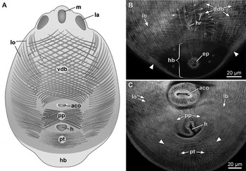
In infective metacercariae, the forebody assumes a spoon-like shape with a distinct ventral concavity. Two sets of diagonal muscles become even more distinct (figs 1F, G and 2A, B): the dorsal set extends over the whole area of the convex dorsal side (fig. 2B, ddb), but the ventral set is less pronounced and covers only the pre-acetabular portion of the ventral concavity (figs 1F and 2A, vdb). Two additional patches of body-wall muscles occupy the posterior part of the ventral side in the same layer as diagonal muscles: one is located between the acetabulum and holdfast and constitutes two sets of short diagonal muscles intersecting each other along the body midline (figs 1G and 2A, C, pp), the other is a transverse group of muscles behind the holdfast, crossing the body from one lateral side to the other (figs 1G and 2A, C, pt). There is yet another set of oblique muscles on the ventral side underneath the diagonal muscles (figs 1G, 2A,C, lo): these muscles originate from the lateral margins of the ventral concavity on either side of the body and slant in the medio-posterior direction, teminating some distance from the midline. Longitudinal muscle fibres in infective metacercariae are wider and shorter (25–30 μm long) than corresponding cercarial muscles and are distinctly spindle-shaped (fig. 1F, lb). These muscle fibres are joined into short rows 2–4 fibres long, which are tightly packed with almost no interspace between adjacent rows. Distal portions of fibres at the ends of each row insert on the fibres of adjacent rows.
Diagrammatic and confocal images of the body-wall musculature of infective metacercariae. (A) Ventral body wall with longitudinal and circular muscles omitted for clarity. (B) Dorsal postacetabular area showing the boundary between the forebody and hindbody (arrowheads). (C) Ventral postacetabular area with a U-pattern of body-wall muscles (arrowheads). Abbreviations: aco, acetabular opening; ddb, dorsal diagonal body-wall muscles; ep, excretory pore; h, holdfast; hb, hindbody; hr, holdfast retractors; la, lappet; lb, longitudinal body-wall muscles; lo, lateral oblique muscles; m, mouth; pp, postacetabular patch of diagonal muscles; pt, posterior transverse muscles; vdb, ventral diagonal body-wall muscles.

The hindbody first appears in pre-infective metacercariae as a small cone-shaped dorsal protrusion at the posterior end of the forebody, with the excretory opening located at the tip of the cone. At this point, however, it has no distinguishable traits in its musculature. In infective metacercariae, the transition between the forebody and hindbody becomes clearly demarcated by differences in body-wall musculature (fig. 2B). Longitudinal body-wall muscles on the hindbody are somewhat thinner than those on the forebody and most diagonal body-wall muscles do not enter the hindbody; at the transition to the hindbody they are spaced much closer to each other and change their angle to lie almost as circular muscles (fig. 2B, arrowheads). Only a few very sparse diagonal muscles, oriented as circular muscles, are found in the body wall of the hindbody, thus forming a second internal layer of the circular body-wall musculature underneath the longitudinal muscles.
The parenchymal musculature consists primarily of dorsoventral muscles. In cercariae, most of these are located in the pre-acetabular region, but in metacercariae they become more evenly distributed over the body. Over the course of metacercarial development the dorsoventral muscles become ever more numerous, and in infective metacercariae they occupy almost all of the space between internal organs in the interior of the forebody (see figs 5C, 6H, dv). The hindbody lacks dorsoventral muscles.
Cercarial tail
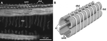
The musculature of the tail stem (fig. 3A, B) is composed of two types of longitudinal muscles (striated and smooth) and transverse-diagonal muscles. The striated musculature comprises two pairs of longitudinal muscle bundles (one ventrolateral and one dorsolateral) that span the entire length of the tail stem and continue into the furcae. In each bundle the medial myofibrils are oriented longitudinally (fig. 3A, lms) and the lateral myofibrils are arranged in a spiral fashion (fig. 3A, sms). In cross-section the myofibrils within the bundles form a U-shaped configuration. The spacing of the striations is about 1 μm. Three pairs of longitudinal smooth muscles (fig. 3A, B, smt) run dorsomedially and ventromedially between dorsolateral and ventrolateral striated muscles, respectively, and extend the entire length of the tail stem but do not continue into the furcae. Transverse-diagonal muscle bundles lie outside the longitudinal muscles. Each of these bundles consists of four myofibrils. On the lateral sides of the tail stem these muscles are oriented perpendicular to its longitudinal axis (fig. 3A, B, ttd), but on the ventral and dorsal sides they turn in a diagonal direction and insert on the next transverse-diagonal muscle on the contralateral side (figs 3A, B, dtd). As a result, these diagonal portions of the muscles form a criss-crossing pattern on the dorsal and ventral sides of the tail stem (fig. 3B). At the distal end of the tail stem, the left and right bundles of dorsal and ventral striated muscles pass into corresponding, left and right, furcae. About two-thirds of the length of the furcae these muscles separate into individual fibres, which spread evenly around the circumference of the furcae. These distal fibres of longitudinal muscles appear to be smooth rather than striated. Transverse-diagonal muscles in the furcae consist of single fine strands and, like those on the tail stem, form a criss-crossing pattern on the dorsal and ventral sides.
Cercarial tail musculature. (A) Confocal image showing a lateral side of the tail stem with the anterior end to the right side. (B) Three-dimensional diagram of a portion of the tail stem. Abbreviations: dtd, diagonal portions of transverse-diagonal muscles; lms, longitudinally oriented myofibrils of striated tail muscles; sms, spirally oriented myofibrils of striated tail muscles; smt, smooth tail muscles; st, striated tail muscles; ttd, transverse portions of transverse-diagonal muscles.

Anterior organ and oral sucker
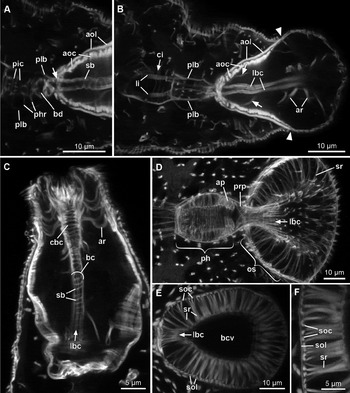
The anterior organ in cercariae (fig. 1A, ao) is pear-shaped and comprises two anatomically distinct, rostral and caudal regions. The caudal region has a conical shape, with the base of the cone facing forward (fig. 1A, cr); the protruding rostral region is enlarged into a sphere and covered with the tegument (fig. 1A, rr). The mouth is more or less in a terminal position, with a slight ventral shift, and opens into a very narrow buccal canal. The inner muscle layer of this canal is formed by two thin diagonal muscles spiralling around the canal in a double helix (fig. 4A, sb). Outside the diagonal muscles are two dorsolateral bundles of spindle-shaped longitudinal muscle fibres (fig. 4B, lbc); these muscles exit the anterior organ at its caudal end (fig. 4A, arrow), where each forks into two branches that continue posteriorly over the dorsal aspect of the outer pharyngeal wall (fig. 4A, B, plb) to insert at the posterior end of the pharynx. The outer wall of the organ's caudal cone is lined with an outer layer of wide (2.5–3 μm) longitudinal muscles (fig. 4A, B, aol) and an inner layer of even wider (4.5–5 μm) circular muscles (fig. 4A, B, aoc). These muscles do not enter the rostral region of the organ, and the anterior endings of the longitudinal muscles anchor on the body wall at the transition between the rostral and caudal regions (fig. 4B, arrowheads). The outer wall of the rostral region is essentially a continuation of the body wall but the body-wall musculature here is modified: the longitudinal muscles are much thinner and probably just the thin endings of more posterior muscles, there are no diagonal muscles, and the circular muscles are about 2–3 times as wide (about 1 μm) as the other circular muscles in the body wall and lie in closely spaced rows. Radial muscles span the space between the lining of the buccal canal and the body wall in the rostral region (fig. 4B, ar). The caudal region lacks radial musculature, but some radial muscles of the rostral region run posteriorly along the buccal canal to attach to the outer wall in the caudal region (fig. 4B, arrows).
Confocal images of the anterior body end of cercariae and metacercariae. (A) Pharynx and caudal portion of the anterior organ of a cercaria, showing longitudinal buccal muscles exiting the organ (arrow). (B) Anterior organ and pharynx of a cercaria showing radial muscles in the organ's caudal region (arrows) and transition between the rostral and caudal regions (arrowheads). (C) Anterior organ of a 3-day-old metacercaria. (D) Oral sucker, prepharynx and pharynx of a pre-infective metacercaria. (E) Oral sucker of an infective metacercaria with the anterior end to the right side. (F) Wall of the oral sucker of an infective metacercaria with the anterior end towards the top. Abbreviations: aoc, circular muscles in outer wall of anterior organ; aol, longitudinal muscles in outer wall of anterior organ; ap, anterior pharyngeal protrusion; ar, radial muscles of anterior organ; bc, buccal canal; bcv, buccal cavity; bd, bulbous distension of pharynx; cbc, circular muscles of buccal canal; ci, circular oesophageal-intestinal muscles; lbc, longitudinal muscle bundles of buccal canal; li, longitudinal oesophageal–intestinal muscles; os, oral sucker; ph, pharynx; phr, radial pharyngeal muscles; pic, circular muscles of pharyngeal canal; plb, longitudinal buccal muscles flanking pharyngeal wall; prp, prepharynx; sb, spiral muscles of buccal canal; soc, circular muscles in outer wall of oral sucker; sol, longitudinal muscles in outer wall of oral sucker; sr, radial muscles of oral sucker.

In 3-day-old metacercariae the anterior organ (fig. 4C) assumes a cylindrical shape, the mouth elongates longitudinally and the rostral region of the organ becomes smaller. The buccal canal is wider than in cercariae (fig. 4C, bc); the longitudinal muscles of the canal (fig. 4C, lbc) increase in number to 6–8 fibres and spread more evenly around its circumference. As in the cercariae, these muscles continue posteriorly over the dorsal side of the pharynx to its posterior end and this arrangement of longitudinal buccal muscles persists throughout the metacercarial development. Spiral muscles of the canal (fig. 4C, sb) remain only in its posterior part and their helix becomes more tightly wound; in the anterior part of the canal these spiral muscles are replaced by tightly spaced circular muscles (fig. 4C, cbc). Wide circular muscles in the outer wall of the organ's caudal region are replaced by numerous slender muscle fibres with somewhat irregular shapes of myofibrils. The number and thickness of the radial muscles increase (32–36 vs. 24–26 muscles) and they begin to spread into the caudal region of the organ.
In 6- to 8-day-old metacercariae the anterior organ begins to transform into the oral sucker. It becomes wider and shorter and assumes an ovoid shape. Only very thin and sparse circular muscles are now visible in the outer wall of the sucker's rostral region and longitudinal muscles disappear here completely. Spiral muscles in the wall of the buccal canal are completely replaced by circular muscles. Wide longitudinal muscles in the outer wall of the sucker's caudal region begin to show signs of resorption and are eventually replaced by thin longitudinal muscle fibres.
From day 8 to pre-infective metacercariae, the oral sucker gradually becomes cup-shaped, the number of circular and longitudinal muscles in the wall of the now considerably enlarged buccal canal is progressively increasing, and the radial muscles spread even further posteriorly, to occupy almost the entire length of the sucker (fig. 4D, sr).
The oral sucker of infective metacercariae has the shape of an elongated cup (fig. 4E) with a widely open buccal cavity (fig. 4E, bcv). The muscular wall of the buccal cavity is invested with inner circular muscles and very thin and sparse outer longitudinal muscles. The outer wall of the sucker consists of inner circular muscles (fig. 4E, F, soc) and thin outer longitudinal muscles (fig. 4E, F, sol). The space between the canal and the outer wall is spanned by short and powerful radial muscles (fig. 4E, F, sr) distributed evenly over the length of the sucker.
Prepharynx and pharynx
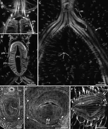
The musculature of the prepharynx (figs 4D, 5A) remains essentially unchanged throughout the development from cercariae to infective metacercariae. It consists of two bilaterally symmetrical sets of diagonal muscles (fig. 5A, dpr) that intersect each other on the dorsal and ventral sides.
Confocal images of the digestive system and holdfast of metacercariae. (A) Prepharynx area of a 10-day-old metacercariae. (B) Pharynx and oesophagus of an infective metacercaria. (C) Oesophagus and intestinal caeca of an infective metacercaria. (D) Holdfast protrusion of a 10-day-old metacercaria with a U-pattern of body-wall muscles (arrowheads). (E) Holdfast protrusion of a pre-infective metacercaria with a U-pattern of body-wall muscles (arrowheads). Anterior is on the right side. (F) Distended distal portion of the holdfast of an infective metacercaria. Anterior is on the right side. Abbreviations: ac, acetabulum; aco, acetabular opening; cd, circular muscles in distal distension of holdfast; ci, circular oesophageal–intestinal muscles; cp, circular muscles in proximal portion of holdfast; dpr, diagonal prepharynx muscles; dv, dorsoventral muscles; es, oesophagus; hc, circular muscles of holdfast; ho, holdfast opening; hr, holdfast retractors; lbc, longitudinal muscle bundles of buccal canal; li, longitudinal oesophageal–intestinal muscles; md, meridional muscles in distal distension of holdfast; os, oral sucker; ph, pharynx; phc, pharyngeal canal; phr, radial pharyngeal muscles; plb, longitudinal buccal muscles flanking pharyngeal wall; pm, meridional muscles of holdfast protrusion; pr, holdfast protrusion; ps, pharyngeal sphincter; s, spines.

The pharynx in cercariae is egg-shaped and has a length of 10–14 μm (fig. 4A, B). The pharyngeal canal has only circular muscles (fig. 4A, pic) and the outer wall is completely devoid of musculature. The anterior end of the pharynx forms a small bulb-shaped distension (fig. 4A, bd) continuous with the prepharynx. Sparse radial muscles span between the wall of the canal and the outer wall of the pharynx (fig. 4A, phr). On days 3 to 7 of metacercarial development, longitudinal muscles begin to develop in the outer pharyngeal wall, and the pharyngeal canal becomes wider. Circular muscles in the outer pharyngeal wall appear by day 8, to the inside of the longitudinal muscles. From day 7 to pre-infective metacercariae, the pharynx becomes larger (from 15 to 40 μm in length) and assumes a cylindrical shape, the radial muscles increase in number (130–150 vs. 26–28 at day 7) and the diameter of the pharyngeal canal also continues to grow. On day 10, a fold begins to grow around the anterior end of the pharynx so that the pharynx forms a protrusion projecting into the prepharynx (fig. 4D, ap). In infective metacercariae the fold around the protrusion becomes much deeper and 14 thick circular sphincter muscles appear in the protrusion underneath the circular muscles of the outer pharyngeal wall (fig. 5B, ps).
Oesophagus and intestinal caeca
In cercariae and early metacercariae, most of the oesophagus and intestinal caeca are lined only with thin circular muscles distributed evenly along their entire length (fig. 4B, ci). Beside circular muscles, there are two pairs of longitudinal muscle bundles (fig. 4B, li) arising from the posterior end of the pharynx, one pair somewhat closer to the dorsal side than the other, but these bundles extend only as far back as the posterior end of the oesophagus. From day 8 these muscle bundles begin to spread progressively further backwards along the intestinal wall outside the circular muscles of the intestine, so that in infective metacercariae they reach almost to the posterior ends of the caeca. As these muscles proceed posteriorly, they bifurcate into branches and occasionally anastomose with each other (fig. 5C).
Holdfast
The developing holdfast becomes clearly evident in 10-day-old metacercariae as a longitudinal slit-like invagination of the body wall, 40–45 μm long by 25–40 μm deep, located 30–50 μm posterior to the acetabulum (fig. 5D, ho). The body surface around the invagination forms an oval protrusion (fig. 5D, pr). Circular body-wall muscles lateral and immediately anterior and posterior to this protrusion converge toward the holdfast opening, to become the meridional muscles of the protrusion (fig. 5E, pm) and then enter the opening as longitudinal muscles of the invagination. Beneath the meridional muscles of the protrusion lies a layer of circular muscles (fig. 5E, hc); this layer continues into the opening to form the circular musculature of the invagination outside the longitudinal muscles. Judging from their position in the body wall, these circular muscles are derived from the longitudinal body-wall muscles. Wide dorsoventral muscles extend from the wall of the invagination to the dorsal body wall, probably functioning as retractor muscles. Longitudinal body-wall muscles running lateral to the holdfast bend around the caudal rim of the protrusion and then run along its contralateral side to form a U-shaped pattern (fig. 5D, E, arrowheads). This characteristic U-shaped arrangement of longitudinal muscles persists through to infective metacercariae (fig. 2C, arrowheads).
In infective metacercariae, the holdfast lumen becomes distended in its distal part (fig. 5F). This sac-like distension is surrounded by a grid-like muscular wall formed by two regular layers of relatively thin muscles. The inner of these two layers consists of meridional muscles (fig. 5F, md), and the outer of circular muscles (fig. 5F, cd). The narrow proximal portion of the holdfast contains only circular muscles (figs 5F, 6H, 7C, cp). These muscles are readily distinguishable from the corresponding muscles of the distended region by being much wider than the latter (1.5–2 μm vs. 0.5 μm), thus providing a clear morphological demarcation between these two regions of the holdfast. Powerful dorsoventral retractors (figs 2B, 5F, 6H, 7D, hr), more numerous than in pre-infective metacercariae, extend from both the proximal and distal regions of the holdfast to the dorsal body wall (fig. 6H).
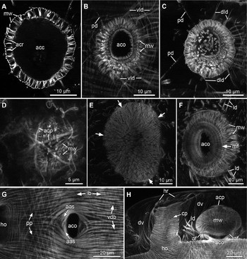
Confocal images of the acetabulum of cercariae and metacercariae, with the anterior end to the right side. (A) Central region of the acetabulum of a cercaria. (B) Acetabular opening and ventrolateral dilators of a cercaria. (C) Acetabulum of a cercaria with deep lateral and posterior dilators. (D) Dorsal side of the acetabulum of a 6-day-old metacercaria with three protractor muscles. (E) Dorsal side of the acetabulum of an infective metacercaria with six sectors of meridional muscles and shorter meridional muscles between sectors (arrows). (F) Ventral side of the acetabulum of an infective metacercaria. (G) Acetabular opening of an infective metacercaria with arch-shaped sphincter muscles. (H) Side view of the acetabulum and holdfast of an infective metacercaria. Abbreviations: aas, acetabular sphincter; acc, acetabular cavity; aco, acetabular opening; acp, acetabular protractors; acr, radial acetabular muscles; af, acetabular fold; cp, circular muscles in proximal portion of holdfast; dld, deep lateral dilators of acetabulum; dv, dorsoventral muscles; ho, holdfast opening; hr, holdfast retractors; lb, longitudinal body-wall muscles; ld, lateral dilators of acetabulum; mf, meridional muscles of acetabular fold; mw, meridional muscles of acetabular wall; pd, posterior dilators of acetabulum; pp, postacetabular patch of diagonal muscles; vdb, ventral diagonal body-wall muscles; vld, ventrolateral dilators of acetabulum.

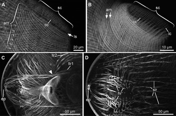
Confocal images of lappets and their retractors in metacercariae. (A) Lappet of a pre-infective metacercaria. (B) Lappet of an infective metacercaria. (C, D) Lappet retractors of an infective metacercaria, with arrowhead indicating the point where two groups of rectractors converge into a single bundle. Abbreviations: am, arch-shaped lappet muscles; cc, circular cushion muscles; cp, circular muscles in proximal portion of holdfast; ep, excretory pore; hr, holdfast retractors; lc, longitudinal cushion muscles; llr1–2, longitudinal lappet retractors; ls, longitudinal strips; sc, secretory cushion; slr, superficial lappet retractors.

Acetabulum
In cercariae, the acetabulum (fig. 1A, ac) is nearly spherical in shape, has a diameter of 20–30 μm and is located slightly posterior to the middle of the body. The musculature of the outer acetabular wall is formed by thin meridional muscles spanning between the outer opening of the acetabulum and its dorsal pole. Meridional muscles are arranged in a radially symmetrical manner with respect to the dorsoventral axis of the acetabulum, and form six sectors. The exact arrangement of these muscles in cercariae and early metacercariae is difficult to trace, but in older metacercariae the position of individual muscles becomes more apparent (fig. 6E). In the dorsal hemisphere of the acetabulum, the meridional muscles in each of the six sectors radiate out in a fan-like fashion, reaching the corresponding muscles of the adjacent sectors in such a way that the tips of the muscles form a pattern of a six-pointed star. Muscles situated between the sectors (fig. 6E, arrows) are shorter than other meridional muscles, and end some distance short of the dorsal pole of the acetabulum.
Numerous short radial muscles in the cercarial acetabulum (fig. 6A, acr) connect the outer wall with the wall of the acetabular cavity. On the ventral side of the acetabulum around its opening, there are two or three relatively thin, circular sphincter muscles lying outside the meridional muscles. No musculature is found beneath the tegument of the acetabular cavity, except a few very fine and irregular meridional muscles. Five pairs of muscles extend from the outer wall of the acetabulum, most likely acting as dilator muscles (fig. 6B, C). Two pairs arise close to the ventral body surface on the lateral sides of the acetabulum (fig. 6B, vld) and extend anterolaterally to terminate on the ventrolateral body wall. Two other pairs originate from the acetabulum somewhat deeper in the body (fig. 6C, dld); these muscles run anterolaterally at a less acute angle than the first two pairs and insert on the lateral sides of the body. A pair of thin muscles arises from the caudal side of the acetabulum and extends posterolaterally to the lateral body wall (figs 1A, 6B, C, pd). Throughout metacercarial development, the number of fibres within these ten bundles is gradually increasing and their arrangement becomes less clear. In infective metacercariae they can be distinguished as two pairs of wide lateral arrays of muscle fibres, one near the ventral surface and the other deeper in the body interior (fig. 6F, H, ld).
In 3-day-old metacercariae, the musculature of the acetabulum remains essentially unchanged, except that 4–5 fine circular (equatorial) muscles develop in the wall of the cavity close to the acetabular opening. At this point, circular muscles lining the wall of the acetabular cavity increase in number; the meridional muscles become broader and are arranged more regularly in three sectors outside of the circular muscles. Three muscles possibly functioning as protractors appear over the dorsal side of the acetabulum (fig. 6D, acp). These muscles intersect each other roughly in the centre of the dorsal hemisphere of the acetabulum at about 30°, and their distal ends anchor on the ventral body wall anteriorly and posteriorly to the acetabulum. These three muscles thus subdivide the dorsal hemisphere of the acetabulum into six sectors, in a similar way as meridional muscles. As development progresses, the number of muscle fibres in these muscles increases, to four or more fibres in infective metacercariae.
In 6- to 8-day-old metacercariae, a circular fold begins to grow around the acetabular opening and the ventral portion of the acetabulum forms a conical papilla, an extension protruding beyond this acetabular fold. In older metacercariae, the fold grows deeper (fig. 6H, af) and its wall becomes invested with meridional muscles (fig. 6F, mf) radiating from the tip of the papilla towards the periphery of the fold.
In pre-infective metacercariae, the circular musculature in the wall of the acetabular cavity and the radial muscles become thicker and more numerous. In infective metacercariae, a powerful sphincter of arch-shaped longitudinal body-wall muscles (fig. 6G, aas) appears around the outer opening of the acetabulum.
Lappets
The musculature of the lappets begins to differentiate by about day 10 of metacercarial development, when two sets of slightly curved longitudinal muscles, dorsal and ventral, appear in the body wall on either side of the oral sucker (fig. 1E, am). These arch-shaped muscles are likely to be derived from longitudinal body-wall muscles, but they form a separate layer between the circular and longitudinal layers. In pre-infective metacercariae, the curvature of the arch-shaped muscles becomes more pronounced (fig. 7A, am). Each set of these muscles, both on the dorsal and ventral sides, comprises about 30 muscles, the width of the set is 50–70 μm. The areas of the body wall between the dorsal and ventral arch-shaped muscles on either side develop into secretory cushions (fig. 7A, sc), evident as elongated oval regions, 40–60 μm long by 10–20 μm wide, with a longitudinally oriented long axis. Both circular and longitudinal muscles in the body wall of the cushions are independent and easily distinguishable from the rest of the body-wall musculature; they are about twice as thin as the corresponding muscles in the adjacent body wall and are confined within the cushion spanning across it, with both distal ends attached at the periphery of the cushion. There are two longitudinal strips that lack musculature (fig. 7A, ls) along the dorsal and ventral margins of the cushion, probably to make room for attachment of the circular cushion muscles and adjacent body-wall musculature. In infective metacercariae these strips disappear and arch-shaped muscles form full circles around the cushion (fig. 7B).
Lappets in infective metacercariae are associated with two sets of retractor muscles. Muscles in the first set (fig. 1G, dlr) are short and run diagonally toward the midline, where they come close to the corresponding muscles of the contralateral side in the area behind the pharynx. The second set consists of three groups of powerful muscles that stretch longitudinally through the body to its posterior end. Muscles in the first two groups run deep in the body, one closer to the midline (fig. 7C, llr1) and the other more laterally (fig. 7C, llr2), then converge into a single bundle immediately posterior to the holdfast opening (fig. 7C, arrowhead) to insert on the body wall in the hindbody around the sphincter of the excretory opening. The third group runs closer to the body surface (fig. 7D, slr); these muscles fan out behind the pharynx, proceed posteriorly in a wide array over much of the dorsum and attach dorsally in the hindbody.
Discussion
Previous light and electron microscopy studies have made clear that some of the cercarial organ systems in diplostomid digeneans undergo substantial reorganization and remodelling during metacercarial development (Szidat, Reference Szidat1924; Erasmus, Reference Erasmus1958; Wiśniewski, Reference Wiśniewski1958; Shigin, Reference Shigin1986; Podvyaznaya & Dobrovolsky, Reference Podvyaznaya and Dobrovolsky2001; Podvyaznaya, Reference Podvyaznaya2013). The present study used the technique of phalloidin staining in combination with confocal microscopy to confirm these observations for musculature in D. pseudospathaceum.
The most obvious and profound changes were observed in the musculature of the body wall and anterior organ. Developmental changes in body-wall musculature can be subdivided roughly into three consecutive phases. The first phase starts when a cercaria is transformed into a diplostomulum, and lasts until day 6 after infection; this phase coincides in time with a slight reduction in body size, when the non-feeding metacercariae use internal resources to provide energy and material for muscle development (Shigin, Reference Shigin1986; Podvyaznaya & Dobrovolsky, Reference Podvyaznaya and Dobrovolsky2001; Podvyaznaya, Reference Podvyaznaya2013). In these early metacercariae, the cercarial musculature remains largely unchanged, but the longitudinal muscle bands become somewhat wider than in cercariae. The adaptive significance of this muscle reinforcement is not entirely clear. It can be related to the need to provide diplostomula with additional muscular force during migration through the host body, but it should be noted that migration is usually completed earlier, within the first 24 h after penetration (Erasmus, Reference Erasmus1959; Shigin, Reference Shigin1986; Höglund, Reference Höglund1991; Podvyaznaya, Reference Podvyaznaya2013).
The process of muscle growth during the first phase is succeeded by the opposite process during the second phase (7-day-old to pre-infective metacercariae), when large muscle bands in the longitudinal musculature appear to break down into individual muscle fibres and show evidence of degeneration. The second phase is associated with active remodelling of the longitudinal body-wall musculature, as shown by electron microscopy (Podvyaznaya & Dobrovolsky, Reference Podvyaznaya and Dobrovolsky2001): during this period the initial cercarial musculature is being replaced with newly developed myoblasts of the definitive musculature. Phalloidin staining does not allow us to differentiate with certainty between degenerating cercarial muscles and developing myoblasts, and individual longitudinal fibres observed in the 8-day-old to pre-infective metacercariae are likely to be a mixture of both.
Diagonal musculature displays even more apparent changes than longitudinal musculature, because degeneration of diagonal cercarial muscles and development of new definitive muscles are clearly separated in time. Cercarial musculature begins to degenerate as early as day 6 of development and disappears almost completely by day 10, but the definitive diagonal musculature starts developing only in pre-infective metacercariae. Of the three body-wall layers, circular musculature seems to be least affected by these transformations and, although circular muscles increase in number over the course of development, it is possible that portions of cercarial musculature persist in the circular muscles of infective metacercariae. In general, the second phase, especially in its later part, is accompanied by a rapid increase in body size and in the number of longitudinal and circular body-wall muscles.
The third phase begins in pre-infective metacercariae with differentiation of the hindbody and is marked by patterning of the definitive body-wall musculature. The definitive musculature, especially that of longitudinal and diagonal muscles, is strikingly different from that of cercariae and early metacercariae. This difference arises from the fact that the body-wall musculature accomplishes two essentially different tasks in cercariae and adult worms. Cercariae use their body-wall musculature primarily for locomotion, but in the adult worms the spoon-shaped forebody functions essentially as a muscular organ to grip the villus in the intestine of the definitive host (see, for example, Öhman, Reference Öhman1965). This ‘gripping’ action is especially clearly reflected in the characteristic arrangement of diagonal body-wall muscles in infective metacercariae. Unlike the cercarial diagonal muscles that constitute a single set of muscles wrapping around the circumference of the body, the diagonal muscles of infective metacercariae are separated into two distinct sets on the dorsal and ventral sides of the body. Similar distinct ventral and dorsal sets were also described for several other digeneans with flattened bodies and ventral concavity (Krupenko, Reference Krupenko2011, Reference Krupenko2014a). It stands to reason that in D. pseudospathaceum these two sets work as antagonists, the ventral set assisting in tightening the forebody around the villus and the dorsal set in loosening the grip. However, the primary role in providing the grip must be played by the third, deeper set of oblique muscles extending from the margins toward the midline of the ventral concavity. These muscles probably bend the lateral margins of the forebody, allowing them to close around the villus. Dorsoventral muscles, which become especially numerous toward the end of metacercarial development, can flatten the forebody and thus also aid the parasite's fixation on the villus. It is very likely that, in infective metacercariae, longitudinal and circular body-wall muscles also form two separate, dorsal and ventral sets, which act as antagonists, but the arrangement of these muscles does not allow us to determine this with complete certainty.
The body-wall musculature of A. cobitidis proterorhini and Cotylurus erraticus (Strigeidae), two other diplostomoids studied by confocal microscopy (Stewart et al., Reference Stewart, Mousley, Koubková, Šebelová, Marks and Halton2003b, Reference Stewart, Mousley, Koubková, Šebelová, Marks and Haltonc), is unusually different from that described in this study for D. pseudospathaceum. Metacercariae and adult forms of these strigeids have no additional diagonal muscles in their body wall similar to those found in infective metacercariae of D. pseudospathaceum. Longitudinal body-wall musculature in metacercariae of A. cobitidis proterorhini and C. erraticus consists of relatively thin and long muscle bundles (Stewart et al., Reference Stewart, Mousley, Koubková, Šebelová, Marks and Halton2003b, their Fig. 2a) rather than of compact rows of short and thick muscle fibres like those of infective metacercariae of D. pseudospathaceum. As the body-wall musculature in metacercariae and adult individuals of these strigeids is similar to that in early metacercariae of D. pseudospathaceum and in strigeid cercariae (Tolstenkov et al., Reference Tolstenkov, Akimova, Terenina and Gustafsson2012b, their Fig. 1), it is likely that, in contrast with D. pseudospathaceum, the body-wall musculature of A. cobitidis proterorhini and C. erraticus do not undergo any substantial remodelling during metacercarial development. Like D. pseudospathaceum, the adult individuals of A. cobitidis proterorhini and C. erraticus parasitize the intestine of birds, but they have a different, cup-like, body shape and employ another mode of attachment to the intestinal mucosa, accomplished primarily by pseudosuckers (Öhman, Reference Öhman1966) rather than by the body-wall musculature. Outside the diplostomoids certain features of similarity to D. pseudospathaceum can be seen in the body wall of adult schistosomatids (Mair et al., Reference Mair, Maule, Day and Halton2000). Significantly, the body wall of Schistosoma mansoni has a compact layer of spindle-shaped longitudinal muscles, essentially identical both in size and arrangement to those of D. pseudospathaceum (Mair et al., Reference Mair, Maule, Day and Halton2000).
Extensive changes in the musculature of the anterior organ in D. pseudospathaceum can be explained by a radical change in its function as it transforms into the oral sucker. The anterior organ is a pear-shaped musculo-glandular structure, the main function of which is penetration of the host's tissues and assistance in migration of the diplostomulum through the host's body (Erasmus, Reference Erasmus1959; Shigin, Reference Shigin1986; Höglund, Reference Höglund1991). The caudal cone-shaped region of the organ houses enlarged terminal portions of penetration gland ducts (Niewiadomska, Reference Niewiadomska1986; Czubaj & Niewiadomska, Reference Czubaj and Niewiadomska1997) and simultaneous contraction of wide circular and longitudinal muscles in its outer wall probably causes massive discharges of secretion at the moment of penetration and during migration. This secretion discharge might also be helped by contraction of diagonal body-wall muscles (Bulantová et al., Reference Bulantová, Chanová, Houžvičková and Horák2011). Contraction of radial or circular muscles in the organ's rostral region causes protrusion or withdrawal of this region; these muscles act in concert with pre-oral spines (Erasmus, Reference Erasmus1958; Niewiadomska, Reference Niewiadomska1986; Höglund, Reference Höglund1991) to help the diplostomulum burrow through the host tissues. Once the diplostomulum has reached its definitive location, the anterior organ begins to transform into the oral sucker, which is an entirely muscular organ. This transformation process is accompanied by changes in the musculature and shape of the organ. Wide muscles in the caudal region degenerate, breaking down into irregular thin fibres, and are gradually replaced by more numerous and thin muscles of the definitive musculature. This process of active muscle remodelling is clearly corroborated by observations made at the ultrastructural level (Podvyaznaya, Reference Podvyaznaya2013). Penetration glands undergo progressive degeneration and disappear completely by the time when metacercariae start actively growing in size (Erasmus, Reference Erasmus1958; Shigin, Reference Shigin1986). The rostral region shrinks in size, eventually becoming indistinct from the caudal region, and the buccal canal grows progressively wider. As penetration gland ducts are receding, the radial muscles spread backwards to occupy the vacated space. The definitive oral sucker is a cup-shaped organ with musculature clearly dominated by radial muscles that dilate the buccal cavity, creating suction during feeding.
Changes observed in the organs of the digestive system (pharynx and midgut) are less dramatic. Curiously, although electron microscopy shows significant metamorphic transformations in the midgut of metacercariae (Podvyaznaya, Reference Podvyaznaya2013), changes in musculature are marked only by progressive spreading of longitudinal muscles posteriorly into caecal branches. The cercarial pharynx is essentially a non-functional, incipient structure with poorly developed musculature. Once the midgut becomes fully functional, the pharynx turns into an efficient muscular pump, which is expressed in the formation of musculature in its outer wall and in the development of the pharyngeal protrusion with a powerful sphincter.
The acetabulum also undergoes only gradual changes, such as the progressive growth of meridional and circular muscles in the wall of the acetabular cavity and development of the acetabular fold with additional meridional muscles. It should also be mentioned that at least some of the acetabular musculature has three- or sixfold radial symmetry: meridional muscles in the acetabular cavity are organized in three sectors and those in the outer wall in six sectors, and three sets of protractor muscles intersect over the dorsal pole of the acetabulum. Sixfold symmetry was also clearly shown for the arrangement of acetabular sensilla (reviewed in Bell et al., Reference Bell, Gibson and Sommerville1996) and their innervation patterns (Niewiadomska & Moczoń, Reference Niewiadomska and Moczoń1984; Niewiadomska et al., Reference Niewiadomska, Czubaj and Moczon1996). The muscle architecture of the acetabulum in D. pseudospathaceum is identical in all essential aspects to that described for another diplostomoid trematode, Cotylurus cornutus (Krupenko, Reference Krupenko2014a). Of the 13 digenean species from 11 different families, whose acetabular musculature has been closely studied by confocal microscopy (Krupenko, Reference Krupenko2010, Reference Krupenko2011, Reference Krupenko2014a, Reference Krupenkob; the present study), sixfold symmetry is evident only in diplostomoid species (D. pseudospathaceum and C. cornutus), suggesting that this character may have a potential taxonomic value.
Our study underlines the complex nature of developmental processes in D. pseudospathaceum metacercariae, and shows their dependence on several developmental, environmental and adaptive factors. Environmental factors have a significant effect on the intensity of developmental processes. The results of our study reinforce previous experimental findings (Sweeting, Reference Sweeting1974; Shigin, Reference Shigin1986) that developmental rates of Diplostomum depend on water temperature and host species. In our experiment metacercarial development was twice as fast as that observed by Podvyaznaya (Reference Podvyaznaya1999, Reference Podvyaznaya2013) when carp under-yearlings were infected with cercariae of D. pseudospathaceum and infected fish were kept at lower temperatures (18–20°C). It should, however, be noted that the early metacercarial development (1–6 days after infection) in our experiment proceeded at almost the same rate as in the experiments of Podvyaznaya (Reference Podvyaznaya1999, Reference Podvyaznaya2013). Light and electron microscopy studies have shown that during this period the larvae do not feed or grow, they actively resorb cercarial glands, and the tegument and gut epithelium undergo profound ultrastructural changes that enhance their absorptive function (Shigin, Reference Shigin1986; Podvyaznaya, Reference Podvyaznaya1999, Reference Podvyaznaya2013). When metacercariae begin to feed, they start growing in size and their morphogenetic processes accelerate. In our study these processes were considerably more intensive than in the experiments of Podvyaznaya (Reference Podvyaznaya1999, Reference Podvyaznaya2013) and metacercariae reached their infective stage faster (20 days vs. at least 40 days).
Another important factor in metacercarial development is transition from consuming endogenous food reserves to active feeding. This factor is largely responsible for reorganization of the foregut musculature, especially of the anterior organ as it transforms into the oral sucker. The midgut also undergoes a significant transformation as the larva switches to active feeding, although this transformation is more apparent at the cellular level rather than in the gross organization of the midgut musculature. Extensive changes occurring in the body-wall musculature are likely to be caused by adaptive factors: specifically, by the differences in the roles this musculature plays in cercariae and adult worms.
Changes observed in diplostomid metacercariae can probably exemplify more general developmental processes in digeneans. For instance, the same process of disintegration of longitudinal bands of body-wall muscles into widely spaced individual fibres as observed in D. pseudospathaceum was also noted in schistosomatids during transformation from miracidia to sporocysts (Bahia et al., Reference Bahia, Avelar, Vigorosi, Cioli, Oliveira and Mortara2006; Bulantová et al., Reference Bulantová, Chanová, Houžvičková and Horák2011). In general, developmental processes in diplostomoid metacercariae appear to be more diverse than was previously thought, involving extensive metamorphic transformations in some organs and more gradual changes in the others. Further study of metacercarial development in different groups of digeneans may help to expand our understanding of these processes and provide insight into the underlying developmental mechanisms in the digenean life cycle.
Acknowledgements
Confocal microscopy was performed at the Taxon Resource Research Centre (Zoological Institute RAS, Saint-Petersburg, Russia).
Financial support
This research received no specific grant from any funding agency in the public, commercial or not-for-profit sectors.
Conflict of interest
None.
Ethical standards
The authors assert that all procedures contributing to this work comply with the ethical standards of the relevant national guides on the care and use of laboratory animals (Poecilia reticulata) and has been approved by the Institutional Animal Ethics Committee (Zoological Institute RAS, St. Petersburg, Russia).