Report
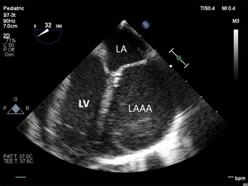
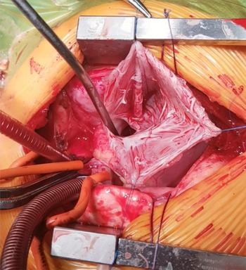
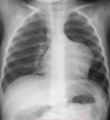
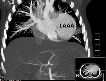
We present the case of a 2-year-old boy with an incidentally detected left atrial appendage aneurysm. The patient had been admitted to the paediatric ward due to lower respiratory tract inflammation. There were no cardiovascular symptoms. The cardiothoracic index was normal in the chest radiography, but there was a remarkable prominence of the superior left heart border. Transthoracic echocardiography revealed a large left atrial appendage aneurysm that compressed the anterolateral wall of the left ventricle. The systolic and diastolic left ventricle functions were preserved. The electrocardiogram showed normal sinus rhythm without any signs of LA overgrowth, and 24-hour Holter electrocardiogram monitoring did not detect any cardiac arrhythmias. Angio-CT demonstrated a 50 × 38 × 49 mm left atrial appendage aneurysm. There was no compression of the pulmonary veins and arteries. Preoperative transesophageal echocardiography confirmed the left atrial appendage aneurysm and free blood flow between it and the left atrium. No thrombus was detected inside the left atrial appendage aneurysm. The patient was qualified for left atrial appendage aneurysm resection. The left atrial appendage aneurysm was approached by median sternotomy and resected under cardiopulmonary bypass. The aneurysm entry was located above the mitral valve and left upper pulmonary vein ostium. The aneurysm sac was excised; afterwards, the left atrial wall was reconstructed using the patient’s own tissues. There were no surgical complications. At the 3-year follow-up, there were no abnormalities detected by transthoracic echocardiography including the normalisation of the left ventricle dimensions and shape.
The left atrial appendage aneurysm is an extremely rare anomaly caused by dysplasia of the pectinate and adjacent muscles of the left atrium. Reference Victor and Nayak1 Most cases are congenital. The lesion is rarely diagnosed during childhood. Reference Zhang, Li and Cao2 Usually, it is detected due to investigation of symptoms such as palpitations, related to atrial arrhythmias, dyspnoea, thoracic pain related to compression of the left coronary artery and cerebrovascular accidents. Sudden death is a feared outcome. Reference Pomerantzeff, Freyre, de Almeida Brandao, Pereira Barreto and de Oliveira3 The diagnosis is based on imaging techniques, in particular, transthoracic and transoesophageal echocardiography. Other diagnostic tools, such as CT, MRI and CT angiography, may put in perspective important extracardiac and intracardiac structures related to the left atrial appendage aneurysm and help in surgical planning. Early surgical intervention is recommended once a diagnosis is made, in order to prevent associated morbidity and mortality. Reference Aryal, Hakim and Ghimire4 Various successful approaches to aneurysmectomy with or without a cardiopulmonary bypass have been described. Reference Victor and Nayak1,Reference Aryal, Hakim and Ghimire4 In this case, a median sternotomy with a cardiopulmonary bypass was selected as the best tactic to performing a complete and precise resection of the left atrial appendage aneurysm, thus preventing inadvertent injury to the adjoining cardiac structures.
In summary, although the left atrial appendage aneurysm is a rare malformation, it causes increased morbidity and mortality. Early and accurate diagnosis enable an appropriate treatment that prevents long-term complications. Reference Aryal, Hakim and Ghimire4
Transoesophageal echocardiogram showing a large left atrial appendage aneurysm overlaying and plateauing the left ventricle free wall (LAAA, left atrial appendage aneurysm; LA, left atrium; LV, left ventricle).

Intraoperative view of the opened aneurysm sac. Notice the relatively small entry to the left atrium.

Preoperative chest radiography. Notice the remarkable bulging of the upper-left edge of the heart outline.

Preoperative CT scan showing the left atrial appendage aneurysm (LAAA, left atrial appendage aneurysm).

Acknowledgements
We thank the anonymous referees for their useful suggestions.
Conflicts of interest
None.