Erythropoietin (EPO) is the hormone necessary for the production of red blood cells that function primarily to transport oxygen from the lungs to tissues. EPO is synthesised in the adult kidney and can be induced by anaemia or hypoxic stress to increase the number of red blood cells and oxygen transport. EPO secreted into the circulation targets the haematopoietic stem cell in the bone marrow to stimulate differentiation along the erythroid lineage and acts as a survival factor for erythroid progenitor cells at the CFU-E (erythroid colony-forming unit) stage. EPO is essential for regulation of haematocrit since the red blood cell lifespan of 120 days requires the production of about two million red blood cells per second. Human EPO was first purified in 1977 from the urine of aplastic anaemia patients (Ref. Reference Miyake, Kung and Goldwasser1). Cloning of the EPO gene led to the production of recombinant EPO (Refs Reference Lin2, Reference Jacobs3) and its clinical use for the past two decades for the treatment of anaemia such as that associated with chronic kidney disease. Increased availability of the recombinant protein has facilitated structural and functional studies of EPO and its receptor, and the discovery of its pleiotropic activity.
Erythropoietin and red blood cell production
EPO protein
Structural studies of human EPO revealed that it is a single polypeptide of 166 amino acids folded into four α-helices linked by two disulphide bridges, between cysteines 6 and 161 and between cysteines 29 and 33 (Refs Reference Lin2, Reference Jacobs3, Reference Lai4). EPO shares structural homology with growth hormone and other members of the haematopoietic class 1 cytokine superfamily (Ref. Reference Wen5). The molecular mass varies from 30 to 34 kDa, depending on the carbohydrate content. The composition of EPO is about 60% protein and 40% carbohydrate, with three N-glycosylation sites at asparagines 24, 38 and 83, and one O-glycosylation site at serine 126 (Ref. Reference Sasaki6). Each N- and O-glycosylation site can accommodate up to four and two sialic acid residues, respectively, although O-linked glycosylation of EPO does not appear to be required for in vivo biological activity (Refs Reference Wasley7, Reference Higuchi8). Nonsialated EPO is rapidly cleared from the circulation via the galactose receptor in the liver (Refs Reference Fukuda9, Reference Tsuda10).
Site of EPO production
During development, definitive erythropoiesis takes place in the fetal liver. EPO is produced primarily in the fetal liver by hepatocytes (Refs Reference Zanjani11, Reference Dame12). In addition to EPO production in fetal liver, EPO gene expression in kidney begins from about 18 weeks in humans, and, at birth, when the site of haematopoiesis switches to the bone marrow, the kidney becomes the predominant EPO-producing organ (Refs Reference Obara13, Reference Juul, Yachnis and Christensen14, Reference Koury15).
EPO expression in fetal liver, adult liver and adult kidney is inducible by hypoxia (Refs Reference Obara13, Reference Kochling, Curtin and Madan16, Reference Bondurant and Koury17). In the mouse during hypoxic induction of Epo, the adult liver can contribute as much as 20–50% of the total Epo produced (Refs Reference Tan, Eckardt and Ratcliffe18, Reference Fandrey and Bunn19). (Note that the nomenclature Epo/EpoR is used in this article when referring specifically to the mouse.) In situ hybridisation and studies in transgenic mice, including mice carrying a reporter gene inserted into a large Epo gene fragment, were useful in localising specific Epo-producing cells; in the kidney these have been identified as the interstitial peritubular cells with neural characteristics (Refs Reference Obara13, Reference Maxwell20, Reference Koury, Bondurant and Koury21). Hypoxic induction of EPO production up to 150-fold or more in the kidney results mainly from an increase in the number of EPO-producing cells rather than from an increase in EPO production per cell (Refs Reference Obara13, Reference Tan, Eckardt and Ratcliffe18, Reference Eckardt22). Regulation of EPO expression in the liver and kidney appears to require different flanking gene elements, which are located immediately 3′ for liver expression and 6–14 kb 5′ for kidney expression (Refs Reference Kochling, Curtin and Madan16, Reference Semenza23).
Regulation of EPO
EPO induction by hypoxia is a critical response for regulation of red blood cell production by anaemic stress or chronic hypoxia. At the cellular level, EPO production is mainly determined by its gene transcriptional activity. The EPO gene has a 3′ enhancer active in hepatocytes that contains a hypoxia-responsive element (HRE) and provides hypoxic induction via interaction with multiple transcription factors including the hypoxia-inducible factor HIF (Refs Reference Semenza and Wang24, Reference Galson25, Reference Ebert and Bunn26, Reference Metzen and Ratcliffe27). HIF is an important regulatory factor for activation of various genes in response to physiological stress or hypoxia, such as those encoding vascular endothelial growth factor (VEGF), EPO and the glucose transporter GLUT-1 (SLC3A1).
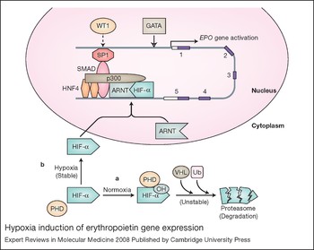
HIF consists of a constitutive β-subunit (also called the arylhydrocarbon receptor nuclear translocator; ARNT) that localises in the nucleus, and an α-subunit [HIF-1α (HIF1A), HIF-2α (EPAS1) or HIF-3α (HIF3A)] found in the cytoplasm (Refs Reference Metzen and Ratcliffe27, Reference Smith, Robbins and Ratcliffe28). At normoxia, the α-subunit is proline hydroxylated, which leads to ubiquitination by interaction with the von Hippel–Lindau protein and rapid degradation by the proteasome. The α-subunit is stabilised by hypoxia and is transported to the nucleus where it forms a dimer with ARNT to affect specific gene activation, depending in part on the α-subunit (Refs Reference Jacobs3, Reference Hu29) (Fig. 1). Induction of transcription through the 3′ HRE in the EPO gene in Hep3G hepatocytes has provided much insight into the regulation of HIF-1 activity by hypoxia (Ref. Reference Ebert and Bunn26). Human genetic mutations that disrupt the normal function of proline hydroxylase or von Hippel–Lindau protein have been identified as causes for abnormally high levels of EPO production that result in polycythaemia (Refs Reference Percy30, Reference Ang31). Manipulating levels of HIF-1α and HIF-2α using RNA interference and genetically modified mouse models indicate that HIF-2α plays a critical role in erythropoiesis and Epo hypoxic induction (Refs Reference Warnecke32, Reference Rankin33, Reference Chavez34, Reference Gruber35), while HIF-1α preferentially activates VEGF and GLUT-1 (Ref. Reference Zelzer36). In patients with excessive erythrocytosis, a gain of function mutation in the HIF-2α gene has been determined to be a genetic lesion in one family, demonstrating that HIF-2α is an important contributor to EPO regulation (Ref. Reference Percy37).
Hypoxia induction of erythropoietin gene expression. (a) At normoxia, the hypoxia-inducible factor HIF-α is hydroxylated at its prolines by prolyl hydroxylase (PHD), which enables interaction with the von Hippel–Lindau (VHL) protein and ubiquitin (Ub); HIF-α is then polyubiquitinated and degraded by the proteasome. (b) Under hypoxic conditions, HIF-α is stabilised and translocated to the nucleus, where it binds its partner ARNT/HIF-1β to activate erythropoietin (EPO) gene transcription via binding to a 3′ enhancer (Ref. Reference Semenza and Wang24). Other factors also contribute to EPO transcription through stabilisation of a macromolecular complex at this 3′ site and reinforcement of enhancer–promoter contact: these include HNF4, p300 (EP300), SMAD3 and SP1 (Refs Reference Ebert and Bunn26, Reference Sanchez-Elsner44, Reference Dame52, Reference Bunn210).The open arrowhead indicates that GATA factors can bind to the 5′ promoter of EPO, and can act as an inducer (GATA4) or repressor (GATA2) in EPO-expressing cells and as a repressor (GATA2/GATA3) in nonexpressing cells (Refs Reference Obara13, Reference Dame52). The dashed arrow indicates that WT1 contributes to tissue-specific EPO expression in hepatocytes (Ref. Reference Dame43). The five exons of the EPO gene (shaded) and the 3′ transcribed untranslated region (open) are shown schematically on the representation of DNA. Abbreviations: ARNT, arylhydrocarbon receptor nuclear translocator; p300, E1A binding protein p300 (EP300); HNF4, hepatocyte nuclear factor 4; SMAD3, SMAD family member 3; SP1, Sp1 transcription factor.

Hypoxic regulation of EPO also requires hepatocyte nuclear factor 4 [HNF4 (HNF4A)], which is constitutively expressed in the major sites of EPO production: the fetal liver and the adult kidney. HNF4 binds to the EPO 3′ enhancer adjacent to the HIF-binding site (Ref. Reference Blanchard38). HIF and HNF4 interact to form a macromolecular complex with transcription coactivator p300 (EP300) for activation of stimulus-specific and tissue-specific gene transcription (Refs Reference Ebert and Bunn26, Reference Ebert and Bunn39). In the fetal liver during early definitive erythropoiesis, the transcription factor GATA4 (see next paragraph) and the retinoic acid receptor RXRA contribute to high EPO gene activation (Ref. Reference Makita, Duncan and Sucov40). RXRA competes with HNF4 for occupancy in the 3′ EPO enhancer, and is no longer necessary later in development as HNF4 activity in the fetal liver increases (Ref. Reference Makita41). The Wilms tumour suppressor, WT1, directly affects EPO expression in hepatocytes and its targeted deletion in mice decreases erythropoiesis in fetal liver (Refs Reference Alberta42, Reference Dame43). Transcription factors SMAD3, which binds to the 3′ EPO enhancer, and SP1, which binds to the promoter region, also cooperate with HIF, HNF4 and p300 in EPO gene transcription and induction by hypoxia (Ref. Reference Sanchez-Elsner44). Together these factors act to stabilise the multifactorial complex interacting with the 3′ enhancer and reinforce the promoter–enhancer contact.
The GATA transcription factors bind the DNA motif GATA, and belong to the zinc finger protein family. GATA1, GATA2 and GATA3 are primarily involved in haematopoiesis, and their expression is also seen in some nonhaematopoietic tissues such as the kidney, urinary tract and nervous system (Refs Reference Ma45, Reference Zhou, Yamamoto and Engel46, Reference Pandolfi47, Reference Hoshino48). GATA4, GATA5 and GATA6 are found in the heart, gut and extraembryonic endoderm (Refs Reference Gao49, Reference Peterkin, Gibson and Patient50, Reference Capo-Chichi51). In fetal liver, GATA4 activates EPO gene expression, recruits chromatin-modifying factors to the EPO promoter to confer an open chromatin configuration, and may participate in the switch of EPO gene expression from fetal liver to adult kidney (Ref. Reference Dame52). GATA1, GATA2 and GATA3 can inhibit EPO expression through binding in the 5′ promoter region (Ref. Reference Imagawa, Yamamoto and Miura53). GATA2 recruits histone deacetylases to downregulate EPO expression and mediates suppression of expression by pro-inflammatory cytokines (Ref. Reference La Ferla54). Transgenic mouse studies showed that a single nucleotide mutation in the GATA box of the Epo gene promoter abolishes the repression by GATA2 and GATA3, and causes ectopic expression (Ref. Reference Obara13). Finally, it was shown that EPO itself regulates expression of GATA1 and GATA2 during erythroid differentiation (Ref. Reference Rogers55) and GATA3 in neural cells and skeletal muscle myoblasts (Refs Reference Yu56, Reference Ogilvie57).
EPO receptor
During erythroid differentiation of haematopoietic stem cells, EPO acts through binding of its receptor (EPOR) on the surface of early erythroid progenitor cells to promote cell survival, proliferation and differentiation down the erythroid lineage (Refs Reference Wu58, Reference Lin59). EPOR is expressed at low levels on early erythroid progenitor cells, the BFU-E (erythroid burst-forming unit) (Ref. Reference Broudy60). It is then upregulated during erythroid differentiation and increases by tenfold or more on the erythroid progenitor cells by the CFU-E stage, which requires EPO signalling for protection against apoptosis. EPO binding to erythroid progenitor cells upregulates expression of its own receptor, which increases EPO response as well as expression of erythroid-specific transcription factors such as GATA1, SCL/TAL1 and EKLF (KLF1), and other erythroid-specific genes (Ref. Reference Chiba, Ikawa and Todokoro61). During late erythropoiesis, EPOR expression is downregulated, and EPO is no longer required for cell survival (Ref. Reference Rogers55). The erythroid precursors enucleate to form reticulocytes that continue to synthesise haemoglobin, and then enter into the circulation to mature into red blood cells. EPO activity in haematopoietic tissue indicates that productive EPO signalling depends on both the level of EPOR cell-surface expression and on the local EPO concentration.
The expression of EPOR extends beyond haematopoietic cells and includes endothelial, neural, muscle, cardiovascular and renal tissues (Refs Reference Ogilvie57, Reference Anagnostou62, Reference Morishita63, Reference Wu64, Reference Juul65). EPO acting as a survival factor and promoting proliferation and differentiation of the CFU-E provides insight on its role in nonhaematopoietic cells. EPO is also associated with cytoprotective and/or proliferative activity in these nonhaematopoietic tissues (Ref. Reference Arcasoy66). Thus, in ischaemic response or tissue repair, in addition to indirectly increasing oxygen delivery through increased erythropoiesis, EPO may directly stimulate nonhaematopoietic progenitor cell proliferation or increase cell survival.
EPOR in erythroid progenitor cells
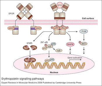
EPOR on the surface of erythroid progenitor cells binds to EPO as a homodimer (Fig. 2). The EPOR polypeptide has a single transmembrane domain and a cytoplasmic domain that lacks tyrosine kinase activity (Ref. Reference Youssoufian67). The human EPOR gene product is encoded as 508 amino acids from eight exons spanning over 6.5 kb (Ref. Reference Noguchi68). The exons vary in length from 29 to 203 amino acids, with the membrane-spanning region encoded in exon 6 and much of the cytoplasmic region encoded in exon 8. The extracellular domain consists of a short α-helix followed by two seven-stranded β-sheet domains (Ref. Reference Syed69). Four of the five cysteine residues in the extracellular domain and a Trp-Ser-X-Trp-Ser motif located proximal to the membrane are conserved across the haematopoietic cytokine superfamily receptors that include the receptors for interleukin (IL)-2, -3 and -4, prolactin and thrombopoietin (Refs Reference D'Andrea and Zhu70, Reference Hilton71). The Trp-Ser-X-Trp-Ser motif contributes to EPOR expression on the cell surface, EPO binding and EPOR activation (Ref. Reference Yoshimura72). With high levels of EPOR expression, only a small fraction is processed into mature EPOR on the cell surface, where it has a half-life of 1 h or less (Ref. Reference Hilton71).
Erythropoietin signalling pathways. The binding of erythropoietin (EPO) to its homodimeric receptor (EPOR) results in a conformation change and transphosphorylation of the associated tyrosine kinase JAK2. JAK2 activation results in phosphorylation of EPOR and STATs. Activated STATs dimerise and translocate to the nucleus to affect specific downstream gene transcription. JAK2 also activates other signal transduction pathways, including the phosphoinositide 3-kinase (PI3K/AKT), mitogen-activated protein kinase (MAPK), and, in nonhaematopoietic cells, nuclear factor (NF)-κB (p50 and p65) pathways. In EPOR, the four conserved cysteines (thin lines) and the proximal conserved Trp-Ser-X-Trp-Ser motif (thick line) in the extracellular domain, as well as the conserved Box1/Box2 regions (heavy lines) in the membrane-proximal cytoplasmic domain, are indicated. Abbreviations: IκB, inhibitor of κB; JAK, Janus kinase; p50 and p65, NF-κB subunits; STAT, ‘signal transducer and activator of transcription’.

As EPOR lacks an intrinsic tyrosine kinase domain, the nonreceptor tyrosine kinase JAK2 is essential for signalling of EPO (Fig. 2) – as is the case for signalling by other ligands of the haematopoietic cytokine receptor superfamily members, such as thrombopoietin and IL-3 (Ref. Reference Parganas73). Studies of JAK2 mutations indicate that JAK2 binding to EPOR at its Box1/Box2 regions (Fig. 2) allows for the progression of JAK2 from an inactive state (when not in contact with EPOR) to an active state with the potential to be activated following EPO stimulation (Ref. Reference Funakoshi-Tago74). EPO binding to the EPOR dimer induces a conformational change that brings the cytoplasmic domains and hence the associated JAK2 proteins in closer proximity to each other, resulting in the transphosphorylation and activation of JAK2 and EPOR (Ref. Reference Livnah75) (Fig. 2). Targeted deletion of Jak2 resembles deletion of Epo or EpoR (Ref. Reference Neubauer76). Mutations in exon 12 of JAK2 that increase JAK2 activity are associated with polycythaemia vera and cytokine-independent growth of bone marrow cells in culture (Refs Reference Baxter77, Reference Levine78, Reference Horn79). These include the substitution of phenylalanine for valine at position 617, detected in approximately 95% of polycythaemia vera patients, and other somatic gain-of-function mutations (Ref. Reference Scott80).
EPO activation of JAK2 via EPOR gives rise to tyrosine phosphorylation of STAT5, its translocation to the nucleus and induction of STAT5 target genes. Complete knockout of Stat5 results in loss of the antiapoptotic proteins and marked decrease in the trans-acting iron-regulatory protein IRP2 in the fetal liver, accompanied with reduction in the transferrin receptor and iron uptake (Ref. Reference Kerenyi81). Mice that lack Stat5 develop a severe embryonic microcytic anaemia and die prior to or shortly after birth. Conversely, activation of Stat5 rescues fetal liver cells from the Jak2- or EpoR-knockout mice and allows erythropoiesis and erythroid progenitor cell proliferation to proceed (Ref. Reference Grebien82).
The EPOR cytoplasmic domain contains tyrosines that provide phosphorylation sites with EPO signalling that act as docking sites for signalling molecules such as the mitogen-activated protein kinase (MAPK), but deletion of the distal cytoplasmic region and associated tyrosines indicate that this region is not required for erythroid mitogenic activity of EPOR (Ref. Reference Miura83). The EPOR C-terminal region also contains a negative-growth-regulatory domain that affects signal transduction or receptor processing (Ref. Reference D'Andrea84), and can hamper EPOR exit from the endoplasmic reticulum and contribute to its degradation (Refs Reference Supino-Rosin85, Reference Nahari86). EPO binding to its receptor mediates endocytosis, and EPO is either resecreted or degraded in lysosomes at a rate determined by its dissociation from its receptor (Refs Reference Gross and Lodish87, Reference Sawyer, Krantz and Goldwasser88). Following EPO binding, EPOR can also be ubiquitinated and the C-terminal portion degraded by proteasomes while still on the cell surface via β-transducin repeat-containing protein (β-TRCP/BTRC) (Ref. Reference Meyer89). Deletion of the C-terminal region that includes the β-TRCP-interacting region and the proximal docking site for the negative regulator SHP1 (a protein tyrosine phosphatase) has been identified in congenital polycythaemia patients (Refs Reference Arcasoy, Harris and Forget90, Reference Kralovics and Prchal91, Reference Kralovics92).
EPOR transcriptional regulation
EPOR is expressed in a cell-restricted fashion and expression depends on the stage of differentiation. The EPOR proximal promoter is characterised by an inverted SP1-binding site (CCGCCC) and an inverted GATA1-binding site (TTATCT) located at positions −151 and −179 from the first codon of the human EPOR gene, respectively (Ref. Reference Noguchi68). The SP1 motif that binds the SP1 transcription factor is required for high-level EPOR promoter activity (Ref. Reference Chin93), and the induction of EPOR by stem cell factor (SCF) has been suggested to be mediated by SP1 (Ref. Reference Sato94). GATA1 binds to the GATA motif and transactivates the EPOR promoter (Refs Reference Chin93, Reference Zon95). In the absence of GATA1, GATA2 can also activate EPOR expression (Ref. Reference Weiss, Keller and Orkin96). However, during erythroid differentiation, GATA1 downregulates GATA2 and EPOR expression follows GATA1 induction by EPO (Ref. Reference Rogers55), indicating that EPO regulates the expression of its own receptor. Additional upregulation of EPOR mediated via antisense EPOR transcripts has recently been suggested (Ref. Reference Zhang97). During terminal differentiation, EPOR is downregulated with decreasing levels of GATA1. CACCC and GC-box motifs are located in the upstream 5′ region, which negatively regulates EPOR promoter activity (Refs Reference Noguchi68, Reference Maouche98). A human EPOR genomic fragment extending 2 kb 5′ and 7 kb 3′ of the coding region was sufficient to provide EPOR expression in haematopoietic tissue in transgenic mice (Ref. Reference Liu, Chin and Noguchi99).
EPOR and development
During embryogenesis, erythropoiesis is initiated in the extraembryonic yolk sac, giving rise to primitive erythropoiesis. In mice, EpoR is expressed in the yolk sac blood islands, but Epo expression is not detected until the shift to definitive erythropoiesis in the fetal liver (Ref. Reference Lee100). In targeted deletion of Epo or EpoR in mice, primitive erythropoiesis proceeds with BFU-E and CFU-E accumulating in the fetal liver (Refs Reference Wu58, Reference Lin59). However, CFU-E require EPO for survival, and progression beyond the CFU-E stage is markedly inhibited, which results in severe anaemia and death around embryonic day E13.5. Close examination of embryos lacking Epo or EpoR also reveals ventricular hypoplasia at embryonic day E12, increased apoptosis in the myocardium and endocardium, and a reduction in the number of cardiac myocytes and endothelial cells (Refs Reference Yu56, Reference Wu64, Reference Yu101). In EpoR-null mice, increased apoptosis is seen in the fetal brain as early as embryonic day E10.5, prior to the onset of anaemia (Ref. Reference Yu56), indicating that EPO may contribute to selective cell survival during organ development. As with erythroid differentiation, EPO may act as an antiapoptotic and mitogenic factor and contribute to progenitor cell survival and proliferation during development. Studies of nonhaematopoietic tissues in animal models and cell culture provide increasing evidence for EPOR expression and EPO activity beyond erythroid cell differentiation (Refs Reference Arcasoy66, Reference Chen, Warin and Noguchi102, Reference Noguchi103) (Fig. 3).
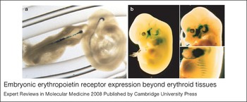
Embryonic erythropoietin receptor expression beyond erythroid tissues. (a) To visualise erythropoietin receptor (EPOR) expression in nonhaematopoietic tissue, a lacZ reporter gene driven by the EPOR promoter was used to produce transgenic mice. In brain, these mice exhibit expression in the neural tube at embryonic day 10.5 (Ref. Reference Liu131). (b) At embryonic day 12.5 (left) and 13.5 (right), reporter gene expression resembles developing skeletal muscle (from Ref. Reference Ogilvie57, with permission from The American Society for Biochemistry and Molecular Biology).

Erythropoietin and vascular endothelium
EPO activity in endothelial cells
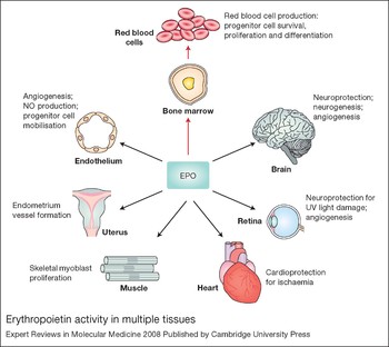
The availability of recombinant EPO as a laboratory reagent provided the opportunity to examine its activity in nonhaematopoietic tissue (Fig. 4). EPO stimulated dose-dependent proliferation and enhanced migration of endothelial cells that expressed the mature erythroid form of EPOR (Refs Reference Anagnostou62, Reference Anagnostou104). EPO treatment of endothelial cell cultures stimulated JAK2, the production of matrix metalloproteinase 2 (MMP2), and the formation of vascular structures (Ref. Reference Ribatti105). EPO protected endothelial cell cultures from anoxia-induced injury, an effect dependent on AKT1 activation and on the maintenance of mitochondrial membrane potential (Ref. Reference Chong, Kang and Maiese106). In vivo, EPO induced a strong proangiogenic response in the chick embryo chorioallantoic membrane (Ref. Reference Ribatti105).
Erythropoietin activity in multiple tissues. Erythropoietin (EPO) acts primarily to regulate erythropoiesis in the bone marrow by stimulating erythroid progenitor cell survival, proliferation and differentiation to produce mature red blood cells. EPO receptor expression on endothelial cells, the endometrium (lining) of the uterus, skeletal muscle myoblasts, the heart, and endothelial cells and neural cells in the retina and brain allows EPO to also act as a survival or mitogenic factor on these nonhaematopoietic cells, providing the potential for a response to EPO in multiple tissues.

Nitric oxide production in EPO-stimulated endothelial cells
Endothelial cells express endothelial nitric oxide synthase (eNOS/NOS3), which produces nitric oxide (NO) to regulate vascular tone, blood pressure homeostasis and blood flow. In endothelial cell cultures, EPO upregulated EPOR and increased endothelial cell sensitivity and response to EPO (Ref. Reference Beleslin-Cokic107). The combination of EPO and hypoxia together induced EPOR, modulated eNOS expression and stimulated NO release in human endothelial cells from umbilical vein and artery (Ref. Reference Beleslin-Cokic107). Animal studies provide evidence for EPO stimulation of NO production in vivo. Rats treated for 14 days with EPO exhibited elevated haematocrit as well as vascular expression of eNOS and basal vasodilation (Ref. Reference Kanagy108). Transgenic mice expressing high levels of human EPO resulting in elevated erythrocytosis had increased eNOS and NO production, and NO-induced relaxation (Ref. Reference Quaschning109). Treatment with the NO synthase inhibitor L-NAME (N-nitro-l-arginine methyl ester) resulted in vasoconstriction, hypertension and death. By contrast, increased erythrocytosis alone without elevated EPO was not associated with increased NO production. Polycythaemia patients with low EPO levels showed no endothelium-dependent vasodilation response to acetylcholine but became responsive with haemodilution (Ref. Reference Defouilloy110). These patients remain responsive to NO and show induced pulmonary vasodilation with inhaled NO. In the transgenic mice with high EPO levels, the increased bioavailability of NO counteracted some of the adverse effects of the very high haematocrit (around 80%) to maintain normotension and to prevent cardiovascular dysfunction (Ref. Reference Quaschning109).
Oestrogen-stimulated EPO production
The angiogenic activity of EPO has been suggested to play a major role in the female oestrus cycle (Ref. Reference Sasaki111). In rodents, oestrogen stimulated Epo production in the uterus and Epo interacted with EpoR in the microvascular endothelial cells in the endometrium to promote angiogenesis (Ref. Reference Yasuda112). Exogenous EPO injected into the uterine cavity of oestrogen-stimulated ovariectomised female mice stimulated blood vessel formation in the endometrium. The endometrial transition from dioestrus to proestrus was blocked by inhibition of endometrial growth and angiogenesis via injection of soluble EPOR that can bind EPO and block its activity. Oestrogen activation of EPO production and the resultant angiogenic response suggest that EPO is one of the oestrogen-regulated angiogenic factors that along with VEGF maintain the cyclic uterine angiogenesis in the oestrus cycle (Ref. Reference Sasaki111).
Embryonic angiogenesis and mobilisation of endothelial progenitor cells
EPO also appears to contribute to embryonic angiogenesis. During mouse development, the vasculature and heart exhibit high levels of Epo and EpoR expression (Refs Reference Wu64, Reference Yu101, Reference Kertesz113). In mice, targeted deletion of Epo or EpoR severely affected angiogenesis and decreased the complexity of the vessel networks, resulting in narrower vessel diameter and fewer branches (Ref. Reference Kertesz113). These changes were observed two days prior to the severe anaemia associated with loss of definitive erythropoiesis in the Epo- or EpoR-null mice, and are consistent with the pattern of Epo and EpoR expression observed in wild-type embryos (Refs Reference Wu64, Reference Yu101, Reference Kertesz113).
This angiogenic activity associated with endogenous EPO during development may relate to the EPO stimulation of endothelial progenitor cell mobilisation or proliferation (Ref. Reference Heeschen114). In adult mice, EPO treatment increased the number of stem and progenitor cells including endothelial progenitor cells in the bone marrow, spleen and peripheral blood (Ref. Reference Heeschen114). Patients with coronary heart disease showed a significant association of EPO and VEGF with the number of circulating endothelial progenitor cells (Ref. Reference Heeschen114), and EPO treatment in patients with renal anaemia or normal volunteers significantly increased mobilisation of circulating endothelial progenitor cells in the peripheral blood (Ref. Reference Bahlmann115). In a mouse model of vascular injury, EPO treatment for three days following wire injury of the femoral artery induced the AKT–eNOS pathway and NO synthesis, increased mobilisation of circulating endothelial progenitor cells, promoted re-endothelialisation, and inhibited neointimal hyperplasia (Ref. Reference Urao116). Mobilisation of endothelial progenitor cells and their recruitment to the pulmonary vasculature and hypoxia activation of NOS in lung were greatly impaired in mice that lack EpoR in the cardiovascular system (Ref. Reference Satoh117). The development of pulmonary hypertension and pulmonary vascular remodelling were accelerated upon exposure to reduced oxygen for 3 weeks compared with wild-type mice. These mice also exhibited poorer response in a model for hind-limb ischaemia using femoral artery ligation. Blood flow recovery, activation of VEGF or its receptor VEGFR2 (KDR), and mobilisation of endothelial progenitor cells were also impaired (Ref. Reference Nakano118). These differences persisted even after transplantation of wild-type bone marrow. Mice expressing EpoR only in haematopoietic tissue also exhibited reduction in VEGF-induced growth of blood vessels. These vascular responses relating to EPO activation of endothelial progenitor cells or loss of EPOR in nonhaematopoietic tissue indicate that activity of endogenous and exogenous EPO extends beyond erythropoiesis and that EPO can mediate significant vascular effects.
EPO and retinopathy
Anaemia in preterm infants is associated with reduced EPO levels (Refs Reference Saito119, Reference Stockman120), and EPO with supplemental iron reduced transfusion needs in infants with birthweights <1000 g without development of severe retinopathy of prematurity (Refs Reference Ohls121, Reference Turker122). However, the progression of retinopathy of prematurity has been linked to extremely low birth weight and exposure to exogenous EPO (Ref. Reference Fortes Filho123). Retinopathy of prematurity begins with reduced vascular growth followed by hypoxia-induced neovascularisation that correlates with elevated VEGF levels (Ref. Reference Aiello124). In a mouse model of retinopathy of prematurity, knockdown of HIF-2α decreased the hypoxic induction of Epo and protected the retina from neovascularisation, suggesting that EPO is the HIF-2α target gene that is responsible for retinopathy of prematurity (Ref. Reference Morita125). Therefore, although early EPO stimulation of endothelial cells as a survival factor has the potential to be protective, as a mitogenic factor EPO may contribute to later development of retinopathy (Ref. Reference Chen126).
In diabetic retinopathy, EPO administration was observed to improve macular oedema, stable vision and fluorescein angiography (Ref. Reference Friedman, Brown and Berman127). However, elevated EPO and VEGF were found in the vitreous fluid of patients with proliferative diabetic retinopathy and branch retinal vein occlusion (Ref. Reference Friedman, Brown and Berman127). A recently discovered polymorphism in the EPO gene promoter is linked to elevated EPO levels in the vitreous body and to diabetic retinopathy, suggesting a possible causal relationship (Ref. Reference Tong128). In a murine model, blocking Epo signalling inhibited ischaemia-induced retinal neovascularisation (Ref. Reference Watanabe129). The vitreous and plasma EPO levels do not correlate, suggesting a local source for elevated EPO in the vitreous fluid. The association of increased circulating endothelial progenitor cells with diabetic retinopathy suggests systemic vasculogenesis contributes to retina neovascularisation rather than local angiogenesis (Ref. Reference Lee, Chae and Kim130).
The mouse model of oxygen-induced retinopathy illustrates that early EPO administration when the endogenous Epo level in the retina is low protected the retina from hyperoxia-induced vaso-obliteration and reduced hypoxia-induced retinal neuron apoptosis (Ref. Reference Chen126). This contrasts with adverse effects observed with later EPO administration when endogenous Epo level is elevated and increased neovascularisation is observed (Ref. Reference Chen126). Therefore, the protective and adverse effects associated with EPO in retinopathy depend on the timing of EPO elevation and indicate that variation in EPO response depends on the local bioavailability of endogenous and exogenous EPO.
Erythropoietin and skeletal muscle
EPOR expression in myocytes
Transgenic mice carrying a reporter gene driven by the EPOR promoter showed reporter gene expression in the developing embryo (Refs Reference Ogilvie57, Reference Liu131). In particular, transgene expression appeared to follow expression of the basic-helix–loop–helix muscle transcription factor MYF5 (Refs Reference Ogilvie57, Reference Patapoutian132) (Fig. 3b). Cultures of primary mouse myoblasts from skeletal muscle demonstrated EpoR expression and a proliferative response to EPO (Ref. Reference Ogilvie57). EPO stimulated expression of EpoR, and the transcription factors MYF5, MYOD1 and GATA3. As observed during erythroid differentiation, EpoR was downregulated during myoblast differentiation and was not detected on mature skeletal muscle. This contrasts with EpoR in myocardium where EpoR expression has been found to persist in adult myocardiocytes (Ref. Reference Calvillo133).
Erythropoietin and heart
Endogenous EPO and heart development
In heart, EPOR is first detected during midgestation (Ref. Reference Wu64) and persists through adulthood (Refs Reference Calvillo133, Reference Wright134). Mice with targeted deletion of Epo or EpoR exhibited apoptosis in endocardium and myocardium, ventricular hypoplasia and a reduction in the number of proliferating cardiomyocytes at embryonic day 12–13 (Refs Reference Yu56, Reference Wu64, Reference Yu101). Although haematopoietic rescue of the EpoR-null genotype resulted in adult mice with no apparent heart morphological defect (Ref. Reference Suzuki135), after myocardial ischaemia–reperfusion these mice showed an increase in infarct size and cardiomyocyte apoptosis compared with wild-type mice (Ref. Reference Tada136). Therefore, endogenous Epo may contribute to heart tissue maintenance or repair in rodents, suggesting the potential for cardioprotection with exogenous EPO administration.
EPO and cardioprotection
Animal models of heart ischaemia or ischaemia–reperfusion injury provide further evidence for the cardioprotective activity of EPO. In rabbits, injection of EPO at the onset of permanent occlusion of the coronary artery reduced infarct size, reduced left ventricle size and improved left ventricle ejection fraction (Ref. Reference Parsa137). After myocardial ischaemia–reperfusion injury in rats, EPO injected intraperitoneally for 7 days reduced loss of cardiomyocytes by 50% (Ref. Reference Calvillo133). In a mouse model for myocardial infarction, a single injection of EPO intraperitoneally following permanent ligation of the left descending coronary artery reduced apoptosis in the area at risk 24 h later and even reduced infarct size at 8 weeks after insult (Ref. Reference Moon138). EPO administration protected against the myocardial ischaemia–reperfusion injury and inflammatory response by phosphoinositide 3-kinase (PI3K) activation of the transcription factor AP-1 (FOS) and increasing eNOS and NO production (Ref. Reference Rui139). In contrast, EPO protection was lost in mice with targeted deletion of cardiac AP-1. The requirement for eNOS in the cardioprotective activity of EPO was demonstrated by the significant reduction in EPO protection in myocardial ischemia–reperfusion injury in eNOS-null mice (Ref. Reference Burger140).
The myocardioprotective effects of EPO may result from a direct antiapoptotic effect on cardiomyocytes and/or from an indirect EPO response by endothelial cells in the heart (Ref. Reference Narmoneva141). In mice, EPO treatment initiated 3 weeks after coronary artery ligation as a model for chronic heart failure induced mobilisation of endothelial progenitor cells from the bone marrow, increased their homing to cardiac microvasculature, and increased myocardial VEGF expression and neovascularisation (Ref. Reference Westenbrink142). In a rat model of postmyocardial infarction heart failure, initiation of EPO treatment 3 weeks after insult promoted neovascularisation, and improved cardiac function and capillary density at 9 weeks, although no difference in infarct size was observed (Ref. Reference van der Meer143). Therefore, EPO cardioprotective activity in rodents may be effective even after the initial 24 h following insult and may improve cardiac function beyond a reduction in infarct size.
Erythropoietin and brain
EPO production and EPOR expression in brain
EPO is produced by astrocytes and neurons and regulated by HIF-2α in a hypoxia-dependent manner (Refs Reference Chavez34, Reference Masuda144), and thus provides a source for EPO on the brain side of the blood–brain barrier. EPO is expressed in human fetal and adult brain and in brains of nonhuman primates and other mammals, and is found in cerebrospinal fluid (Refs Reference Juul65, Reference Marti145, Reference Marti146). Examination of EpoR in the developing mouse revealed EpoR expression in regions associated with neurogenesis (Ref. Reference Yu56); expression localises to the neural tube at midgestation (Fig. 3a) at levels comparable to adult bone marrow (Ref. Reference Liu131). Epo expression follows EpoR expression by ~24 h in neural crest cells and mesenchyme-derived cells (Ref. Reference Tsai147). EpoR was downregulated as neural progenitor cells differentiated to mature neurons (Ref. Reference Chen148) and by birth was about 1% or less than the level at embryonic day 10.5 (Ref. Reference Liu131). EPOR mRNA in human fetal brain is the mature erythroid form but is processed less efficiently than in erythroid cells (Ref. Reference Chin149). In adult brain from humans, nonhuman primates and rodents, the hippocampus, cortex and midbrain exhibit EPO binding (Refs Reference Dame12, Reference Marti146).
EPO and neuroprotection
EPO stimulated neural progenitor cell proliferation, and promoted neural differentiation and mature neuron survival (Refs Reference Yu56, Reference Morishita63, Reference Chen148, Reference Shingo150, Reference Studer151). EPO was neuroprotective against glutamate toxicity and hypoxia (Refs Reference Yu56, Reference Morishita63). In NT2 cells with neural characteristics, EPO induced GATA3, a GATA factor required for normal brain development, and GATA3 transactivated EPOR expression via the GATA1-binding motif (Refs Reference Pandolfi47, Reference Yu56). Hypoxia increased EPOR expression in neural cell cultures, and therefore EPO response (Refs Reference Yu56, Reference Chin149), although EPOR does not appear to be regulated by HIF. During development, EpoR-null mice had smaller brain size with a thinner layer of neuroepithelium prior to death in utero from severe anaemia (Ref. Reference Yu56). Neural cells from these mice proliferated more slowly and were less tolerant to hypoxia. Mice with EpoR expression restricted to haematopoietic tissue survived through adulthood with normal haematocrit but exhibited increased apoptosis in the developing brain, neural cells with increased sensitivity to hypoxia, and decreased neural cell proliferation and increased sensitivity to glutamate toxicity in adult brain (Ref. Reference Suzuki135). Adult mice with knockdown of EpoR brain expression showed defective neural progenitor cell migration to the peri-infarct cortex in response to ischaemic stroke (Ref. Reference Tsai147). The perturbation in neural cell response with loss of EpoR expression suggests that endogenous Epo is neuroprotective in embryonic and adult mice.
In gerbils, infusion of soluble EPOR in brain during mild brain ischaemia resulted in neural degeneration and impaired learning (Ref. Reference Sakanaka152). Rodents show increased EpoR in the periphery of ischaemic brain injury, and EPO administration in brain reduced injury and improved performance in the Morris water maze test (Refs Reference Sadamoto153, Reference Bernaudin154). In mice, the increased EpoR expression in ischaemic stroke was followed by Epo production in brain, and exogenous EPO reduced infarct size (Ref. Reference Bernaudin155). These studies suggest the induction of EPOR followed by endogenous EPO is neuroprotective in response to ischaemic injury and that increasing EPO bioavailability can significantly reduce injury and promote repair. In ischaemic preconditioning, a period of mild brain ischaemia provides resistance to severe ischaemic insult. HIF activation in preconditioning increased VEGF and EPO, which can contribute to tolerance to brain ischaemia (Ref. Reference Bernaudin154). Soluble EPOR directly administered to the brain blocked the protective effect of preconditioning (Refs Reference Prass156, Reference Malhotra157, Reference Ruscher158). EPO administration in the retina mimicked hypoxia preconditioning and protected photoreceptors from ultraviolet light damage (Ref. Reference Grimm159). Protection was also observed in mice expressing high levels of human EPO (Ref. Reference Grimm160).
EPO levels in the central nervous system did not correlate with serum levels without blood–brain barrier dysfunction in patients with traumatic brain injury (Ref. Reference Marti145). In patients administered large doses of EPO intravenously, increase of EPO in the cerebrospinal fluid depended on disruption of the blood–brain barrier (Ref. Reference Xenocostas161). Animals treated with systemic EPO showed brain uptake of less than 0.1% in the absence of injury to the blood–brain barrier (Ref. Reference Banks162). Nevertheless, systemic or intraperitoneal EPO administration in rodent models of brain ischaemia or trauma was neuroprotective (Refs Reference Brines163, Reference Sun164, Reference Yatsiv165), suggesting that the blood–brain barrier was compromised for EPO permeability or that EPO neuroprotective activity is indirect.
EPO signalling in neural cells
EPO stimulates JAK2 activation in both erythroid progenitor cells and in neural cells. Brain ischaemia in transgenic mice with increased EPO in brain resulted in increased activation of Jak2, ERK1/2 (MAPK1/3) and AKT, and induction of Bcl-xL (Ref. Reference Kilic166). Hippocampal neuron cultures showed EPO treatment maintained mitochondrial membrane potential and inhibited cytochrome c release and caspase activity (Ref. Reference Chong, Kang and Maiese167). In mice with targeted deletion of Stat5, hippocampal neurons showed loss of EPO neurotrophic activity but EPO neuroprotection remained (Ref. Reference Byts168). The PI3K signalling pathway was implicated in EPO protection of hippocampal CA1 neurons in a rat model for ischaemic stroke (Ref. Reference Zhang169). While EPO signalling in neural cells is similar to that in erythroid progenitor cells, the participation of NF-κB is only associated with EPO neuroprotection (Ref. Reference Digicaylioglu and Lipton170) (Fig. 2). Epo antisense RNA decreased Jak2/Stat5 and NF-κB in hypoxia preconditioning and blocked the protective activity of hypoxia preconditioning (Ref. Reference Liu171).
EPO neuroprotection and angiogenesis
In brain, EPOR is expressed on both endothelial and neural cells (Ref. Reference Chen, Warin and Noguchi102). The mild brain phenotype in mice that lack EpoR in neural cells may result from EPO response by endothelial cells in the brain (Ref. Reference Wang172). In a rodent model of ischaemic stroke, EPO treatment increased VEGF, brain-derived neurotrophic factor (BDNF), neurogenesis and angiogenesis. EPO treatment of endothelial cells cocultured with neural progenitor cells increased matrix metalloproteinase production by endothelial cells and neural cell migration (Ref. Reference Wang173). EPO stimulation of neural progenitor cells without hypoxic stress increased VEGF production modestly and induced endothelial expression of VEGFR2, resulting in enhanced angiogenesis by VEGF (Ref. Reference Wang174). These studies suggest the possibility that EPO neuroprotection may result from a direct effect of EPO on neural progenitor cells and neurons or may be mediated through other cell types such as endothelial cells. Endothelial cells can also increase eNOS activity in response to EPO, and therefore can also affect oxygen delivery to the brain beyond red blood cell production.
Therapeutic potential in brain diseases
High-dose EPO administration provided long-term neuroprotection and neurogenesis in a neonatal model of ischaemic stroke in rodents (Ref. Reference Gonzalez175). Juvenile rats subjected to acute brain injury and treated with EPO for 2 weeks showed prevention of cognitive dysfunction and brain atrophy 3 and 9 months after injury (Ref. Reference Siren176), providing support for use of EPO for progressive neurodegenerative disorders. A proof-of-concept study for EPO treatment in ischaemic stroke was initiated and indicated the potential for EPO benefit (Ref. Reference Ehrenreich177). Similarly, EPO showed promise for improving clinical outcome in a pilot study for EPO treatment in multiple sclerosis (Ref. Reference Ehrenreich178). A Phase II study of 39 schizophrenic patients demonstrated an association between EPO treatment and improved cognitive function (Ref. Reference Ehrenreich179). Elucidation of the mechanisms for EPO neuroprotective effects in brain disorders to reduce injury and possibly improve psychopathology will give new insight for potential treatment.
Other forms of EPO and its receptor
Modifications of EPO protein
Recombinant human EPO (rhEPO), which is about 40% carbohydrate and 60% protein, is produced as a combination of several glycoforms that are distinct from human serum EPO (Ref. Reference Skibeli, Nissen-Lie and Torjesen180). Although not required for in vitro binding, carbohydrate moieties provide in vivo stability to the EPO protein and resistance to thermal inactivation (Refs Reference Fukuda9, Reference Tsuda10). The addition of N-linked carbohydrates decreased receptor-binding activity but increased rhEPO serum half-life and in vivo activity (Ref. Reference Elliott181). Prolonged in vivo activity reduced the frequency required for therapeutic EPO administration in adult patients with chronic kidney disease and a single dose in anaemic preterm neonates accelerated effective erythropoiesis (Refs Reference Ling182, Reference Warwood183). Other approaches to extend the in vivo half-life of EPO include homodimerisation by linking EPO via a 17 amino acid flexible peptide, or multimerisation to dimers and trimers via chemical crosslinking; these increased the biological activity of EPO in vitro and in vivo (Refs Reference Sytkowski184, Reference Sytkowski185). A fully synthetic EPO of 166 amino acids similar to the native protein with two branched polymer moieties covalently attached significantly increased EPO activity in vitro and extended the half-life in vivo (Refs Reference Kochendoerfer186, Reference Topf187). A third generation of EPO includes modification by polyethelene glycone to also extend in vivo half-life and, like the first generation of pharmacological EPO, was shown to reduce anaemia in patients with renal disease (Refs Reference Topf187, Reference Macdougall188).
Non-EPO-based erythroid-stimulating agents
Small peptides, or EPO mimetics with sequences unrelated to EPO, can activate the EPOR homodimer (Refs Reference Livnah75, Reference Wrighton189). An antibody that targets the extracellular domain of human EPOR was also used as the basis to develop an EPO mimic that was active in vivo in a mouse model expressing human EPOR and appeared to bind in a symmetric manner to EPOR distinct from the two asymmetric binding sites for EPO (Ref. Reference Liu190).
An alternate therapeutic strategy for anaemia is to activate the hypoxic regulatory mechanisms associated with activation of EPO production, a treatment approach that was shown to induce endogenous EPO production in rhesus macaques (Refs Reference Bernhardt191, Reference Hsieh192).
Nonerythroid forms of EPO and its receptor
Efforts to segregate EPO activity in erythropoiesis from its nonhaematopoietic protective effects have led to investigation of alternative forms of EPO. Excessive EPO can result in high haematocrit and high blood viscosity, which in an animal model can lead to degenerative processes and increased vascular permeability in liver and kidney, degeneration of nerve and muscle fibres and decreased neuromuscular junctions, and a general reduction in life expectancy (Ref. Reference Heinicke193). The short half-life of EPO stripped of sialic acid (asialoerythropoietin) is not sufficient to increase erythropoiesis in vivo but has been reported to be neuroprotective in animal models (Ref. Reference Erbayraktar194). Other EPO modifications such as carbamylation have been proposed to result in EPO that acts specifically in nonhaematopoietic cells without stimulation of erythropoiesis (Ref. Reference Fiordaliso195). Carbamylated EPO did not bind the classical EPOR or stimulate haematopoietic activity in select animal models (Ref. Reference Leist196). Neuroprotective activity observed with carbamylated EPO appears to be associated with upregulation of the extracellular signalling molecule SHH (sonic hedgehog) (Ref. Reference Wang197). The structure of the receptor for carbamylated EPO and the role of EPOR in the receptor complex have not yet been determined.
A specific nonhaematopoietic form for the receptor to native EPO has been proposed consisting of a heteromeric complex between EPOR and the common cytokine β receptor (Ref. Reference Brines198). Beyond this initial report, the precise structure for this alternative EPO receptor heterocomplex has not yet been elucidated. Interestingly, this alternative form of EPO receptor may not be necessary for EPO neuroprotective activity since neural cells that lack the common cytokine β receptor exhibit EPO protection and, unlike EPOR, the common cytokine β receptor is barely detected in astrocytes and neurons in the hippocampus, which exhibits EPO protection in a neurodegenerative rodent model (Refs Reference Um, Gross and Lodish199, Reference Nadam200). Nevertheless, efforts continue in searching for a nonhaematopoietic-specific EPO receptor and in developing agents like asialoerythropoietin and peptide mimetics capable of stimulating an EPO response in nonhaematopoietic cells/tissue without increasing erythropoiesis.
Adverse effects associated with EPO therapy
EPO, anaemia and chronic renal disease
The potential for adverse effects of EPO action was suggested in patient studies in which exogenous EPO was used to elevate haematocrit to bring it within normal range. For example, EPO therapy in two trials of chronic renal disease was able to correct anaemia, but normalising haematocrit did not reduce the risk of cardiovascular events (Refs Reference Drueke201, Reference Singh202). Treatment of patients with EPO to reach a target haemoglobin level of 11.3 g/dl or 13.5 g/dl showed an increase incidence of composite events (death, myocardial infarction, hospitalisation for congestive heart failure and stroke) in the high haemoglobin group, and more patients in this group had one or more serious events (Ref. Reference Singh202). In a separate study involving patients with a milder anaemia, correction of anaemia to the 13 to 15 g/dl range compared with a subnormal range of 10.5 to 11.5 g/dl was associated with improved quality of life but did not change the incidence of the first cardiovascular event or combined adverse events, and was related to increased hypertensive episodes, headaches and the number of patients requiring dialysis (Ref. Reference Drueke201). Studies such as these illustrate the difficulty in determining that appropriate target haemoglobin level in EPO therapy and the potential for increased adverse effects using target haemoglobin levels in or close to the normal range.
Anaemia in cancer patients and EPO therapy
Several clinical studies have examined EPO therapy to treat anaemia in cancer patients and these are reviewed in greater detail elsewhere (Ref. Reference Jelkmann203). For example, EPO treatment in anaemic cancer patients receiving nonplatinum chemotherapy was associated with reduced transfusion requirements, increased haemoglobin and improvement of quality of life (Ref. Reference Littlewood204). In a specific cancer type, patients receiving chemoradiotherapy for squamous cell carcinoma of the oral cavity and oropharynx followed by resection of the primary tumour bed and neck dissection, showed increased cancer control and survival rates with EPO treatment without supplemental iron (Ref. Reference Glaser205). The associated benefit of EPO treatment in cancer patients with anaemia led to efforts to maintain haemoglobin in the normal range.
By contrast to earlier studies, EPO treatment to increase or maintain haemoglobin levels close to or in the normal range drew attention to potential adverse effects. In patients with metastatic breast cancer irrespective of chemotherapy, a trial to maintain normal haemoglobin concentration was terminated early due to significant increase in disease progression and thrombotic and vascular events (Ref. Reference Leyland-Jones206). In anaemic patients with head and neck cancer undergoing radiotherapy, a double-blind placebo-control trial using EPO therapy to normalise haemoglobin resulted in bringing haemoglobin into the normal range (>14 g/dl in women and >15 g/dl in men), but also showed decreased benefit in cancer control and survival in the EPO treatment group compared with placebo (Ref. Reference Henke207). Despite imbalance of risk factors and heterogeneity of the study population, these studies illustrate the adverse effects associated with high haemoglobin targets in cancer patients, particularly in metastatic breast cancer and head and neck cancer.
An evaluation of clinical trials comparing EPO treatment with placebo or standard of care for anaemia in cancer patients underscored the associated risk of venous thromboembolism with EPO administration and increased mortality in select solid cancers (Ref. Reference Bennett208). The recommendations for EPO treatment for low haemoglobin level (approaching or <10 g/dl) for cancer patients with chemotherapy-related anaemia are reviewed elsewhere (Ref. Reference Rizzo209). While EPO has been shown to be useful in increasing haemoglobin level in cancer patients with chemotherapy-associated anaemia, awareness of potential adverse events raises several questions and concerns, including the appropriate target haemoglobin level, the role of supplemental iron, specific tumour-type response to EPO, variation in individual response, and the direct effect of EPO in stimulating nonhaematopoietic tissue response or cancer cell growth (Ref. Reference Jelkmann203).
Conclusions
CFU-Es are highly sensitive to EPO due to their markedly elevated level of EPOR expression. EPO itself induces expression of its receptor in CFU-E via induction of the erythroid transcription factor GATA1. By contrast, EPOR expression in nonhaematopoietic cells lacks the transactivation by GATA1 characteristic of erythroid gene expression. As a consequence, EPOR levels and EPO response are more modest in endothelial cells, cardiomyocytes and neurons. Nevertheless, the survival and proliferative activities of EPO that are required for red blood cell formation appear to extend to other EPOR-expressing cells, resulting in EPO protective activity associated with stress or ischaemia in nonhaematopoietic tissue such as heart and brain. Tissue-specific induction of EPOR may promote nonhaematopoietic EPO responses such as cardioprotection or neuroprotection. Identification of the spatial and temporal expression of EPOR will provide insight into the full range of tissues that may exhibit a protective response to EPO.
Erythroid, endothelial and other cellular responses determine the overall effects of exogenous EPO administration. To what extent can the nonerythroid EPO response such as cardioprotection or neuroprotection be utilised without increasing haematocrit? Conversely, the nonhaematopoietic responses to EPO raise further concern about adverse events in treatment of anaemia with high-dose EPO. Although initially considered primarily an erythroid cytokine, EPO function as a survival and proliferative factor for progenitor cells beyond the erythroid lineage is apparent. The extent to which exogenous EPO can be used to promote repair in ischaemic stroke or cardiovascular events as indicated by culture studies and animal models warrants further validation in clinical studies.
Acknowledgements and funding
We thank Alan N. Schechter and Blanche Alter for insightful discussions, and the peer reviewers for their helpful comments and suggestions. Current funding is provided by the Intramural Research Program of the National Institute of Diabetes and Digestive and Kidney Diseases.