Introduction
Tracheopathia chondro-osteoplastica is a rare, benign disorder characterised by numerous cartilaginous and bony submucosal nodules protruding into the tracheo-bronchial lumen. It was first described in 1857 by Wilks, a physician at Guy's Hospital. The autopsy findings in a 38-year-old man who had died from tuberculosis revealed bony deposits in the larynx, trachea and bronchi.Reference Wilks1 The disease characteristically affects the anterior and lateral tracheal wall and occurs in patients over 50 years of age, with no gender predominance.Reference Park, Shin, Lee, Jeon and Lee2 We report an unusual cause of progressive dyspnoea and stridor in a male smoker aged 37 years who required an urgent tracheostomy and laser debulking of his sub-glottic stenosis.
Case report
A 37-year-old man who had been a life-long heavy smoker and an intravenous drug user presented to the ENT department with a 15-year history of gradually worsening dyspnoea and noisy breathing.
He had initially been referred by his general practitioner, at 22 years of age, to a respiratory physician due to wheezing on exertion. A diagnosis of asthma had been made and the patient had been treated with bronchodilators and inhaled steroids, but with little improvement. He had later been jailed for drug-related offences and had been lost to follow up.
Eight years later, he had undergone emergency treatment for a perforated duodenal ulcer. Post-operatively, he had developed stridor on extubation in the recovery unit. Fibre-optic laryngoscopy had identified a stenotic segment in the sub-glottic region. This had resolved spontaneously, but unfortunately the patient again did not attend for follow up.
Five years later, at the age of 35 years, the patient had re-presented to the ENT department with worsening dyspnoea. At this time, a computed tomography (CT) scan of his neck had demonstrated an area of tracheal stenosis 1.5 cm below the level of the vocal folds. The maximum transverse diameter of the stricture had been 5 mm and the maximum anteroposterior diameter 9 mm. The length of the stricture had been 2.8 cm. The tracheal rings and cricoid cartilages had been of normal calibre. Again, the patient had failed to attend for follow up.
Two years later, the patient re-presented to the ENT department with dyspnoea at rest and inspiratory stridor. He was admitted to hospital.
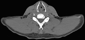
A repeat CT scan revealed malformation in the configuration of the anterior tracheal rings, involving the cricoid bone and with narrowing of the trachea. The length of the stricture, the maximum transverse diameter and anteroposterior diameters remained unchanged from his previous CT scan (Figures 1 and 2).
Axial computed tomography scan of normal trachea at the level of the piriform fossa.

Axial computed tomography scan showing sub-glottic stenosis.

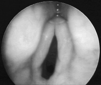
Fibre-optic laryngoscopy showed marked sub-glottic stenosis. Investigations failed to identify any systemic cause, including Wegener's granulomatosis or sarcoidosis. There was no significant past history of prolonged endotracheal intubation.
Microlaryngoscopy with biopsy and laser de-bulking under general anaesthetic were scheduled. However, per-oral intubation failed due to the extent of the patient's sub-glottic stenosis, which necessitated an urgent tracheostomy. A biopsy of a hard, sub-glottic lesion was undertaken (Figure 3), and CO2 laser debulking was performed.
Endoscopic, microlaryngoscopic photograph showing sub-glottic stenosis.

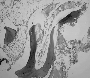
Histological analysis revealed mucosal tissue covered by respiratory epithelium and non-keratinising squamous epithelium with fragments of calcified and bony material, consistent with tracheopathia chondro-osteoplastica (Figure 4).
Photomicrograph showing bony nodule with trabecular bone separated by fatty marrow (H&E; ×medium magnification).

At the time of writing, decannulation had not been possible despite further laser debulking and surgical debridement procedures using the Medtronic Straightshot M4 Microdebrider (Medtronic, Watford, England).
Discussion
The progression of tracheopathia chondro-osteoplastica is slow, and over 90 per cent of cases are diagnosed only as post-mortem findings.Reference Prakash, McCullough, Edell and Nienhuis3 Symptoms may be absent or non-specific, and include cough, hoarseness, haemoptysis, wheezing and dyspnoea.Reference Hayes4
The precise aetiology of the condition remains unknown; several hypotheses have been suggested but none validated. In 1863, Virchow described tracheopathia chondro-osteoplastica as an exostosis and enchondrosis arising from the normal tracheal rings.Reference Virchow5 In 1910, Aschoff suggested that cartilaginous metaplasia of the subepithelial connective tissue may be the cause.Reference Aschoff6 In 1968, Sakula related the condition to end-stage primary amyloidosis, but this has not been proven.Reference Sakula7 More recently, an infective association with atrophic rhinitis and pharyngitis in conjunction with atypical microorganisms including Klebsiella ozaenae and Mycobacterium avium has been reported.Reference Baugnee and Delaunois8, Reference Harma and Suurkari9 Importantly, nodule formation in the trachea submucosa has been confirmed, based on histopathological evidence, to be related to the interaction between transforming growth factor beta-1 and bone morphogenetic protein-2.Reference Tajima, Yamakawa, Katagiri and Sasaki10
• Tracheopathia chondro-osteoplastica is a rare, benign disorder characterised by numerous cartilaginous and bony submucosal nodules protruding into the tracheo-bronchial lumen
• The precise aetiology remains unknown; several hypotheses have been suggested but none validated
• Endoscopic laser debulking can be employed to manage significant airway stenosis
• Significant tracheal stenosis requiring tracheostomy is rarely necessary; however, the presented case represents the first report of sub-glottic stenosis caused by tracheopathia chondro-osteoplastica which required an urgent tracheostomy
The differential diagnosis includes tuberculosis-related calcificating lesions, sarcoidosis, relapsing polychondritis, neoplasm and tracheo-bronchial calcinosis.Reference Meyer, Dossing and Broholm11 At microlaryngoscopy, the lesions have been described as having the appearance of a rock garden or cobblestones, and biopsy is required in order to obtain a definitive histological diagnosis and to exclude neoplasm.Reference Clee, Anderson and Johnston12 However, some authors consider the presence of multiple calcified, submucosal nodules with sparing of the posterior tracheal wall, on neck and thorax CT scans, to be pathognomonic.Reference Bottles, Nyberg, Clark and Hinchcliffe13, Reference Onitsuka, Hirose, Watanabe, Nishitani, Kawahira and Matsuura14 Histopathological assessment has demonstrated foci of bone marrow with active haematopoiesis.Reference Pounder and Pieterse15
A review of the literature did not reveal any specific treatment for tracheopathia chondro-osteoplastica. Non-specific, symptomatic treatment, including prophylactic antibiotics and cough suppressants, assists in minimising complications. Endoscopic laser debulking and external beam irradiation have been attempted in order to manage significant airway stenosis.Reference Prakash, McCullough, Edell and Nienhuis3 Tracheostomy may be required if the symptoms are severe enough to require bypass of the obstruction.
Conclusion
Tracheopathia chondro-osteoplastica is an extremely rare, idiopathic condition. It should be considered in the differential diagnosis of sub-glottic or tracheal stenosis. Slow disease progression with a benign course over time is most common; however, significant tracheal stenosis may occur, although tracheostomy is rarely necessary.