Introduction
Surgical experience plays a major role in the success of operations. This fact has been demonstrated in locoregional surgical control techniques in colorectal cancer surgeryReference Martling, Holm, Rutqvist, Moran, Heald and Cedemark1 and in the audiometric results of functional stapes surgery.Reference Levenson2 This experience starts at the beginning of the training for otology residents. It requires extensive knowledge of temporal bone anatomy and surgical simulation of cadaveric temporal bone drilling.
The shortage of organ donors and reluctance towards donating organs, as well as the new regulations for patient safety and the occupancy costs of operating theatres,Reference Roberts, Bell and Duffy3–Reference Beard, Jolly, Newble, Thomas, Donnelly and Southgate5 which further limit surgical drilling simulation, have forced the scientific community to create three-dimensional (3D) models that accurately resemble cadaver models. The virtual 3D models have two major drawbacks: cost and lack of feedback force, the latter of which is not yet comparable to that encountered during actual temporal bone drilling.
We aimed to develop a synthetic model of temporal bone that would allow otology residents to learn the drilling technique. We designed a temporal bone resin prototype by stereolithography,Reference Bakhos, Velut, Robier, Al Zahrani and Lescanne6 manufactured using rapid prototyping from Digital Imaging and Communication in Medicine (‘DICOM’) data. We validated the resemblance of the resin prototype to a cadaveric temporal bone by calculating the measurements and distances of the various elements; this showed no significant differences between the two temporal bones.Reference Bakhos, Velut, Robier, Al Zahrani and Lescanne6
Interest in the use of the prototype in teaching surgical techniques has not been validated. The aim of this study was to have experienced otologists (who were not involved in the development of the project) evaluate the temporal bone prototype in a surgical scenario.
Materials and methods
Manufacture of prototype
High-resolution images of a cadaveric temporal bone without malformation were acquired using a Philips spiral computed tomography scanner from the neuroradiology department. A Digital Imaging and Communication in Medicine file, which comprised 179 slices, was obtained with the following protocol: definition of 1024 × 1024, current of 200 mA, voltage of 140 kV, field of view 160°, overlapping slices of 0.27 mm and slice thickness of 0.55 mm.
In order to manufacture a stereolithography prototype, it was necessary to convert the Digital Imaging and Communication in Medicine imaging file into a machine ‘.stl’ file. Computer processing of the images and conversion of the Digital Imaging and Communication in Medicine image files in .stl required an iMac computer with an Intel Core 2 (T7400; 2.16 GHz, 2 GB, ATI Radeon X1600 video random access memory of 256 MB). We used two software programs. The 3D-Doctor (version 3.5; Able Software, Lexington, Massachusetts, USA) was used for image processing and selecting the areas of interest, and Magics rapid prototyping software (Materialise, Leuven, Belgium) was used to prepare the files before conversion to .stl file format.
Twenty-five identical resin prototypes of right temporal bone were manufactured by rapid prototyping, from the .stl file, using a stereolithography machine (SLA® 5000) and a white resin (Somos® 14120). This right temporal bone prototype has been validated in a previous study, in which the distances and diameters correlated with those of the scanner images of the cadaveric temporal bone model.Reference Bakhos, Velut, Robier, Al Zahrani and Lescanne6
One person painted the prototypes in coloured acrylic for simple identification of the different structures during dissection. The dura mater was painted pink, the lateral sinus and petrosal sinus were blue, the intrapetrous internal carotid artery was red, and the facial canal was injected with yellow paint (Figure 1).
Temporal bone prototype shown fixed in place.

The conformation allowed surgical training in the posterior cavities and surgical practice performing a mastoidectomy (Figure 2), followed by a translabyrinthine approach.
The third portion of the facial nerve of the drilling prototype; the lateral semicircular canal has been partly opened. 1 = incus; 2 = posterior tympanotomy; 3 = third portion of the facial nerve; 4 = lateral semicircular canal; 5 = lateral sinus; 6 = dura mater; 7 = external auditory meatus; 8 = mastoid tip
Validation of prototype
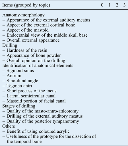
Twenty-five otologists outside the service, who had no initial involvement in the project, received a right temporal bone prototype and an evaluation grid (Table I) to complete after drilling of the resin model. This grid comprised 20 items, each scored from 0 to 3. The value 0 corresponded to ‘poor’, 1 to ‘medium’, 2 to ‘good’ and 3 to ‘excellent’. The criteria analysed were grouped into five topics: anatomy-morphology, drilling, identification of anatomical elements, stages of drilling and others. The experts could write comments at the bottom of the survey.
Right temporal bone prototype evaluation grid
A score of 0 corresponds to ‘poor’, 1 to ‘medium’, 2 to ‘good’ and 3 to ‘excellent’
An otology expert was defined as a surgeon in otology with more than 10 years' experience, who was accustomed to performing cochlear implant surgery, cholesteatoma surgery and middle-ear surgery. The experts were: professors, at French (n = 18), Algerian (n = 1) or Belgian (n = 1) universities; associate professors (n = 3); or known experimental otology surgeons in private practice with publications in the field of otological surgery (n = 2).
In order to assess the otologists' satisfaction with the prototype, we calculated the average rating for each item and each group of items (rounded to two decimal places). An overall satisfaction rating (percentage) was calculated, and all scores greater than or equal to 2 (good or excellent) were considered satisfactory.
Results
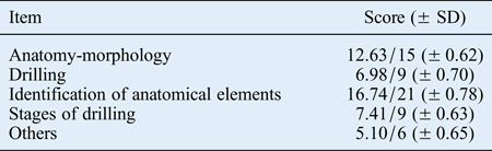
Twenty-five prototypes and questionnaires were sent to French-speaking experienced otologists, who were not involved in the project. Twenty-three returned their questionnaires, yielding a response rate of 92 per cent. Based on these 23 questionnaires, we calculated the average scores for each group of items (rounded to 2 decimal places); scores for the 5 topics assessed are shown in Table II.
Average score for each category of items
SD = standard deviation
Concerning anatomy and morphology (for which each item was rated out of 3), the average ratings for appearance were: 2.70 (± 0.47 standard deviation (SD)) for the external auditory meatus, 2.52 (± 0.73 SD) for the aspect of the external cortical bone, 2.65 (± 0.49 SD) for the aspect of the mastoid, 2.22 (± 0.80 SD) for the endocranial view of the middle skull base and 2.54 (± 0.50 SD) for the overall external appearance of the prototype.
From the perspective of drilling (for which items were rated out of 3), the hardness of the resin was estimated at 2.35 (± 0.65 SD), the appearance of bone powder at 2.17 (± 0.83 SD) and the overall opinion on the drilling at 2.46 (± 0.58 SD).
For the identification of the anatomical elements (rated out of a score of 3), the sigmoid sinus was rated 2.83 (± 0.39 SD), the antrum 2.39 (± 0.89 SD), the sino-dural angle 2.48 (± 0.59 SD), the tegmen antri 2.61 (± 0.50 SD), the short process of the incus 2.26 (± 0.92 SD), the lateral semicircular canal 2.13 (± 0.92 SD) and the mastoid portion of the facial canal 2.04 (± 0.88 SD).
Regarding the stages of drilling (rated out of 3), the quality of the masto-antro-atticotomy was scored as 2.59 (± 0.58 SD), the drilling of the external auditory meatus as 2.52 (± 0.67 SD) and the quality of the posterior tympanotomy as 2.30 (± 0.63 SD).
For the ‘other’ criteria (rated out of 3), the benefit of coloured acrylic was scored as 2.43 (± 0.73 SD), and the usefulness of the prototype for drilling of the temporal bone as 2.67 (± 0.56 SD).
The overall prototype score (for the 23 returned questionnaires), calculated from the average scores for each group of items, was 48.87 out of 60 (minimum = 32, maximum = 58). The total number of scores greater than or equal to 2 for the 20 items (as completed by the 23 otologists) was 422 out of 460, yielding a satisfaction rate of 92 per cent.
The major limitations raised by the otologists concerned the facial nerve, the ossicular chain and labyrinth, and the resin used. Ten experts considered the colour of the facial nerve to be excessively vivid for drilling, especially in the third segment. Three otologists found the oval and round windows too difficult to identify, and five experts were disappointed with the ossicular fixity and by the anatomy of the bony labyrinth, the latter of which made identification of the posterior semicircular canal difficult. Nine experts found the resin dust too thick, thus blocking the suction, and found the synthetic bone excessively soft. Six experts expressed the desire to have prototypes with anatomical variants and malformations, including aplasia. Improvements proposed by the evaluators predominantly concerned the resin, the ossicles and the facial nerve.
Discussion
The evaluation of our model revealed a satisfaction rate of 92 per cent, with average scores for groups of items ranging from 2.04 to 2.83 (out of 3). These scores and the reviewers' comments confirm the suitability of the prototype for temporal bone drilling. Additional prototypes will therefore be developed to resolve the shortage of cadaveric material.
A review of the literature revealed that other specialists have similarly developed prototypes for surgical training, primarily in orthopaedics, neurosurgery, ophthalmology and maxillofacial surgery.Reference Paiva, Amorim, Bezerra and Masini7–Reference Uhlig and Gerding9 A neurosurgery team developed a prototype of the cervical spine through the process of stereolithography, using the scans of a patient with Ewing's sarcoma in the cervical spine, to perform a surgical simulation.Reference Paiva, Amorim, Bezerra and Masini7 This simulation allowed the surgical team to understand more completely the patient's disease in order to better plan the spinal surgery approach. The operation was uneventful with no complications.
In dental surgery, a prototype for training in standard surgical procedures, using various materials, was developed from Digital Imaging and Communication in Medicine images.Reference Lambrecht, Berndt, Christensen and Zehnder8 These prototypes aimed to allow training prior to surgery.
In ophthalmology, the need for surgical technique training arose because of a marked expansion in the use of lasers for treatment of ophthalmic diseases. A German team uses a pig eye model maintained in a dummy orbit made from black anodised aluminium.Reference Uhlig and Gerding9 This prototype has allowed the teaching of direct and indirect ophthalmoscopy, and various surgical techniques, to undergraduate and graduate students, using the yttrium aluminium garnet (‘YAG’) laser and ultrasonography methods. After this training, the students are able to make a correct medical diagnosis after ophthalmoscopy, and to easily handle ultrasonography and laser techniques without requiring supervision by a senior doctor. There is interest in these newly developed prototypes because they allow surgeons to practice identical surgical procedures as often as necessary because models from rapid prototyping can be manufactured at an infinite rate.
With regard to otology, the shortage of cadaveric temporal bones posed a training problem. Training is essential for drilling, given the anatomical complexity and the technical expertise needed to perform an intervention. Various types of 3D models have been developed by different surgical teams.
There are 3D virtual models available in the form of films on the internet or in the form of reconstruction through computerised anatomical reconstruction.Reference Wang, Merchant and Sorensen10, Reference Sorensens, Dobrezeniecki, Larsen, Frisch, Sporring and Darvann11 Imaging provides 3D anatomical information but does not allow surgical training. Other virtual models of the temporal bone have been developed that do allow virtual surgical simulations,Reference Fried, Uribe and Sadoughi12 as a result of the rapid development of computational techniques for image processing.Reference Neri, Sellari Franceschini, Berrettini, Caramella and Bartolozzi13–Reference Zirkle, Roberson, Leuwer and Dubrowski15 Such simulation allows unlimited repetition of the drilling procedure, with the ability to locate anatomical landmarks during the training.Reference Wiet, Stredney, Sessanna, Bryan, Welling and Schmalbrock16 These 3D models allow simple manipulation to be conducted from a computer outside university hospitals,Reference Wiet, Stredney, Sessanna, Bryan, Welling and Schmalbrock16, Reference Kakizawa, Hongo and Rhoton17 and at any time of day, which is not possible in an anatomy laboratory. The major disadvantages of these models are the absence of return forceReference Mason, Applebaum, Rasmussen, Millman, Evenhouse and Panko18 and the purchase price of the models.Reference Uhl, Plaisant, Ami and Delmas19
There are physical temporal bone prototypes such as Pettigrew temporal bones, on which ossicular surgery and posterior ear surgery can be practised.20, Reference Mills and Lee21 A bleeding impression during drilling is created using a colourful irrigation stagnant in the remaining mastoid cells. We did not incorporate a bleeding simulation system in our synthetic model, and this has been criticised. Another drawback of our prototype compared with the Pettigrew models was the rigidity of the ossicular chain, which did not permit the surgeon to practice ossicular surgery. Amendments will be necessary to perfect our prototype and broaden its use. Such future improvements may be possible with the development of stereolithography techniques, particularly in terms of manufacturing precision and the possibility of using several composites.
The latest models are manufactured from liquid resin, polymerised and then solidified by stereolithography, or created from selective laser sintering. The models of temporal bones made from polyamide nylon and glass beads are less realistic in drilling simulation than those produced by stereolithography or sintering.Reference Mori, Yamamoto, Oyama, Ueno, Nakao and Honma22 A review of the literature shows that the disadvantage of the models manufactured by sintering and stereolithography is the production of a bone powder that regularly obstructs the suction.Reference Suzuki, Ogawa, Kawano, Hagiwara, Yamaguchi and Ono23 Our model yields a drilling simulation resembling that of cadaveric temporal bones, with a powder that is similar to powdered bone; however, the powder was considered too heavy by some reviewers. This characteristic can be improved with modification of the resins, at a more expensive production cost.
The lowest prototype score was related to the poor identification of the third portion and the second genu of the facial nerve. Overall, however, the otologists' responses indicate that our prototype is an effective training tool, with a satisfaction rate of 92 per cent. This model is among a few to have been submitted for evaluation by independent experts, in contrast to the models listed above.
Changes and further improvements can be made because the design and production of prototypes are straightforward, and stereolithography advances may eventually allow the production of a mobile ossicular chain.
• The shortage of cadaveric temporal bones has prompted the creation of physical prototypes
• These alternatives offer a solution for temporal bone dissection training but must be evaluated by otology experts
• A synthetic model of temporal bone was evaluated
• Evaluation demonstrated that the prototype was a good training tool for drilling and otological surgery
• However, improvements in resin quality, facial nerve positioning and ossicular mobility are needed, as is the design of pathological prototypes
Validation of the temporal bone prototype has been achieved; the next goal will be to demonstrate the model's usefulness in teaching the temporal bone drilling process to aspiring ENT students. A previous study has emphasised that the interest in these 3D virtual models lies in the understanding of the temporal bone anatomy.Reference Venail, Deveze, Lallemant, Guevara and Mondain24 The utility of these models in the surgical training of inexperienced otologists has not been evaluated. We plan to evaluate the benefit of our prototype in surgical skills training by administering a questionnaire such as the one developed by an American team.Reference Lawrence and Tomolo25 It would evaluate the skills of otology residents who have been trained using our prototypes versus those of residents who have received no such training, and would be completed by confirmed experts in otology. The interest in this prototype is in drilling technique training. The implications of the prototype for surgical practice could be discussed during a training day, such as the annual days of formative internship assessment advocated by the Accreditation Council for Graduate Medical Education.Reference Nadeau and Tysinger26 The method for evaluating our prototype based on the quality of its contribution to the training of otology residents remains to be explored.
Conclusion
Our synthetic temporal bone, designed using a stereolithography technique, was validated by a panel of French-speaking experts in otology. The model appears to be a good training tool for drilling and otological surgery. However, some improvements are needed, especially regarding the quality of the resin, the positioning of the facial nerve and ossicular mobility. We will evaluate the usefulness of our prototype in the training of aspiring otologists.
Acknowledgements
We would like to thank Sandy Oba (House Ear Institute, Los Angeles, USA) for the proofreading. We would also like to thank the French-speaking otology experts: D Ayache (Fondation Rothschild, Assistance Publique-Hôpitaux de Paris), S Bobin (Kremlin-Bicêtre, Assistance Publique-Hôpitaux de Paris), P Bordure (Centre hospitalier universitaire Nantes), JC Chobaut (Centre hospitalier universitaire Besançon), V Darrouzet (Centre hospitalier universitaire Bordeaux), O Deguine (Centre hospitalier universitaire Toulouse), C Debry (Centre hospitalier universitaire Strasbourg), B Frachet (Avicenne, Assistance Publique-Hôpitaux de Paris), B Fraysse (Centre hospitalier universitaire Toulouse), B Gratacap (Clinique du docteur Jean Causse, Colombiers), JP Lavieille (Nord, Centre hospitalier universitaire Marseille), P Herman (Lariboisière, Assistance Publique-Hôpitaux de Paris), J Magnan (Nord, Centre hospitalier universitaire Marseille), C Martin (Centre hospitalier universitaire Saint Etienne), S Moreau (Centre hospitalier universitaire Caen), D Portman (Institut Portman, Bordeaux), S Schmerber (Centre hospitalier universitaire Grenoble), T Somers (Centre hospitalier universitaire Anvers, Belgium), O Sterkers (Beaujon, Assistance Publique-Hôpitaux de Paris), E Truy (Centre hospitalier universitaire Lyon), T Van Den Abbeele (Robert Debré, Assistance Publique-Hôpitaux de Paris), F Venail (Centre hospitalier universitaire Montpellier), C Vincent (Centre hospitalier universitaire Lille), A Uziel (Centre hospitalier universitaire Montpellier), and N Yahi (Bachir Mentouri, Kouba-Alger, Algeria).