Cyanotic CHDs causing the right to left shunt in the cardiac structures are characterised by decreased blood oxygen level. Early diagnosis and surgical treatment at the optimum age are crucial, and new surgical techniques for the management provide a prolonged life expectancy for these patients. The changes in the peripheral vascular structures subsequent to cyanotic CHD have been previously reported. But those in the blood vessels of the central nervous system are not well known. Here, we present a case with surgically treated cyanotic CHD, having an extensive enlargement in the cranial and spinal vasculature and presenting with diffuse cerebral and spinal dysfunction occurring in the late period of follow-up.
Case report
A 24-year-old female patient who was diagnosed at birth with double-inlet double-outlet right ventricle, left ventricle hypoplasia, and left atrial isomerism had undergone total cava-pulmonary connection procedure performed at the age of eight. She was admitted with progressive neurological deterioration occurring over a period of 6 months.
She had a normal motor and mental development, a higher educational level, and only suffered from respiratory problems related to hypoxemia until the age of 23. Her first neurologic complaint was a new onset headache which was due to a temporoparietal brain abscess detected on MRI. Antimicrobial treatment including metronidazole, vancomycin, and ceftriaxone was given for the abscess in another hospital. One month later, she started experiencing difficulty walking, pain, and allodynia in her limbs. Diagnostic workup for her pain in the extremities including peripheral arterial and venous Doppler ultrasonography was normal. Electroneuromyographic studies showed sensory and motor axonal polyneuropathy predominant in lower limbs which might be related to ceftriaxone and metronidazole. Therefore, these antibiotherapies were discontinued. In the follow-up period, vancomycin was also stopped because of renal insufficiency and brain abscess was treated surgically with a burr hole aspiration method resulting in clinical and radiological recovery.
After the operation, she was able to walk again without assistance. But 2 months later, she was hospitalised in the ICU because of heart failure treated with vascular stenting applied to the level of pulmonary bifurcation and anastomosis. Afterward, her walking ability and cognitive functions gradually deteriorated over 1 month. On admission to our neurology clinic, neurological examination revealed impaired orientation and cooperation, and attention deficit (with a mini-mental state examination score of 20/28) as well as neck flexion weakness, quadriparesis predominant on the right side with the presence of the Babinski sign, and impaired joint position sense. She was using antiplatelet therapy (acetylsalicylic acid 100 mg per day). Her blood oxygen saturation level was between 83 and 87% without any changes before and after the onset of the symptoms. Furthermore, she was cyanotic and moderately hypertensive.
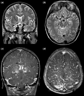
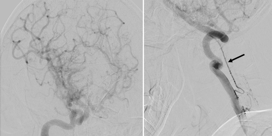
Brain MRI showed enlargement of cerebral arterial and venous system notably on deep venous structures, expanded perivascular spaces, and diffuse leptomeningeal enhancement following gadolinium injection probably as a consequence of the retarded motion of blood in the enlarged vessels (Fig 1). Cerebral angiography confirmed these findings, showing also the enlargement of the punctured femoral artery. Moreover, a similar enlargement of spinal arteries was seen on cerebral angiography (Fig 2). However, the ophthalmic examination did not find any abnormalities of retinal vessels.
Intracranial vascular enlargement is seen in T2 weighted (a, b) and diffuse leptomeningeal contrast enhancement in T1 weighted (c, d) magnetic resonance images.

Enlargement of cranial and spinal vasculature (arrow) is showed by digital subtraction angiography.

Positron emission tomography was also performed to better understand the functionality of the brain parenchyma and showed bilateral hypometabolic activity on the parietal and occipital lobes, predominant on the left side. However, brain perfusion MRI did not detect any hypoperfused area. Furthermore, somatosensorial evoked potential study results revealed an impairment of the posterior cord conduction and autonomic involvement compatible with the presence of myelopathy. Her antiplatelet therapy has been continued with the same dosage and physiotherapy was started to improve functionality. Six months later, the patient experienced an acute ischaemic infarct in the anterior and middle cerebral artery border zone, presenting with an epileptic seizure. She was admitted to another hospital where she died.
Discussion
Chronic hypoxemia causes secondary erythrocytosis leading to chronically elevated viscosity and increased nitric oxide scavenging. These changes cause endothelial dysfunction, impaired tissue perfusion, and vascular remodelling.Reference Cordina and Celermajer1 In this condition, increased vessel wall shear stress is thought to cause vasodilatation.Reference Cordina and Celermajer1 In patients with cyanotic CHD enlarged, tortuous vessels are reported in the coronary,Reference Fyfe, Perloff and Niwa2,Reference Niwa, Perloff and Bhuta3 pulmonary,Reference Niwa, Perloff and Bhuta3 and retinal vasculature.Reference Mansour, Bitar and Traboulsi4 But the involvement of the blood vessels in the central nervous system is not well known. In our case, we may suggest that cerebral and spinal vascular structural changes occurred by the same adaptive mechanism to chronic hypoxia.
Furthermore, a series of shunt operations (Norwood, Glenn, and Fontan procedures) recomposing the blood circulatory system are performed to the patients with cyanotic CHDs related to single ventricle heart in the early years of their life. These surgical procedures rerouting venous blood directly to the lungs also cause high central venous pressure that leads to low perfusion and congestion.Reference Gewillig and Brown5 It has been suggested that vascular stiffness and high central venous pressure may also lead to impaired cerebral perfusion.Reference Saiki, Kurishima and Masutani6 We did not detect any focal perfusion defect on brain perfusion MRI. But global hypoperfusion of the brain and spinal cord might also be present as the perfusion imaging reveals only relative defects. We hypothesised that subacute worsening of cardiac functions following central nervous system infection and vascular stenting of the pulmonary artery may increase central venous pressure and central nervous system blood volume load. These changes may disrupt cerebral autoregulation and contribute to increased intraluminal pressure. As a result of this haemodynamic deterioration, we suggest that blood brain barrier disruption led to the neuronal dysfunction in our case.
Conclusion
The radiological changes of the vascular structures of the central nervous system occurring in operated patients with cyanotic CHDs have not been previously well-described. To our knowledge, this is the first case with cyanotic CHD having diffusely enlarged vessels in the cranial and spinal, arterial and venous vessels that lead to cerebral and spinal dysfunction. Nowadays, most of these patients having cyanotic CHD reach adulthood, thanks to the advances in cardiological care and emerging surgical technologies. We think that the clinicians need to be familiar with the neuroradiological changes of the vasculature occurring as a consequence of altered cardiac physiology and circulatory haemodynamics. Further research is also needed to recognise these changes and to enlighten the relevant neurological conditions.
Acknowledgements
The authors thank Kivanc Yalin MD for enlightening them on cardiological perspective and Professor Oguzhan Coban and Faruk Ugur Dogan MD.
Financial Support
The authors received no financial support for this study.
Conflict of Interest
The authors declare that they have no conflict of interests.
Ethical Standards
All procedures performed in studies involving patients were in accordance with the ethical standards of the institutional and/or national research committee and with the 1964 Helsinki declaration and its later amendments or comparable ethical standards. Ethics committee approval was not applicable as the data were analysed retrospectively and had no effect on treatment.