INTRODUCTION
Neospora caninum (Apicomplexa: Sarcocystidae), a tissue cyst-forming coccidian closely related to Toxoplasma gondii, is a major cause of infectious bovine abortion worldwide, causing important economic losses to the cattle industry (Dubey et al. Reference Dubey, Buxton and Wouda2006, Reference Dubey, Schares and Ortega-Mora2007; Dubey and Schares, Reference Dubey and Schares2011). Neospora caninum has a heteroxenous life cycle, in which dogs (Canis familiaris) and other canids – coyotes (Canis latrans), dingos (Canis lupus dingo) and grey wolves (Canis lupus) – are currently recognized as definitive hosts (McAllister et al. Reference McAllister, Dubey, Lindsay, Jolley, Wills and McGuire1998; Dubey et al. Reference Dubey, Jenkins, Rajendran, Miska, Ferreira, Martins, Kwok and Choudhary2011; King et al. Reference King, Brown, Jenkins, Ellis, Fleming, Windsor and Slapeta2012) and cattle and other ungulates are natural intermediate hosts (Dubey et al. Reference Dubey, Schares and Ortega-Mora2007; Dubey and Schares, Reference Dubey and Schares2011). Transplacental transmission of N. caninum is very efficient in cattle (Dubey et al. Reference Dubey, Buxton and Wouda2006). In cattle, abortion may occur either due to primary infection, following the ingestion of oocysts shed by definitive hosts (exogenous transplacental transmission), or following the reactivation of a pre-existing chronic infection during pregnancy (endogenous transplacental transmission) (Dubey et al. Reference Dubey, Buxton and Wouda2006, Reference Dubey, Schares and Ortega-Mora2007; Dubey and Schares, Reference Dubey and Schares2011). Dogs may also serve as intermediate hosts and, although rare, this infection can cause fatal cases of canine neosporosis associated with neurological signs (Barber et al. Reference Barber, Payne-Johnson and Trees1996; Dubey, Reference Dubey2003; Reichel et al. Reference Reichel, Ellis and Dubey2007). Transplacental transmission from dam to pup, although more variable than in cattle, appears to be efficient in the dog and the most severe cases of neosporosis occur in congenitally infected pups (Barber et al. Reference Barber, Payne-Johnson and Trees1996; Dubey, Reference Dubey2003; Reichel et al. Reference Reichel, Ellis and Dubey2007).
Different factors, including the timing of transmission during pregnancy and the immune status of the cow and fetus, have been directly related to the occurrence of abortion. Additional factors, such as virulence of the isolates, may also play an important role in the outcome of abortion in cattle and clinical neosporosis in the dog. Little is known regarding the variability of N. caninum virulence among isolates. In contrast to T. gondii, although it is ubiquitously distributed in a wide host range, viable N. caninum isolates are difficult to obtain (Dubey and Schares, Reference Dubey and Schares2011) and the number of isolates obtained to date is limited. Most of these have been isolated from clinically affected hosts and, therefore, might be selected towards more virulent isolates. Different studies have demonstrated intra-specific variability in mice among N. caninum isolates obtained from clinically affected hosts (Atkinson et al. Reference Atkinson, Harper, Ryce, Morrison and Ellis1999; Quinn et al. Reference Quinn, Miller, Ryce, Windsor and Ellis2002; Collantes-Fernandez et al. Reference Collantes-Fernandez, Lopez-Perez, Alvarez-Garcia and Ortega-Mora2006). Additionally, bovine isolates obtained from asymptomatic calves have exhibited marked differences in parasite burdens, immunoglobulin IgG isotype kinetics, pathogenicity and vertical transmission in well-established cerebral and pregnant BALB/c mouse models, with some isolates showing high virulence (Rojo-Montejo et al. Reference Rojo-Montejo, Collantes-Fernandez, Regidor-Cerrillo, Alvarez-Garcia, Marugan-Hernandez, Pedraza-Diaz, Blanco-Murcia, Prenafeta and Ortega-Mora2009b ; Pereira Garcia-Melo et al. Reference Pereira Garcia-Melo, Regidor-Cerrillo, Collantes-Fernandez, Aguado-Martinez, Del Pozo, Minguijon, Gomez-Bautista, Aduriz and Ortega-Mora2010; Regidor-Cerrillo et al. Reference Regidor-Cerrillo, Gomez-Bautista, Del Pozo, Jimenez-Ruiz, Aduriz and Ortega-Mora2010). Recent studies in pregnant cattle have confirmed the influence of the N. caninum isolate on the outcome of infection (Rojo-Montejo et al. Reference Rojo-Montejo, Collantes-Fernandez, Blanco-Murcia, Rodriguez-Bertos, Risco-Castillo and Ortega-Mora2009a ; Caspe et al. Reference Caspe, Moore, Leunda, Cano, Lischinsky, Regidor-Cerrillo, Alvarez-Garcia, Echaide, Bacigalupe, Ortega Mora, Odeon and Campero2012).
The aim of this study was to test the pathogenicity of 6 isolates obtained from naturally infected dogs using a well-established pregnant BALB/c mouse model that allows the evaluation of the vertical transmission rate of neosporosis and the morbidity and mortality of the progeny (Lopez-Perez et al. Reference Lopez-Perez, Collantes-Fernandez, Aguado-Martinez, Rodriguez-Bertos and Ortega-Mora2008). The present study examined four isolates obtained from oocysts shed in the feces of naturally infected dogs (Nc-Ger2, Nc-Ger3, Nc-Ger6 and Nc-6Arg) and two isolates obtained from infected tissues of dogs with neurological signs (Nc-Bahia and Nc-Liv).
MATERIALS AND METHODS
Ethics statement
All protocols involving animals were approved by the Animal Research Committee of the Complutense University, Madrid, Spain, according to the proceedings described in the Regulation of Internal Regime for Animal Research Committee (published at BOUC, no. 2, on 9 February 2006) and Spanish and EU legislations (Law 32/2007, R.D. 1201/2005 and Council Directive 2010/63/EU). The ethics committees consented to this procedure.
Isolate cultures and preparation of N. caninum isolates for BALB/c inoculation
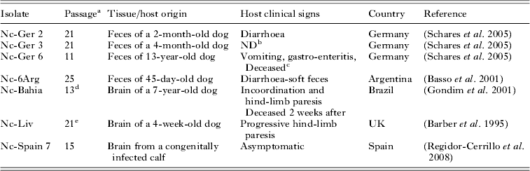
In this study we evaluated six canine N. caninum isolates whose host and geographical origins are detailed in Table 1. The bovine Nc-Spain 7 isolate was also included as an inter-assay control group for the BALB/c pregnant model, as it had been previously characterized under similar conditions (Regidor-Cerrillo et al. Reference Regidor-Cerrillo, Gomez-Bautista, Del Pozo, Jimenez-Ruiz, Aduriz and Ortega-Mora2010). Cryopreserved isolates were reactivated and grown in MARC-145 cells, as described previously (Regidor-Cerrillo et al. Reference Regidor-Cerrillo, Gomez-Bautista, Pereira-Bueno, Aduriz, Navarro-Lozano, Risco-Castillo, Fernandez-Garcia, Pedraza-Diaz and Ortega-Mora2008). The tachyzoites used for the mouse inoculations were recovered when the parasites were still intracellular to preserve their invasion capability (at least 80% of undisrupted parasite vacuoles in the cell monolayer). The inocula were prepared as described previously (Pereira Garcia-Melo et al. Reference Pereira Garcia-Melo, Regidor-Cerrillo, Collantes-Fernandez, Aguado-Martinez, Del Pozo, Minguijon, Gomez-Bautista, Aduriz and Ortega-Mora2010). The tachyzoite number was determined by Trypan blue exclusion followed by counting in a Neubauer chamber and the parasites were resuspended in PBS at 2×10⁶ tachyzoites/200 μL. Mice were inoculated within an hour of tachyzoite collection. All isolates were passaged in cell culture between 11 and 25 times after their isolation in cell culture from oocysts and bovine tissues (Nc-Ger2, Nc-Ger3, Nc-Ger6, Nc-6Arg and Nc-Spain7), after passage in mice and re-isolation (Nc-Liv) or from cultures provided in the laboratory (Nc-Bahia) (Table 1).
Summary of isolate name, host, geographical origin and passage number in the cell cultures of isolates included in this study

a Total passages in cell culture after isolation.
b Not determined.
c Dog splenectomized 47 days before sampling.
d Numbers of passages in our laboratory after being kindly provided by L. F. Gondim.
e Number of passages after re-isolation in cell culture. Nc-Liv was previously passaged in a mouse.
Experimental design: mating and pregnancy of mice and N. caninum inoculations
Pathogenicity studies were performed using a pregnant BALB/c mouse model as described previously (Lopez-Perez et al. Reference Lopez-Perez, Risco-Castillo, Collantes-Fernandez and Ortega-Mora2006, Reference Lopez-Perez, Collantes-Fernandez, Aguado-Martinez, Rodriguez-Bertos and Ortega-Mora2008). Briefly, 6-week-old inbred BALB/c mice, weighing 18–22 g, were purchased from a commercial supplier (Harlan Iberica, Barcelona, Spain). The mice were free of common viral, parasitic and bacterial pathogens according to the results of routine screening procedures performed by the producer. Female mice were housed in groups of 10 in a controlled environment with 12 h-light and 12 h-dark cycles and were provided with rodent food and water ad libitum. At 8 weeks of age, the oestrus cycles were synchronized as a result of the Whitten effect (Whitten, Reference Whitten1957), after which two female mice were placed with a male for 4 nights. The last day that the females were housed with males was determined as day 0 of pregnancy. The female mice were randomly assigned into eight experimental groups with 12 mice in each group.
At day 7 of gestation, the mice were subcutaneously (s.c.) inoculated with 2×10⁶ tachyzoites per mouse of each N. caninum isolate or sterile PBS (sentinel control group).
Samples from dams and progeny and clinical data collection
After the tachyzoite inoculation, the pregnant mice and pups were examined daily for clinical signs compatible with neosporosis, including rough hair coats, rounded back, inactivity, anorexia and neurological signs such as head tilting, walking in circles, ataxia, limb weakness and paralysis (Pereira García-Melo et al. Reference Pereira Garcia-Melo, Regidor-Cerrillo, Collantes-Fernandez, Aguado-Martinez, Del Pozo, Minguijon, Gomez-Bautista, Aduriz and Ortega-Mora2010). The appearance of lethargy followed by neurological signs was considered for culling. Pregnant mice were identified on day 18 of pregnancy by weighing and were individually housed. The pregnancy rate was established when pregnant mice gave birth. The litter size was defined as the number of pups delivered per dam. The offspring were also weighed every 2–3 days, beginning at day 15 post-partum (PP). The dams were maintained until day 30 PP and the pups were evaluated until the end of the experiment at day 50 PP. Early pup mortality was defined as the number of pups that died within 48 h after birth. Pup mortality was considered as the number of pups that died from day 3 PP to day 50 PP. Only pregnant mice continued until the end of the experiment, and non-pregnant infected mice were culled. All the mice were euthanased in a CO2 chamber.
Blood samples were collected from the dams and pups by cardiac puncture immediately after euthanasia and the recovered sera were preserved at −80 °C for ELISA analysis. At necropsy, brains and lungs from the pups and dams were also collected and preserved at −80 °C until further analysis by PCR. Some of the tissue samples from the pups could not be collected due to cannibalism by the dams.
Parasite DNA detection by nested-PCR
Only 50% of the mice that succumbed to infection were analysed by PCR to confirm N. caninum infection. All of the dams and a representative group of pups that survived to day 50 PP (at least 50% of the number of pups randomly selected from each litter) were analysed by nested-PCR. The vertical transmission rate was determined by the PCR detection of N. caninum DNA in the brains or lungs of the pups.
Genomic DNA was extracted from 20 mg of brain or lung tissue from mice using the commercial kit Maxwell® 16 Mouse Tail DNA Purification Kit developed for the automated Maxwell® 16 System (Promega, Madison, Wisconsin, USA) according to the manufacturer's instructions. Detection of parasite DNA was performed by a nested-PCR against the ITS-1 region of N. caninum, as described previously (Collantes-Fernandez et al. Reference Collantes-Fernandez, Lopez-Perez, Alvarez-Garcia and Ortega-Mora2006; Pereira Garcia-Melo et al. Reference Pereira Garcia-Melo, Regidor-Cerrillo, Collantes-Fernandez, Aguado-Martinez, Del Pozo, Minguijon, Gomez-Bautista, Aduriz and Ortega-Mora2010). The concentration of DNA was determined by spectrophotometry and adjusted to 50–100 ng μL−1 and 250–500 ng of each extracted DNA sample was used for PCR parasite detection.
Immune response
Neospora caninum-specific IgG1 and IgG2a antibodies in the dams and pups were assessed by ELISA as described previously (Collantes-Fernandez et al. Reference Collantes-Fernandez, Lopez-Perez, Alvarez-Garcia and Ortega-Mora2006). Briefly, an ELISA was based on soluble N. caninum tachyzoite antigen (0·25 μg in 100 μL per well), using diluted serum samples (1:100) and an anti-mouse IgG1 or IgG2a antibody (1:5000; Southern Biotechnology, Birmingham, AL, USA). Specific IgG responses against recombinant rNcGRA7 and rNcSAG4 proteins were also determined by ELISA. Briefly, 96-well plates were coated with 0·1 μg per well of rNcGRA7 or rNcSAG4 as previously described (Jimenez-Ruiz et al. Reference Jimenez-Ruiz, Bech-Sabat, Alvarez-Garcia, Regidor-Cerrillo, Hinojal-Campana and Ortega-Mora2013). The serum samples were applied at a 1:100 dilution and an anti-mouse IgG peroxidase-conjugated antibody (Sigma, USA) was used at a 1:3000 dilution.
The serum samples were analysed in duplicate and the average O.D. value was converted into a relative index percent (RIPC) using the following formula: RIPC = (O.D. sample − O.D. negative control)/(O.D. positive control − O.D. negative control)×100. The antibody isotype balance was evaluated by the IgG1/IgG2a ratio.
Statistical analysis
Differences in pregnancy, early pup mortality, pup mortality and vertical transmission rates were analysed with the Chi-square (χ 2) test or Fisher's exact F-test. In addition, pup morbidity and mortality were analysed by the Kaplan–Meier survival method to estimate the percentage of healthy or surviving animals at each time point (days PP) (Bland and Altman, Reference Bland and Altman1998). To compare the health and survival curves between infected groups, the log-rank statistical test was applied (Bland and Altman, Reference Bland and Altman2004). The median survival time or the day at which 50% of the pups were clinically affected or had died was calculated (Bland and Altman, Reference Bland and Altman1998). When significant differences were found, a multiple comparison test was used to examine all possible pair-wise comparisons in the χ 2 and Kaplan–Meier tests. A value of P<0·05/k was considered statistically significant for pairwise comparisons, with k corresponding to the number of groups. Finally, a one-way ANOVA test followed by a Tukey's multiple comparison test was employed to compare litter sizes, pup body weights and IgG1, IgG2a, rNcGRA7 and rNcSAG4 antibody responses. In addition, Spearman's rank correlation coefficient (ρ) was applied to analyse the association between the parasite detection rates in dams and their progeny (vertical transmission rate). Statistical significance for all analyses was established at P<0·05. All statistical analyses were performed with Statgraphics Plus v.5.1 (StatPoint, Inc., Herndon, VA, USA) and GraphPad Prism 5 v.5.01 (San Diego, CA, USA) software.
RESULTS
The results from the Nc-Spain 7 infected mice are shown in Tables 2 and 3 and Figs 1–3.
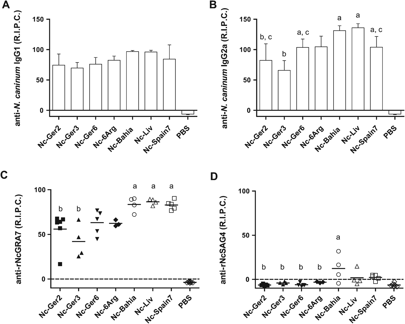
The humoral immune responses of dams inoculated with N. caninum isolates evaluated by ELISA. Columns represent the average RIPC of the anti-N. caninum IgG1 (A) and IgG2a (B) levels determined for each group and the error bars are S.D. The scatter-plot graphs represent the RIPC values of specific antibodies against rNcGRA7 (C) and rNcSAG4 (D) in the dams from each group. The horizontal lines represent the average values for each group. a,b,c; Different letters over the boxes and scatter-plots indicate significant differences between the N. caninum isolates, according to the 1-way ANOVA test followed by the Tukey's multiple comparison test. Note that the absence of letters above boxes or scatter plots indicates the absence of statistically significant differences between this group and any other infected group, according to Tukey's multiple comparison test.

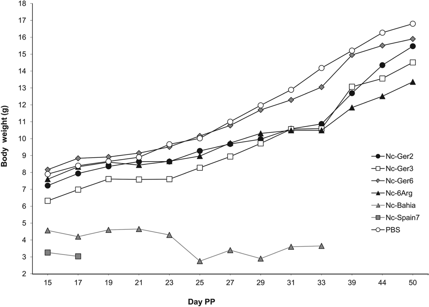
Body weight progression of pups born to dams that were infected on day 7 of pregnancy with 2×10⁶ tachyzoites of each N. caninum isolate included in this study (five canine isolates; Nc-Ger2, Nc-Ger3, Nc-Ger6, Nc-6Arg and Nc-Bahia and one bovine isolate; Nc-Spain7) and the PBS control group. Nc-Liv data are not represented because only two pups survived infection at day 15 PP. Each point represents the average body weight of all animals in a group. Note that the pups progressively succumbed to infection in the clinically affected groups, particularly in the Nc-Bahia and Nc-Spain 7 groups.

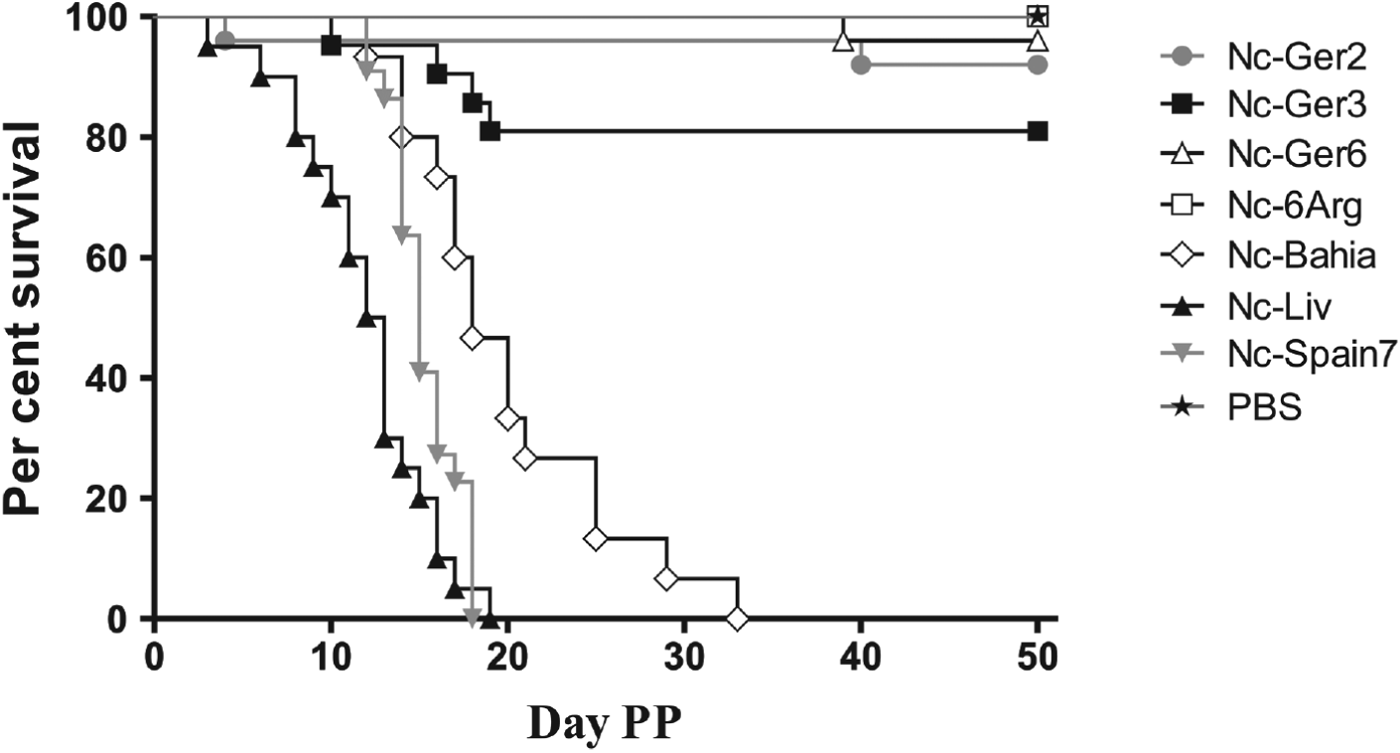
Kaplan–Meier survival curves for the pups born to dams that were infected on day 7 of pregnancy with 2×10⁶ tachyzoites of the different N. caninum isolates included in this study and the uninfected group. Each point represents the percentage of survival animals at that day.

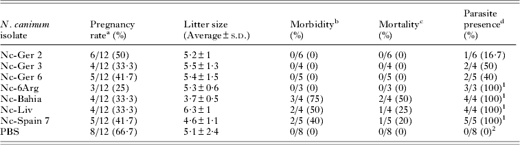
The effects of Neospora caninum infection on dams: pregnancy, morbidity and mortality rates, litter sizes and frequencies of parasite PCR-detection in dam brains

a Number of pregnant mice/no. of females (percentage).
b Number of dams with clinical sings (lethargy, ataxia and limb weakness or paralysis)/no. of dams (percentage).
c Number of dead dams/no. of dams (percentage).
d Number of nested-PCR positive dams at day 30 PP/no. of dams in the group (percentage).
1,2 Percentages determined for infected groups followed by unlike superscripts differ significantly in a χ 2 Multiple-Comparison Test.
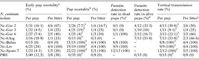
The effects of Neospora caninum infection on early pup and pup mortality and parasite PCR-detection rate in pup brains

a Number of stillborn and dead pups at day 2 PP/no. of total pups born (% early pup mortality).
b Number of dead pups from day 3 PP onwards/no. of pups alive by day 2 (% pup mortality).
c PCR-positive in pups that succumbed to infection from day 3 PP/no. of analysed pups (% parasite detection).
d PCR-positive pups that survived infection at day 50 PP/no. of analysed pups (% parasite detection).
e Litters with at least one PCR-positive pup (% vertical transmission rate).
1,2 Percentages for infected groups followed by unlike superscripts differed significantly in a χ 2 multiple comparison test.
3 Vertical transmission rate for Nc-6Arg infected group only differs significantly from the PBS control group in a χ 2 multiple comparison test.
Evaluation of N. caninum infection in dams
Pregnancy rate and litter size
In this study, the average pregnancy rate was 40·6%. The pregnancy rates varied from 25% for the females inoculated with the Nc-6Arg isolate to 66·7% for those inoculated with PBS (Table 2), although no significant differences were found between the groups (P = 0·56; χ 2 = 5·837). Additionally, no significant differences in litter size were found among the inoculated groups (P = 0·45; 1-way ANOVA followed by Tukey's multiple comparison test) (Table 2).
Morbidity and mortality
Skin lesions consisting of dermal nodules at the sites of the parasite inoculation (interscapular region) were observed in the dams from several inoculated groups, beginning at 7 days post-inoculation (dpi), and these resolved by 14 dpi. The percentage of dams with skin lesions was 50% for the groups infected with the Nc-Bahia and Nc-Liv isolates, which developed ulcerative or non-ulcerative dermal nodules, followed by the mice inoculated with Nc-6Arg (33%). Skin lesions were not detected in the remaining groups inoculated with Nc-Ger2, Nc-Ger3 and Nc-Ger6. Clinical signs such as rough hair coats, rounded back, anorexia and lethargy were also observed in the dams infected with the Nc-Bahia and Nc-Liv isolates from day 10 PP onwards. Two dams from the Nc-Bahia group and one dam from the Nc-Liv group were euthanased prior to day 30 PP due to the severity of the neurological signs (ataxia, limb weakness and paralysis). No clinical signs were observed in the other N. caninum infected groups or the PBS control group (Table 2).
Parasite DNA detection
The frequency of N. caninum DNA detection in brains from the dams is shown in Table 2. High percentages (⩾80%) of PCR-positive dams were detected in the groups infected with the Nc-6Arg, Nc-Bahia and Nc-Liv isolates. The percentage of detection in the dams inoculated with the other 3 isolates (Nc-Ger2, Nc-Ger3 and Nc-Ger6) varied from 16·7 to 50%. Thus, there were significant differences in the parasite detection frequency among the dams from different inoculated groups (P<0·001; χ 2 = 26·79) and the groups with the highest parasite detection frequencies (Nc-6Arg, Nc-Bahia and Nc-Liv) showed significant differences compared with the PBS control group (considering P⩽0·0062 with Fisher's exact test) (Table 2).
Antibody responses
Serum IgG1 and IgG2a levels were significantly increased at day 30 PP in all N. caninum infected groups in comparison to the PBS-inoculated group (P<0·001; 1-way ANOVA followed by Tukey's multiple comparison test). Significant differences in IgG1 levels were not found between isolates (Fig. 1A). The highest IgG2a levels were detected in the dams infected with the Nc-Bahia and Nc-Liv isolates (P<0·001; 1-way ANOVA followed by Tukey's multiple comparison test) (Fig. 1B). The Nc-Ger3 group showed the highest IgG1/IgG2a ratios compared with those of the Nc-Bahia, Nc-Liv and Nc-6Arg isolate groups (P<0·0045; 1-way ANOVA followed by Tukey's multiple comparison test).
All of the mice inoculated with N. caninum developed specific antibodies against rNcGRA7 (P<0·001; 1-way ANOVA followed by Tukey's multiple comparison test). Similarly to the IgG2a responses, the highest anti-rNcGRA7 levels were observed with the Nc-Bahia and Nc-Liv isolates and the lowest levels were detected with Nc-Ger2 and Nc-Ger3 (Fig. 1C). For rNcSAG4, only the dams inoculated with the Nc-Bahia and Nc-Liv isolates developed specific antibody levels that exceeded the cut-off. The Nc-Bahia group showed significantly higher anti-rNcSAG4 IgG levels than the Nc-Ger2, Nc-Ger3, Nc-Ger6 and Nc-6Arg groups (P<0·001; 1-way ANOVA followed by Tukey's multiple comparison test) (Fig. 1D).
Evaluation of N. caninum infection in the offspring
Early pup mortality
Early pup mortality rates are shown in Table 3. The differences among the groups were not significant (P = 0·1939, χ 2 = 9·91).
Morbidity, analysis of body weights and mortality in pups
Clinical signs such as the appearance of lethargy followed by neurological signs were displayed by a reduced number of pups from the Nc-Ger2 (7·69%), Nc-Ger3 (19·05%) and Nc-Ger6 (4%) groups. No clinical signs were observed in the offspring of the uninfected and Nc-6Arg groups throughout the experiment. All pups from the Nc-Bahia and Nc-Liv groups succumbed to infection by day 33 PP due to the severity of their clinical signs (Fig. 3). The detected significant differences in the morbidity rates among the N. caninum infected groups were similar to those obtained for the pup mortality rates between the groups (see below), because all mice that showed clinical signs had succumbed to infection by day 50 PP.
With regard to weight analysis, the offspring of the Nc-Bahia and Nc-Ger3 groups had significantly lower body weights than those from the control and the Nc-Ger6 groups throughout the experiment. Similar differences with the control and Nc-Ger6 groups were also detected in the Nc-6Arg group from day 31 PP (P<0·001; 1-way ANOVA test followed by Tukey's multiple comparison test). Significant differences among the N. caninum-infected groups were also observed between the Nc-Bahia group, which had the lowest average body weights, and the Nc-Ger2, Nc-Ger3 and Nc-6Arg infected groups. The Nc-Liv group was excluded from the body weight analysis because only two pups survived at day 15 PP (Fig. 2).
The pup mortality rate varied among all infected groups (Fig. 3). All mice from the Nc-Bahia and Nc-Liv isolate groups succumbed to infection before day 35 PP, whereas the remaining N. caninum-infected groups had mortality rate of 19% or less (Table 3). A comparison of the pup mortality rates showed significant differences among the groups (P<0·001, χ 2 = 149·7), and the pup mortality rates in the groups infected with the Nc-Bahia and Nc-Liv isolates were significantly higher than in those of the remaining infected groups (P<0·001, with Fisher's exact test). When the median survival time was analysed, similar significant differences were found among the groups (P<0·001, χ 2 = 222·4, logrank test). Moreover, the median survival time detected in the pups infected with the Nc-Liv isolate was significantly lower than that of the Nc-Bahia infected group. The remaining groups had a median survival time between 43·5–50 days and there were no significant differences detected between them.
Vertical transmission
Neospora caninum DNA was detected in the brains from all infected pups that died before day 50 PP (100%), confirming that the observed clinical signs were due to N. caninum infection. Parasite DNA was not detected in the mice that succumbed to infection from the Nc-Ger2 and Nc-Ger3 groups, likely because only the lungs were available for analysis (Table 3). Dams from the Nc-Bahia and Nc-Liv groups transmitted the infection to 100% of the analysed pups. The parasite detection rates in the groups infected with isolates from oocysts varied from 9·1 to 53·8% (Table 3).
Significant differences in vertical transmission rates were detected among the groups (P<0·001, χ 2 = 55·92) (Table 3); where the Nc-Bahia and Nc-Liv groups showed a significantly higher vertical transmission rate than the groups infected with Nc-Ger2, Nc-Ger3 and Nc-Ger6 (P⩽0·0046, with Fisher exact test). Interestingly, a high vertical transmission rate (>50%) was determined in the Nc-6Arg infected group, although clinical signs were not reported in the infected pups. A significant correlation was also identified between parasite detection in the brains of dams and the vertical transmission rate in pups (P = 0·048; ρ coefficient = 0·7770).
Antibody responses in pups
Neospora caninum-specific antibodies were detected in all pups from the Nc-Ger2, Nc-Ger3, Nc-Ger6 and Nc-6Arg groups that survived the infection until day 50 PP. Similar immunoglobulin isotype profiles were observed in the pups and dams. The highest IgG1 and IgG2a levels were detected in the pups inoculated with Nc-6Arg and Nc-Ger6 (P<0·001, one-way ANOVA followed by Tukey's multiple comparison test).
DISCUSSION
The aim of this work was to study the virulence and vertical transmission capacity of six N. caninum isolates obtained from clinical cases of naturally infected dogs in a well-established pregnant BALB/c mouse model (Lopez-Perez et al. Reference Lopez-Perez, Risco-Castillo, Collantes-Fernandez and Ortega-Mora2006, Reference Lopez-Perez, Collantes-Fernandez, Aguado-Martinez, Rodriguez-Bertos and Ortega-Mora2008). Our results demonstrated differences in the pathogenicity, vertical transmission and immunological responses of the N. caninum isolates included in this study. Notably, a clear dichotomy of pathogenicity was observed between the isolates obtained from oocysts (Nc-Ger2, Nc-Ger3, Nc-Ger6 and Nc-6Arg) and those obtained from the tissues of dogs with neurological signs (Nc-Bahia and Nc-Liv). Systemic clinical signs attributed to N. caninum infection were observed in a variable proportion of the dams infected with the Nc-Bahia and Nc-Liv isolates, whereas the remaining isolates were unable to produce systemic signs in the dams. Skin lesions, which were likely due to parasite replication at the inoculation site, were also more severe and more frequent in the dams inoculated with N. caninum isolates that produced systemic signs. The pups infected with the Nc-Liv and Nc-Bahia isolates showed 100% mortality. Even though neurological signs were not evident, all the Nc-Liv infected pups were weak and did not survive the infection as previously described (Regidor-Cerrillo et al. Reference Regidor-Cerrillo, Gomez-Bautista, Del Pozo, Jimenez-Ruiz, Aduriz and Ortega-Mora2010). The pups infected with Nc-Bahia had the lowest average body weights during the course of infection, evident neurological signs and rapidly succumbed to infection. By contrast, pups infected with the isolates obtained from oocysts (Nc-Ger2, Nc-Ger3, Nc-Ger6 and Nc-6Arg) showed higher median survival times, the lowest morbidity and mortality rates (⩽19%) and, except for the offspring infected with the Nc-Ger3 isolate, average body weights that were similar to that of the control throughout the experimental period. The finding that all N. caninum isolates from oocysts showed a low-moderate virulence is in contrast to previous studies, in which a representative number of bovine isolates obtained from asymptomatic calves were assayed under the same experimental conditions to reveal a broader range in pathogenicity among the N. caninum isolates (Regidor-Cerrillo et al. Reference Regidor-Cerrillo, Gomez-Bautista, Del Pozo, Jimenez-Ruiz, Aduriz and Ortega-Mora2010). Attenuation of isolate virulence in vivo by prolonged in vitro passage has been previously reported (Bartley et al. Reference Bartley, Wright, Sales, Chianini, Buxton and Innes2006). In this study, isolates from oocysts were used under a similarly controlled number of cell culture passages after isolation to minimize the potential contribution of in vitro maintenance to the low–moderate virulence demonstrated by these isolates in mice.
The isolation source might constitute a likely explanation for the differences in virulence observed between the isolates from oocysts and tissue infections, because the isolates from neurologically affected dogs might be biased toward highly virulent isolates. Neurologically affected cases of neosporosis (in young and older dogs) have been associated with severe parasitism and, likely, active tachyzoite proliferation that overcomes well-established humoral immune responses (Barber et al. Reference Barber, Payne-Johnson and Trees1996; Dubey, Reference Dubey1999). Higher transmission rates and higher incidence of disease were observed in the offspring of bitches with high serum titres (Barber and Trees, Reference Barber and Trees1998; Heckeroth and Tenter, Reference Heckeroth and Tenter2007). The Nc-Liv and Nc-Bahia isolates included in this study were obtained from a congenitally infected 5-week-old pup and a 7-year-old dog, respectively that showed high antibody levels, high parasite loads in their tissues and neurological signs such as paresis. These dogs ultimately succumbed to infection despite chemotherapeutic treatment (Barber et al. Reference Barber, Holmdahl, Owen, Guy, Uggla and Trees1995; Gondim et al. Reference Gondim, Pinheiro, Santos, Jesus, Ribeiro, Fernandes, Almeida, Freire, Meyer and McAllister2001). These cases may have resulted from infections with highly virulent isolates that were able to escape immune response. Conversely, oocyst shedding in the feces suggests the recent consumption of N. caninum infected tissues by dogs and occurs prior to seroconversion against tachyzoite antigens (Schares et al. Reference Schares, Heydorn, Cuppers, Conraths and Mehlhorn2001a ). Dogs often remain seronegative after the consumption of infected tissues in experimental studies, even when oocyst shedding through the feces has stopped (Dijkstra et al. Reference Dijkstra, Eysker, Schares, Conraths, Wouda and Barkema2001; Gondim et al. Reference Gondim, Gao and McAllister2002, Reference Gondim, McAllister and Gao2005; Rodrigues et al. Reference Rodrigues, Gennari, Aguiar, Sreekumar, Hill, Miska, Vianna and Dubey2004; Pena et al. Reference Pena, Soares, Ragozo, Monteiro, Yai, Nishi and Gennari2007; Cavalcante et al. Reference Cavalcante, Monteiro, Soares, Nishi, Alves Neto, Esmerini Pde, Sercundes, Martins and Gennari2011). These results suggest that the enteric cycle does not induce a strong immune response that would place selective pressure on multiplying N. caninum in the gut of the definitive host. This may avoid a selection of parasites towards higher virulence. Notably, to date, only 11 isolates were reported to have been obtained from oocysts (Basso et al. Reference Basso, Venturini, Venturini, Hill, Kwok, Shen and Dubey2001, Reference Basso, Herrmann, Conraths, Pantchev, Vrhovec and Schares2009a , Reference Basso, Schares, Barwald, Herrmann, Conraths, Pantchev, Vrhovec and Schares b ; Schares et al. Reference Schares, Heydorn, Cuppers, Conraths and Mehlhorn2001b , Reference Schares, Pantchev, Barutzki, Heydorn, Bauer and Conraths2005), and four of these isolates were characterized by bioassays in the present study and found to have a low–moderate virulence.
Little is known about the inherent factors of N. caninum that are responsible for differences in virulence. Recently, the invasion and proliferation capacities of bovine isolates were associated with pathogenicity in mice (Regidor-Cerrillo et al. Reference Regidor-Cerrillo, Gomez-Bautista, Sodupe, Aduriz, Alvarez-Garcia, Del Pozo and Ortega-Mora2011). High multiplication rates of the parasite in host tissues may allow them to circumvent the immune response, thus facilitating dissemination, transplacental transmission and producing pathology. In this study, the parasite DNA detection rates in pup and dam brains varied from 10 to 100%, depending on the isolate. The isolates with the highest (100%) parasite DNA detection rates (Nc-Bahia and Nc-Liv) also produced the most severe clinical signs and the highest vertical transmission and pup mortality rates. The results for Nc-Liv (100% parasite detection, transmission and pup mortality) were consistent with the results of previous studies (Quinn et al. Reference Quinn, Miller, Ryce, Windsor and Ellis2002; Regidor-Cerrillo et al. Reference Regidor-Cerrillo, Gomez-Bautista, Del Pozo, Jimenez-Ruiz, Aduriz and Ortega-Mora2010). These observations suggest that, similar to bovine isolates, the more virulent isolates can spread more widely and achieve higher parasite burdens in infected mouse brains, leading to more severe lesions (Regidor-Cerrillo et al. Reference Regidor-Cerrillo, Gomez-Bautista, Del Pozo, Jimenez-Ruiz, Aduriz and Ortega-Mora2010). This is supported by the increased levels of specific antibody responses against the recombinant proteins rNcGRA7 and rNcSAG4 in the dams. It has been postulated that, for highly virulent isolates, a higher parasite burden or a more rapid conversion to the bradyzoite stage increases the exposure of mice to these proteins and, consequently, results in strong anti-rNcGRA7 and anti-rNcSAG4 responses (Aguado-Martinez et al. Reference Aguado-Martinez, Ortega-Mora, Alvarez-Garcia, Rodriguez-Marco, Risco-Castillo, Marugan-Hernandez and Fernandez-Garcia2009; Jimenez-Ruiz et al. Reference Jimenez-Ruiz, Bech-Sabat, Alvarez-Garcia, Regidor-Cerrillo, Hinojal-Campana and Ortega-Mora2013). The most virulent N. caninum isolates in this study (Nc-Bahia and Nc-Liv) induced the highest antibody levels against rNcGRA7 and rNcSAG4.
Interestingly, N. caninum DNA was detected in 100% of the dams inoculated with the Nc-6Arg isolate and a high vertical transmission rate was observed in their progeny (>50%). However, clinical signs and mortality were not observed in the dams or pups. These results suggest a distinctive behaviour for this isolate in mice, specifically a high capacity to spread widely, persist in dams, cross the placenta and infect the offspring, but the low tachyzoite yields achieved in the tissues are likely insufficient to cause disease. Recent studies have demonstrated that dendritic cells (DC) can act as systemic carriers (‘Trojan horses’) for N. caninum dissemination and that the mechanisms used by the parasite for tissue spreading and transmigration across biological barriers vary among isolates (Collantes-Fernandez et al. Reference Collantes-Fernandez, Arrighi, Alvarez-Garcia, Weidner, Regidor-Cerrillo, Boothroyd, Ortega-Mora and Barragan2012). Further studies to evaluate the potential differences in migration capacity, intracellular growth, invasion, proliferation and tachyzoite–bradyzoite conversion rates in vitro may help explain the differences in virulence that are observed among N. caninum isolates during in vivo experiments.
With regard to the pregnant BALB/c model used in this study, an advantage is the use of a limited number of animals because transmission and virulence are evaluated in the offspring and the litter size normally allows determination of these parameters with sufficient statistical power. This model was previously used to characterize the pathogenicity of N. caninum isolates and in vaccine trials (Debache et al. Reference Debache, Alaeddine, Guionaud, Monney, Muller, Strohbusch, Leib, Grandgirard and Hemphill2009; Rojo-Montejo et al. Reference Rojo-Montejo, Collantes-Fernandez, Regidor-Cerrillo, Alvarez-Garcia, Marugan-Hernandez, Pedraza-Diaz, Blanco-Murcia, Prenafeta and Ortega-Mora2009b ; Regidor-Cerrillo et al. Reference Regidor-Cerrillo, Gomez-Bautista, Del Pozo, Jimenez-Ruiz, Aduriz and Ortega-Mora2010; Jimenez-Ruiz et al. Reference Jimenez-Ruiz, Alvarez-Garcia, Aguado-Martinez, Salman, Irache, Marugan-Hernandez and Ortega-Mora2012). The results observed for the Nc-Spain 7 and Nc-Liv infections in the present study were similar to those previously described using the same pregnant mouse model (Regidor-Cerrillo et al. Reference Regidor-Cerrillo, Gomez-Bautista, Del Pozo, Jimenez-Ruiz, Aduriz and Ortega-Mora2010). Pup mortality rates of 95 and 100% were recorded for the pups delivered from dams infected with Nc-Spain7 and Nc-Liv at days 30 and 22 PP, respectively, thus confirming their reproducibility (Regidor-Cerrillo et al. Reference Regidor-Cerrillo, Gomez-Bautista, Del Pozo, Jimenez-Ruiz, Aduriz and Ortega-Mora2010). Moreover, recent studies that compare isolates in an early pregnant bovine model (intravenous inoculation of N. caninum tachyzoites at day 70 of pregnancy) revealed similar pathogenicity results as those observed in this pregnant mouse model, although cattle and mice exhibit many differences in physiology and immune responses (Rojo-Montejo et al. Reference Rojo-Montejo, Collantes-Fernandez, Blanco-Murcia, Rodriguez-Bertos, Risco-Castillo and Ortega-Mora2009a ; Caspe et al. Reference Caspe, Moore, Leunda, Cano, Lischinsky, Regidor-Cerrillo, Alvarez-Garcia, Echaide, Bacigalupe, Ortega Mora, Odeon and Campero2012).
This paper describes marked differences in virulence between oocyst isolates and isolates from the tissues of dogs suffering from canine neosporosis in a pregnant BALB/c mouse model. Oocysts appear to play a key role in the epidemiology of neosporosis and horizontal transmission in cattle might be an important route for maintaining and spreading the infection within herds (French et al. Reference French, Clancy, Davison and Trees1999; More et al. Reference More, Bacigalupe, Basso, Rambeaud, Beltrame, Ramirez, Venturini and Venturini2009; Dubey and Schares, Reference Dubey and Schares2011; Eiras et al. Reference Eiras, Arnaiz, Alvarez-Garcia, Ortega-Mora, Sanjuanl, Yus and Dieguez2011). A recent study associated epidemic abortion on herds with the recent exposure of cattle to N. caninum, as evidenced by low avidity antibodies, and microsatellite genotyping showed a uniform allele pattern among the N. caninum isolates infecting different aborted foetuses of a single herd. This suggested that horizontal transmission, most likely via oocysts, was associated with epidemic abortion (Basso et al. Reference Basso, Herrmann, Conraths, Pantchev, Vrhovec and Schares2009a ). However, there is evidence that postnatal transmission of N. caninum may have occurred in herds but without causing an abortion storm (Dijkstra et al. Reference Dijkstra, Barkema, Bjorkman and Wouda2002; Hall et al. Reference Hall, Reichel and Ellis2005). Therefore, further studies are urgently needed to elucidate the effect of differences in N. caninum isolate virulence on the occurrence of bovine abortion in the field. Further studies including both isolates from oocysts and isolates from clinical cases are necessary to gain a better understanding of the differences in the clinical presentation of infection with N. caninum in the field.
ACKNOWLEDGEMENTS
We gratefully acknowledge all the members of the SALUVET group for their excellent collaboration. We thank Diana Williams (Liverpool School of Tropical Medicine, Liverpool, UK) for kindly providing the Nc-Liv isolate and Luis Fernando Pita Gondim (University Federal of Bahia, Brazil) for the Nc-Bahia isolate. We also thank Dr Magdalena Rambeaud for her critical review of this manuscript.
FINANCIAL SUPPORT
This work was funded by the Spanish Ministry of Economy and Competitiveness (Ref. AGL2010-22191/GAN) and by MinCyT through PICT Prestamo BID 2007-01204, of Argentina. The funding body had no role in the study design, data collection and analysis, decision to publish, or preparation of the manuscript.