Introduction
Buschke–Lowenstein tumour (BLT), also known as giant condyloma acuminatum (GCA) is a rare verrucous lesion. It is usually associated with low-risk human papillomavirus infection, types 6 or 11, and rarely with high-risk types. Reference Aydogdu, Uzun and Mirapoglu1 The significant risk factors include immuno-compromised conditions such as human immunodeficiency virus infection, poor hygiene and sexual or perinatal transmission. Reference Purzycka-Bohdan, Nowicki and Herms2 About 0·1% of the general population is affected, often adults in their forties, with a male-to-female ratio of 2·7:1. Reference Trombetta and Place3
The primary method of treatment is surgical or laser excision. Other modalities, including cryotherapy, immunotherapy, chemotherapy and radiation therapy, are used as adjuvant treatment or when excision is morbid or contraindicated. Reference Pennycook and McCready4
Case Report
A 24-year-old female on antiretroviral therapy presented with a slow-growing painless mass over the vulva for 6 months. There was no history of discharge or bleeding per vagina or changes in bladder–bowel habits.
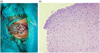
Examination under anaesthesia revealed an exophytic growth approximately 12 × 10 cm, involving the entire perineum. The mass extended into the lower 2 cm of the vagina involving the anterior and right lateral vaginal walls, both the urethral and anal openings, while sparing the rectal mucosa. The overlying skin was ulcerated and had blood-stained and malodorous discharge. Multiple bilateral inguinal nodes were palpable, the largest measuring approximately 2 × 1 cm in size on the right side (see Figure 1a).
(a) An exophytic mass over vulva involving urethra, lower vagina and perianal region. (b) Histopathological examination reveals stratified squamous epithelium with parakeratosis.

A biopsy of the tumour revealed proliferative stratified squamous epithelium with parakeratosis and papillomatosis but no intraepithelial malignancy indicating a BLT (see Figure 1b).
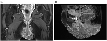
Pelvic magnetic resonance imaging depicted a 3 × 7 × 9 cm altered signal intensity lesion in the perineum, lower part of the vagina and anal canal, along with a few necrotic bilateral inguinal lymph nodes, the largest measuring 2 × 1 5 cm on the right side (see Figure 2a and 2b).
(a, b) Coronal and sagittal section of MRI showing massive exophytic lesion over vulva.

Due to the extensive nature of the lesion, surgical resection was not feasible. Thus, the patient was advised definitive external beam radiation therapy (EBRT). The treatment plan and associated risks were explained to the patient and a written informed consent was taken.
EBRT was planned on the Cobalt-60 teletherapy unit using conventional X-ray-based simulation in a supine frog-leg position using 6 megavoltage photons. A curative dose of 45 Gray (Gy) in 25 fractions to the perineum covering the entire mass with a 15 × 12 cm X-Y field was delivered.
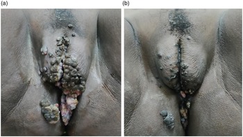
Assessment after 25 fractions resulted in a good clinical response with a reduction in size to approximately 6 × 5 cm; hence, a boost of 10·8 Gy in 6 fractions was given by a small perineal field of 10 × 9 cm (see Figure 3a).
(a) Regression of growth after radiation therapy with 45 Gy in 25 fractions. (b) Minimal residual lesion following curative dose of radiation delivery.

Upon the conclusion of radiation, the patient experienced a near complete response with a residual lesion approximately 2 × 1 cm (see Figure 3b). Topical podophyllin application was given for the residual lesion involving the perianal region. She is on 3 monthly follow-up. Nine months following the conclusion of therapy, the patient is asymptomatic and disease free.
Discussion
BLT is uncommon at such a young age. Judge et al. reported the first case of an extensive GCA in a 50-year-old white female. Two case series of BLT in patients younger than 50 years have been documented to date. Reference Chu, Vezeridis and Libbey5
Sobrado et al. demonstrated an excellent response for vulvar BLT and giant perianal condylomas using an EBRT dose of 45 Gy at 1·8 Gy per fraction. Reference Creasman, Haas and Fox6 A patient with urethral warts resistant to other forms of treatment was the first reported to use EBRT with 25 Gy using 2·5 Gy fractions for two and a half weeks. Reference Sobrado, Mester and Nadalin7
In this particular case, the patient received curative EBRT resulting in a favourable response with minimal skin toxicity. Close observation is essential due to a propensity for recurrence or malignant transformation. EBRT using photons is an effective alternative for treating resistant BLT lesions with acceptable outcomes, followed by local adjuvant therapies for residual lesions. The optimal dose and fractionation are not clearly defined in literature. Although earlier studies cautioned against radiotherapy for the risk of secondary malignant transformation, in recent literature, the use of EBRT has resulted in reasonable local control and significant downstaging of the disease. Reference White, Donnellan and Leong8,Reference Danoff, Holden and Thompson9
Conclusion
BLTs not amenable to surgery or topical therapy can be offered radiotherapy with favourable outcomes. Patients need close observation to detect recurrence or malignant transformation. Further studies with a larger number of patients and long-term follow-up are required.
Acknowledgements
None.
Financial Support
None.
Conflicts of Interest
None.