Introduction
Eleginops maclovinus is a large protandric fish that normally lives in coastal areas and sub-Antarctic waters near ocean beaches, in estuaries and in river mouths. They are distributed from Valparaiso to the Beagle Channel in the Pacific Ocean, and from the Beagle Channel to Uruguay in the Atlantic Ocean, and can also be found in the Falkland Islands (Guzmán & Campodónico, Reference Guzmán and Campodónico1973; Pequeño et al., Reference Pequeño, Lamilla, Lloris and Rucabado1995; Pavés et al., Reference Pavés, Pequeño, Bertrán and Vargas2005; Licandeo et al., Reference Licandeo, Barrientos and González2006; Martin & Bastida, Reference Martin and Bastida2008). This is a hermaphrodite-protandrous species with a primitive form of spermatozoa with a total length of 44.0 µm, a head length of 2.1 µm and head width of 2.5 µm. The midpiece has a length of 0.7 µm and its tail measures 41.2 µm long (Valdebenito et al., Reference Valdebenito, Cosson, Contreras, Sánchez, Pinheiro, Risopatrón, Farías and Figueroa2017). There is no information on the ontogeny of this species. Ontogeny is a process that occurs over different stages, and begins to activate successive mitotic replications and transformations that originate in specialized, complex and functional cells that give life to individuals (Kunz, Reference Kunz2004; Avery et al., Reference Avery, Killen and Hollinger2009; Akiyama et al., Reference Akiyama, Tero and Kobayashi2010). The success of this process depends on the quality of the gametes and the numerous events that occur before and during embryonic development. Bobe & Labbé (Reference Bobe and Labbé2010) analyzed the molecular and cellular characteristics that affect the quality of male and female gametes, before, during and after fertilization in fish. ‘Before’ fertilization, they can be affected by mechanisms that are responsible for the initiation of sperm motility, morphology and quality of the plasmatic membranes of spermatozoa; the physical and chemical parameters of the ovary or coelomic fluid, and the appearance of the oocyte during its first stages of development (Kjørsvik et al., Reference Kjørsvik, Mangor-Jensen and Holmefjord1990; Bromage et al., Reference Bromage, Bruce, Basavaraja and Rana1994; Penney et al., Reference Penney, Lush, Wade, Brown, Parrish and Burton2006; Pavlov & Emel´yanova, Reference Pavlov and Emel´yanova2008); the volume of the egg and the lipid droplets of species like Seriola lalandi (Moran et al., Reference Moran, Smith, Gara and Poortenaar2007), the composition of lipids and fatty acids in fish eggs and semen of species like Oncorhynchus mykiss (Vassallo-Agius et al., Reference Vassallo-Agius, Watanabe, Yoshizaki, Satoh and Takeuchi2001), Dexten (Giménez et al., Reference Giménez, Estévez, Lahnsteiner, Zecevic, Bell, Henderson, Piñera and Sánchez-Prado2006) and Ictalurus punctatus (Sink & Lochmann, Reference Sink and Lochmann2008) and post-ovulatory ageing of oocytes under in vivo or in vitro conditions (Mommens et al., Reference Mommens, Storset and Babiak2015). ‘During’ embryonic development, the characteristics and abnormalities of the first blastomeres have been used as criteria for discrimination between good and bad quality embryos (Kjørsvik et al., Reference Kjørsvik, Mangor-Jensen and Holmefjord1990, Reference Kjørsvik, Hoehne-Reitan and Reitan2003; Bromage et al., Reference Bromage, Bruce, Basavaraja and Rana1994; Shields et al., Reference Shields, Brown and Bromage1997; Penney et al., Reference Penney, Lush, Wade, Brown, Parrish and Burton2006; Pavlov & Emel´yanova, Reference Pavlov and Emel´yanova2008; Avery et al., Reference Avery, Killen and Hollinger2009; Bobe & Labbé, Reference Bobe and Labbé2010; Vásquez et al., Reference Vásquez, Llanos-Rivera and Castro2010; Valdebenito et al., Reference Valdebenito, Sánchez, Effer and Ubilla2012; Effer et al., Reference Effer, Sánchez, Ubilla, Figueroa and Valdebenito2012). In Hippoglossus hippoglossus, different characteristics of the blastomeres (symmetry, cell size, adhesion between blastomeres, margins and inclusions) are positively correlated with the percentage of hatched larvae (Shields et al., Reference Shields, Brown and Bromage1997). In Scophthalmus maximus the symmetry of blastomeres is positively correlated with hatching rates, larval tolerance towards stress and larval survivability, metamorphosis and pigmentation (Kjørsvik et al., Reference Kjørsvik, Hoehne-Reitan and Reitan2003). In Limanda ferruginea, eggs with abnormal blastomeres present the highest mortalities when compared with eggs with normal blastomeres (Avery & Brown, Reference Avery and Brown2005). In Zebrasoma scopas, Pavlov & Emel´yanova (Reference Pavlov and Emel´yanova2008) used symmetry, blastomeres adhesion, cellular division, cleavage synchronization, and the formation of syncytia as criteria to evaluate fish egg quality and, in Gadus morhua, Avery et al. (Reference Avery, Killen and Hollinger2009) related egg mortality, hatching success, larval deformations and mortality to abnormal cleavage patterns.
In Patagonian blennie, there have been no previous studies on symmetry of the first blastomeres. Due to the importance of this species for artisan fisheries and possible production in Chilean aquaculture, it is important to study their first stages of embryonic development, to provide preliminary information for the creation of diagnostics tools to measure gamete and/or embryo quality. This research morphologically describes the first blastomeres in Patagonian blennie (Eleginops maclovinus).
Materials and methods
Procurement of gametes and artificial fertilization
Oocytes were collected in July 2015, from broodstock of Patagonian blennie (E. maclovinus) held for 2 years under experimental breeding at the Quillaipe Experimental Station (41°330´53.01´´S; 72°46´41.25´´W) in Fundación Chile, Los Lagos Region in the south of Chile. The fish were kept in fibreglass ponds 5 m3 at a rate of water exchange 0.5 times per hour, fed with fresh fish at a daily rate of 0.5% of body weight by day and under a natural photoperiod. The average weight and length of the males were 258.3 ± 12.4 g and 27.0 ± 5.2 cm and females were 1540 ± 250 g and 45.3 ± 6.2 cm. The fish were sedated with Benzocaine® (10 ml/100 l of water), and gametes were then extracted using the method described by Groison et al. (Reference Groison, Fauvel, Suquet, Kjesbu, Le Coz, Mayer and Cosson2010): the urogenital pore was disinfected with alcohol and slight abdominal pressure was applied. The semen was then collected with a 5 ml syringe, with graduations every 0.5 ml to determine the volume of semen taken from each male. The semen extracted was only used if it presented maximum motility, and when the volume obtained was greater than 1 ml. The semen collected was transported to the Reproduction Laboratory of the Catholic University of Temuco (UCT), Temuco, Chile, using oxygenated containers at a constant temperature of 4°C and in the absence of light. For motility evaluation, a pool was prepared with the semen of five males which presented maximum motility; for the other evaluations, the semen of each male was considered individually. Fertilization was carried out at a temperature of 10.5°C, with a ‘pool’ of eggs from five females and semen from eight males, using the standard fish farming methods for reproduction. Eggs were then grouped into a Zoug jar type incubator at 10.7 ± 0.5°C in the dark registering a fertility average of 89.3 ± 4.3% evaluated in four groups of 100 eggs each.
Determination of the first cleavages
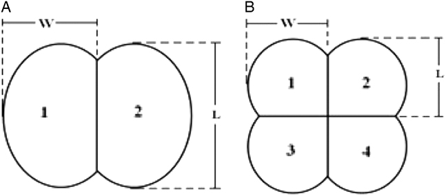
Eggs were analyzed every 30 min from fertilization until 5 h after fertilization, then under a stereoscopic Olympus SZ60 (JAPAN) magnifying lens with a cold light (Olympus LG-PS2), equipped with a Micropublisher 3.3 RTV digital camera (CANADA). In total, 100 embryos from each sampled time period were observed and photographed at ×56 magnification, and then they were classified according to their morphology and frequency of appearance. The images were processed using the software Q Capture Pro 7.0 (CANADA), in which the diameter of the unfertilized blastodisc, and the length and width of each blastomere were measured (Fig. 1) following the criteria proposed by Valdebenito et al. (Reference Valdebenito, Sánchez, Effer and Ubilla2012).
Criteria used to determine length (L) and width (W) of blastomeres, and the designation of number of each blastomere in embryos with two (A) or four blastomeres (B) (Effer et al., Reference Effer, Sánchez, Ubilla, Figueroa and Valdebenito2012).
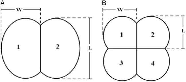
For the morphometric description for embryos with two and four blastomeres, we proceeded in the same way for each blastodisc and evaluated symmetry by comparison of the proportions of the lengths (L1/L2, L1/L3, L1/L4, L2/L3, L2/L4, L3/L4) and widths (W1/W2, W1/W3, W1/W4, W2/W3, W2/W4, W3/W4; Fig. 1B ), using the highest value as the denominator of both of the proportions analysis.
In total, 100 additional blastodisc (BC: control blastodiscs) with four blastomeres, symmetrical cleavages and normal appearance were selected visually following the criteria proposed by Shields et al. (Reference Shields, Brown and Bromage1997). Then morphometric symmetry was determined, as described in the previous paragraph, to compare the blastodiscs characterized visually as abnormal or asymmetrical.
Statistical analysis
Values for proportions of length and width of the symmetrical or normal blastomeres and for the asymmetrical or abnormal blastomeres, were analyzed using the statistical software GraphPad Prism® version 5.0 (GraphPad Software, San Diego CA, USA). A Dunnet´s multiple comparisons test was done to evaluate any statistically significant differences between the proportions of length and width of the blastodisc sampled from the control group (BC). The level of significance was fixed at a P-value <0.05 (n=100 replicates).
Results
Determination of cleavages
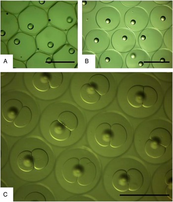
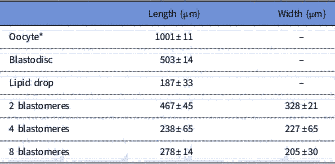
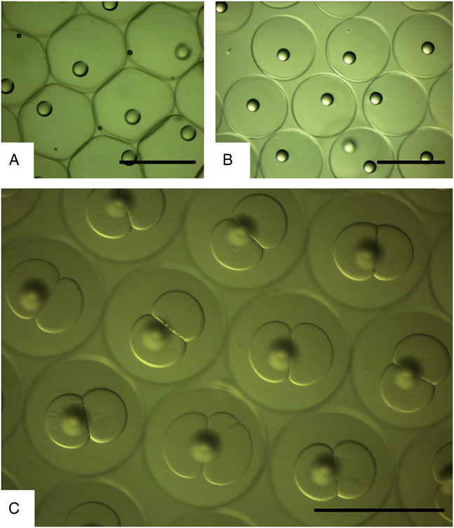
The viable eggs were free, pelagic and transparent. Before fertilization the oocytes presented a somewhat turgid aspect (Fig. 2a ) (maximum average diameter of 987 ± 41 µm) and after fertilization and hydration, the eggs were pelagic and their diameters increased to 1001.5 ± 11 µm (the differences were not statistically significant) presented spherical shape (Fig. 2b , Table 1) and had a smooth chorion of 8.7 ± 1.1 µm thickness and one oil globule with 187.4 ± 33.8 µm diameter. After fertilization and in contact with water, they developed a small perivitelline space that, 105 min after fertilization, had an average size of 12.6 ± 3.2 µm. After 2 h the fully developed blastodisc and was 503 ± 33 µm in diameter and had a regular shape disc suspended in the yolk mass (Fig. 2b and Table 1).
Microphotography of E. maclonivus eggs before (A) and 105 min after fertilization (B). Embryos with first cleavage ended at 3.5 h (C), incubated at 10.7°C. Magnification is ×56 and references bars in all images represent 1000 µm.
Morphometric parameters (average ± standard deviation) of oocytes, blastodisc and blastomeres in Patagonian blennie (E. maclovinus)

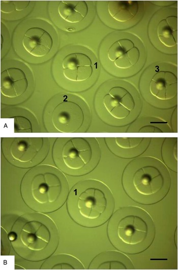
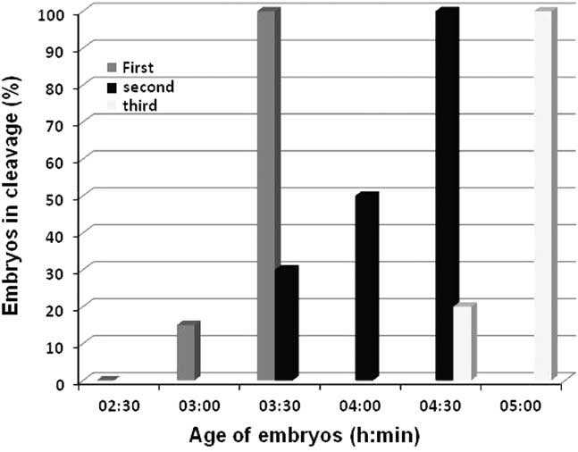
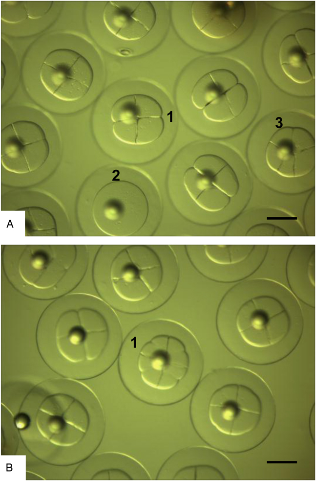
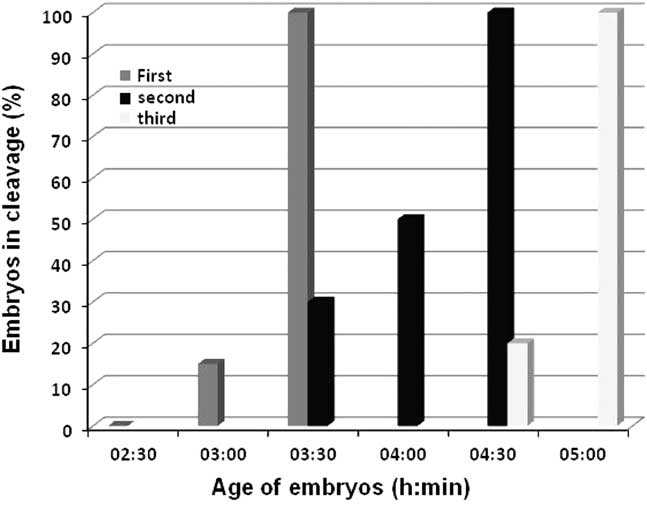
After 3 h of incubation (hours post fertilization, hpf), cleavage was observed in 15% of the eggs. At 3.5 h (3.5 hpf), 100% of the eggs had completed the first cleavage (Fig. 2c). At 4.5 h incubation (4.5 hpf), 100% of the eggs had ended the second cleavage and ended the third cleavage at 5 h (Figs 3a, b and 4).
Microphotography of E. maclonivus embryos 4.5 hpf. (A) With four blastomeres (1), without fertilization (2) and odd number of blastomeres. (B) With four and eight blastomeres (1). Magnification is ×56 and references bars in all images represent 200 µm.
Percentage of Patagonian blennie (E. maclonicus) embryos with cleavages, up to 2.5 h of incubation at 10.7°C.
After the first cleavage, the blastomeres were symmetrical in shape in most embryos and had a length of 467.4 ± 45 µm and a 328.3 ± 21 µm width. The length/width ratio was 1.4 (Fig. 3).
Morphological characterization
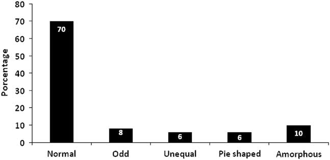
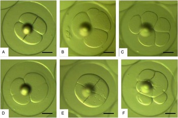
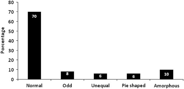
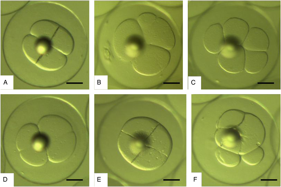
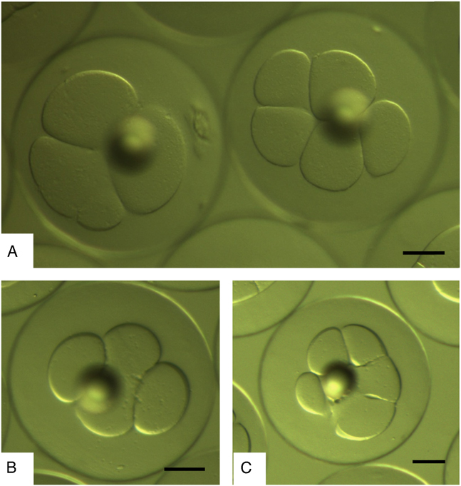
Figure 5 shows the five groups of blastomeres classified by morphology and frequency of appearance in embryos of E. maclovinus, from which four groups were considered to be abnormal morphologically (Figs 5–7). In total, 70% of the analyzed embryos presented normal cells (Category 1: G1) (Fig. 5a ); 8% had odd blastomeres (G2) (Fig. 5b, c ); 6% had unequal cells (G3) (Fig. 5d ); 6% had ‘pie-shaped’ blastomeres (G4) (Fig. 5e ) and 10% had amorphous blastomeres (G5) (Fig. 5f ).
Morphological categories and appearance percentages of the observed groups of blastomeres in Patagonian blennie (E. maclovinus). N=100.
Microphotographs of different types of embryos in Patagonian blennie (E. maclovinus). (A) Normal (G1). (B, C) with odd number of blastomeres (G2). (D) Unequal blastomeres (G3). (E) ‘Pie-shaped’ (G4) and (F) amorphous blastomeres (G5). Magnification ×56 and references bars in all images represent 200 µm.
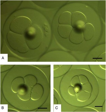
Microphotographs of different embryos with odd number of blastomeres. Magnification ×56 and references bars in images (A) and (B) represent 200 µm. In image (C) bar represents 300 µm.
Morphometric characterization
The mean diameter of the 70 blastodisc symmetrical of E. maclovinus was 503.8 ± 14.2 µm and was 107.0 ± 15.5 µm high in the central region. After the first cleavage, two symmetrical blastomeres with ratios L1/L2 and W1/W2 that did not differ significantly from 1.0 (1.00 ± 0.005 and 0.99 ± 0.04, respectively) were formed. The means of length and width from each blastomeres were 467.3 ± 45.6 µm and 328.9 ± 21.1 µm, respectively. The ratio L/W was 1.43 ± 0.19. The mean values obtained after second segmentation for length and width in the four cells were 238.8 ± 65.7 µm and 227.7 ± 65.3 µm respectively with a L/W ratio of 1.06 ± 0.09 from four blastomeres. The third cleavage ends 5 h after fertilization and the fourth cleavage ends 6 h after fertilization, forming an embryo of 16 blastomeres. In the group with four asymmetric blastomeres, ratios L/W (1.13 ± 0.7) were statistically different from that of the Normal group and the proportions differed significantly at 1.
Discussion
There are no antecedents about the ontogenetic development of the Patagonian blennie (Eleginops maclovinus). The beginning of the development in this species is similar to characteristic processes for marine fish. They have telolecithal eggs with partial cleavage and form a small perivitelline space similar to that observed by Bustos & Landaeta (Reference Bustos and Landaeta2005) in the early development of hake south (Merluccius australis) in Chile, Cuartas et al. (Reference Cuartas, Rosas, Velásquez and Cabrera2003) for corocoro rayao (Haemulon bonarienses) of mangroves in Venezuela, Horie et al. (Reference Horie, Utoh, Yamada, Okamura, Zhang, Mikawa, Akazawa, Tanaka and Hideo2002) in common Japanese conger (Conger myriaster) and Moran et al. (Reference Moran, Smith, Gara and Poortenaar2007) in yellowtail kingfish (Seriola lalandi). In the central region of the egg there is a lipid conspicuous drop similar to that observed by Ebanks (Reference Ebanks2013) in cobia (Rachycentron canadum), Pavlov & Emel’yanova (2008) in Zebrasoma scopas and Moran et al. (Reference Moran, Smith, Gara and Poortenaar2007) in Seriola lalandi.
In Patagonian blennie, the first cleavage occurred at 3.5 h, similar to that recorded for Conger myriaster by Horie et al. (Reference Horie, Utoh, Yamada, Okamura, Zhang, Mikawa, Akazawa, Tanaka and Hideo2002) who observed the first segmentation at 4 h after fertilization, describing typical discoidal cleavage and two blastomeres of equal size. Similarly the 16 blastomeres were observed at 6 h, as in Patagonian blennie.
Most embryos were observed to be symmetrical (70%). This finding is similar to that described by Moran et al. (Reference Moran, Smith, Gara and Poortenaar2007) in Seriola lalandi, who found 36% abnormalities in embryos, pointing that the most predominant abnormalities were asymmetrical cell cleavage and indistinct margins (odd number of blastomeres). Vásquez et al. (Reference Vásquez, Llanos-Rivera and Castro2010) found levels of 3–13% of abnormal embryos in wild specimens of sardine (Strangomera bentincki) in southern Chile and Westernhagen et al. (Reference Westernhagen, Dethlefsen, Cameron, Berg and Fürstenberg1988) reported for cod, plaice and flounder a percentage of deformed embryos of 18–31%, 23–28% and 19–30%, respectively.
Pavlov & Emel´yanova (Reference Pavlov and Emel´yanova2008) studied the symmetry of the first blastomeres in Z. scopas, identifying some asymmetrical embryos, asynchrony of cleavages, syncytia, and other aberrations of development attributed to oocyte post-ovulatory ageing. Likewise, Mommens et al. (Reference Mommens, Storset and Babiak2015) described malformations in embryos and larvae of Atlantic salmon that were directly related to the ageing of oocytes after ovulation in the abdominal cavity of females, as this species does not spawn spontaneously in ponds. Lahnsteiner & Patarnello (Reference Lahnsteiner and Patarnello2005) evaluated the quality of eggs based on the shape of the lipid drop, egg and yolk, correlating them with larval survivability.
Five groups were identified based on blastomere morphology from which four groups presented blastomeres that had visually abnormal cleavage patterns. These patterns originated from different factors such as: (1) elevated temperatures during the incubation phase (Pavlov & Moksness, Reference Pavlov and Moksness1994); (2) overmaturity or ageing of eggs (Valdebenito et al., unpublished data); and/or (3) triploidy induction (Rani, Reference Rani2005). Conversely, patterns of anomalies have been described as marginal, doughnut, triple or pie shaped and, in some cases, patterns of asymmetry can be combined (Avery & Brown Reference Avery and Brown2005; Avery et al. Reference Avery, Killen and Hollinger2009). Embryos with unequal pie-shaped blastomeres (Fig. 4C, E ) have been reported by Avery et al. (Reference Avery, Killen and Hollinger2009) in Gadius morhua, which produces viable larvae without significant differences in mortality compared with eggs with symmetrical blastomeres.
Lyman-Gingerich & Pelegri (Reference Lyman-Gingerich and Pelegri2007), attributed some asymmetries in the first blastomeres in zebrafish to mutants genes: Cobblestone mutants do not initiate mitosis and generate giant blastomeres and embryos with odd numbers of cells. Barrette mutants affect nuclear division and the pattern of membrane deposition. Golden gate mutants show abnormal DNA bridges indicative of defects in chromosome segregation during mitosis and mutation in aura results in defects in the deposition of adhesive membranes at the furrow.
This research provided the first antecedents of embryonic development in Patagonian blennie and establishes a solid basis for creation of an early evaluation tool for embryo quality in this species, demonstrating that a high percentage of early embryos (70%) showed a ‘normal’ characteristic that ensures a high survival in later stages of development.
Acknowledgements
The authors are thankful to Ms Aurora Sambolin for reviewing the English version.
Financial support
We are grateful to Quillaipe Experimental Station of Fundación Chile and to projects FONDECYT 1151315, 1180387 and MEC 80140066 for financial support.
Conflicts of interest
There are no conflicts of interest.