Introduction
Interferon-γ is a dimerised, soluble cytokine which is the only member of the type II class of interferons.Reference Ealick, Cook, Vijay-Kumar, Carson, Nagabhushan and Trotta1 It is secreted by T-helper 1 cells (Th1 cells), Cytotoxic T cell (Tc cells), dendritic cells and natural killer cells. It is the hallmark cytokine of Th1 cells.Reference Schroder, Hertzog, Ravasi and Hume2 Interferon gamma has antiviral, immunoregulatory and anti-tumour properties; it alters transcription in various genes, producing a variety of physiological and cellular responses.Reference Abbas, Murphy and Sher3 These responses include increased antigen presentation of macrophages, activation of lysosomes in macrophages and suppression of T-helper 2 cells (Th2 cell) activity. In addition, interferon-γ causes normal cells to increase expression of class I major histocompatibility complex molecules, promotes adhesion and binding required for leukocyte migration, and promotes natural killer cell activity. Interferon-γ is important in acute infections: intracellular microbial infection has been reported to induce predominantly type 1 T cell responses, characterised by production of a large amount of type 1 T-cell cytokines, namely interferon-γ, tumour necrosis factor α (TNF-α) and interleukin 2.Reference Carter and Dutton4–Reference Zinkernagel and Hengartner6 These type 1 T cell responses promote phagocytic cell mediated, proinflammatory responses and effectively eradicate microbes.Reference Carter and Dutton4–Reference Zinkernagel and Hengartner6 However, excessive type 1 T cell responses could cause excessive inflammation and tissue damage.Reference Carter and Dutton4–Reference Zinkernagel and Hengartner6
Animal models of otitis media with effusion have suggested that the inflammatory stimulus is bacterial endotoxin, which stimulates interferon-γ and TNF-α production, resulting in increased mucin production and mucous hyperplasia.Reference Morikawa, Zhang, Nonaka and Morikawa7, Reference Fischer, Rochelle, Voynow, Akley and Adler8 On the other hand, resolution following macrolide antibiotic treatment has been reported to be due to modification of the host's immunological response, in the form of interference with the activity of T helper cells, resulting in suppression of the gene expression and release of immunoregulatory cytokines.Reference Lasisi, Arinola and Bakare9
Most of the research on immunoregulatory cytokines in otitis media has addressed otitis media with effusion. However, suppurative otitis media is the predominant presentation in developing countries, and is one of the most common and intractable ear diseases. We have found that the concentration of immunoglobulin (Ig) G in serum and middle-ear secretions differs significantly, comparing patients with acute suppurative otitis media and chronic suppurative otitis media (CSOM).Reference Lasisi, Arinola and Bakare9 This suggests that IgG could be used in monitoring and predicting chronicity in suppurative otitis media.
The aim of this study was to determine the concentrations of interferon-g (a T helper 1 cell infectious-type cytokine) in children with acute suppurative otitis media, and to determine the significance of interferon-γ concentration to the nature of otorrhoea and the outcome of otitis media.
Methods
Patients
Ethical approval for the study was obtained from the institution review board of the University of Ibadan and the University College Hospital Ibadan. Informed consent was obtained from patients' parents, including consent for publication.
Following these steps, recruitment was commenced. The inclusion criteria were children under the age of 12 years with acute suppurative otitis media (using three months as a cut-off limit for acute otitis media).Reference Lasisi, Olaniyan, Muibi, Azeez, Idowu and Lasisi10 All the patients were recruited from the general out-patient department and the otorhinolaryngology out-patient clinic of the University College Hospital, Ibadan.
A clinical history was taken from all patients. Examination of the ear, nose and throat was then performed using a hand-held otoscope, Shirom lamp and head mirror.
Patients were offered suction ear toileting, topical wick dressing or nasal decongestants for two to four weeks. Oral antibiotics for one week was added to the treatment regime if otorrhoea persisted after four weeks of topical dressing.
All patients were followed up for six to 10 months. The criteria for resolution of otitis media and commencement of tympanic membrane healing were taken as cessation of otorrhoea and an otoscopic finding of neo-membrane formation closing the tympanic membrane perforation.
Sample collection
At recruitment and before treatment commencement, middle-ear secretions were collected from patients via Pasteur pipette aspiration into a microfuge bottle, and stored at −80°C.
Interferon-γ assay
The wash buffer, substrate solution, interferon-γ standard and calibrator diluent were reconstituted as specified by manufacturers' instructions. One hundred microlitres of assay diluent was added into each well, followed by 100 µl of the standard, sample and control. The solution was then incubated for 2 hours at room temperature. The aspirate was washed 4 timiddle ear secretions, and 200 µl of conjugate was added to each well. The solution was incubated for 2 hours at room temperature. The aspirate was then washed 4 timiddle ear secretions and 200 µl of substrate solution was added. The solution was incubated at room temperature for 30 minutes, protected from light. The enzyme-linked immunosorbent assay method was employed to assay interferon-γ, using the Thermolabsystems Multiskan® Ex system (R&D Systems Europe Ltd, Abingdon, Oxon, UK). The colour change was read at 450 nm immediately. The standard curves for the various classes of immunoglobulins were plotted on semi-log graph paper, and the concentrations of the test and control samples were read off the standard curve.
Statistics
The main outcome variable was the interferon-γ concentration in the middle-ear secretions of patients with resolved acute otitis media or CSOM.
Data were initially explored using Stata software. Variables were analysed by unpaired t-test both for equal and unequal variance, using the variance ratio function of the Stata software to determine the appropriate use of Satterthwaite's correction for the degrees of freedom. The level of statistical significance was set at p < 0.05 for all analyses.
The Pearson test was used to assess the correlation between mean middle-ear secretion interferon-γ concentrations and mean middle-ear secretion concentrations of IgG, IgA and IgE (assayed and reported earlier for these same patients).Reference Lasisi11
Results
The study commenced with the enrolment of 358 cases of acute suppurative otitis media. Nine-month follow up was achieved in 304 patients (85 per cent). Of these patients, acute suppurative otitis media resolved in 187, while otorrhoea persisted beyond three months (i.e. CSOM) in 117. The 304 patients completing follow up comprised 173 boys and 131 girls, aged between four months and nine years (mean age 6.6 years; standard deviation (SD) 1.32). On initial examination, 171 patients had purulent otorrhoea and 133 had mucoid otorrhoea.
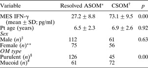
Patients' middle-ear secretion interferon-γ concentrations ranged from 12 to 126 pg/ml. Those whose acute suppurative otitis media resolved had a mean interferon-γ concentration of 27.2 pg/ml (SD 8.8), while those who progressed to CSOM had a mean interferon-γ concentration of 73.1 pg/ml (SD 9.5) (Table I).
Patients with resolved ASOM vs CSOM: comparison

Total n = *187, †117, ‡173, **131, §171 and #133. ASOM = acute suppurative otitis media; CSOM = chronic suppurative otitis media; MES = middle-ear secretion; IFN-γ = interferon-γ; SD = standard deviation; pt = patient; OM = otitis media
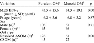
Children with purulent otitis media had a mean middle-ear secretion interferon-γ concentration of 43.5 pg/ml (SD 15.6), while those with mucoid otitis media had a mean concentration of 74.3 pg/ml (SD 19.1) (Table II).
Patients with purulent vs mucoid otitis media: univariate analysis

Total n = *171, †133, ‡173, **131, §187 and #117. OM = otitis media; MES = middle-ear secretion; IFN-γ = interferon-γ; SD = standard deviation; pt = patient; ASOM = acute suppurative otitis media; CSOM = chronic suppurative otitis media
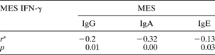
We undertook univariate analysis using unpaired t-testing to compare mean interferon-γ concentration results. This revealed significant differences between those patients whose acute suppurative otitis media resolved and those who progressed to CSOM (p = 0.00), and also between patients with purulent versus mucoid otitis media (p = 0.00). The age and sex distributions were not statistically significant, comparing resolved acute suppurative otitis media versus CSOM (Table I), and comparing purulent versus mucoid otitis media (Table II). Pearson correlation testing revealed significant inverse correlation between middle-ear secretion interferon-γ concentration and middle-ear secretion concentrations of IgG (p = 0.01), IgE (p = 0.03) and IgA (p = 0.00) (Table III).
Middle-ear secretion concentrations of IFN-γ vs immunoglobulins: univariate analysis
*Pearson correlation coefficient. MES = middle-ear secretion; IFN-γ = interferon-γ; Ig = immunoglobulin
Discussion
The main finding from this study was that middle-ear secretion of interferon-γ was associated with a tendency to chronicity in suppurative otitis media. In addition, we found a higher middle-ear secretion concentration of interferon-γ in mucoid otitis media compared with purulent otitis media.
These findings are similar to those of Chen et al. Reference Chen, Liang, Zheng, Liu, Liu and Long12 and others.Reference Yellon, Doyle, Whiteside, Diven, March and Fireman13–Reference Maxwell, Leonard and Kreutzer15 Chen et al. reported that the middle-ear secretion interferon-γ concentration of patients with chronic otitis media was much higher than that of those with acute otitis media. They concluded that high expression of interferon-γ in the middle-ear effusion may be a marker for suppurative otitis media which follows a chronic course, and that the interferon-γ in the middle-ear effusion may be produced in the tympanic cavity, rather than being a simple transudate from blood.
Similarly, Yellon et al. Reference Yellon, Doyle, Whiteside, Diven, March and Fireman13 and Himi et al. Reference Himi, Suzuki, Kodama, Takezawa and Kataura14 concluded that a higher concentration of interferon in middle-ear effusions may be a marker for otitis media chronicity. Maxwell et al. Reference Maxwell, Leonard and Kreutzer15 found that middle-ear secretion tumour necrosis factor α (TNF-α) levels were significantly lower in children who had undergone multiple tube insertions (p = 0.02). Higher levels of TNF-α were noted in those children who subsequently developed episodes of otitis media after tube placement (p = 0.02). Maxwell et al. concluded that cytokines and their inhibitors were present in a large number of middle-ear effusions and may be important in the regulation of inflammatory processes in chronic otitis media with effusion.
Interferon-γ is produced by T lymphocytes in response to viral or bacterial challenge. It has been shown to activate macrophages, stimulate cytotoxic cell activity, and increase B cell differentiation and antibody production; however, it also has the potential to cause tissue damage.Reference Yellon, Leonard, Marucha, Craven, Carpenter and Lehmann16–Reference Hotomi, Samukawa and Yamanaka18 The cytotoxic and osteoclast- and fibroblast-stimulating activity of interferon-γ may be responsible for the persistence of otorrhoea seen in some patients.Reference Himi, Suzuki, Kodama, Takezawa and Kataura14–Reference Nassif, Simpson, Izzo and Nicklaus17 In an earlier study comparing immunoglobulin levels in acute suppurative otitis media versus CSOM, significant differences were found in serum IgG concentrations (p = 0.04) and in middle-ear secretion concentrations of IgG (p = 0.02) and IgE (p = 0.04); these results suggested that immunoglobulins were a factor in suppurative otitis media chronicity.Reference Lasisi, Arinola and Bakare9, Reference Lasisi, Arinola and Olayemi19
In this study, we found a correlation between middle-ear concentrations of interferon-γ and immunoglobulins, suggesting that the interferon-γ in middle-ear secretions is secreted from the middle-ear mucosa. Chen et al. Reference Chen, Liang, Zheng, Liu, Liu and Long12 and othersReference Himi, Suzuki, Kodama, Takezawa and Kataura14–Reference Yellon, Leonard, Marucha, Craven, Carpenter and Lehmann16 believe that the interferon-γ found in middle-ear effusions may be produced by the tympanic cavity, rather than being a simple transudate from blood. In addition, other authors have found a correlation between the middle-ear secretion concentrations of interferon-γ and other cytokines and immunoglobulins.Reference Yellon, Doyle, Whiteside, Diven, March and Fireman13–Reference Nassif, Simpson, Izzo and Nicklaus17 We found an inverse correlation between middle-ear secretion interferon-γ concentrations and those of IgG, IgE and IgA; in explanation, we hypothesise that a high middle-ear secretion interferon-γ concentration may result in mucosal damage, leading to reduced secretion of protective immunoglobulins in the middle ear and thus leading to chronic otitis media.
• The immunoregulatory roles of interferon-γ have been reported in patients with otitis media with effusion; however, they remain to be determined in cases of acute suppurative otitis media
• This study aimed to determine the levels of interferon-γ in middle-ear secretions in children with acute suppurative otitis media, and to assess the significance of this cytokine in the outcome and nature of otorrhoea
• High middle-ear concentrations of interferon-γ appeared to be a factor in the chronicity of suppurative otitis media; further research in this area may lead to the development of agents that assist control of this condition
Similarly, interferon-γ may facilitate the production of TNF-α, which has been implicated in the increased secretion of mucin by airway epithelium, hence promoting persistence of otorrhoea. Such mucin secretion induction has been reported to occur via an intracellular pathway that appears to involve endogenously produced nitric oxide.Reference Yellon, Leonard, Marucha, Craven, Carpenter and Lehmann16–Reference FitzGerald, Green, Stafford, Birchall and Pearson21 Mucin secretion could potentially be enhanced by cyclic guanosine monophosphate via activation of phosphokinase G, a serine kinase involved in such biological responses as calcium signalling, neutrophil degranulation, potassium and chloride channel functioning, and mucin secretion.Reference Yellon, Leonard, Marucha, Craven, Carpenter and Lehmann16–Reference Storgaard, Larsen, Blegvad, Nodgaard, Ovesen and Andersen20
One major significance of our findings is that otitis media chronicity may be associated with a high middle-ear secretion concentration of interferon-γ. However, this study did not show a causal relationship between raised middle-ear secretion interferon-γ concentration and acute suppurative otitis media. The clinical implication of our findings is that assessment of middle-ear aspirate interferon-γ concentration may enable patients to be better informed regarding the likely time course of their ear infection, and may identify children requiring longer term follow up. It is scientifically possible that control of otitis media chronicity may be improved by the use of agents which can counteract the effects of interferon gamma (among other Th1 cytokines).
We did not study the role of the size and the bacterial load of the adenoids in affecting the outcome of suppurative otitis media. These are also important factors, which we hope to investigate in future research.
Conclusion
We conclude from this study that a high middle-ear secretion concentration of interferon-γ is a factor in the chronicity of suppurative otitis media. Further research in this area may lead to the development of agents aiding the treatment of suppurative otitis media.
Acknowledgements
We would like to express our appreciation for the helpful advice of Professors A O Osoba and R A Bakare of the Department of Medical Microbiology and Parasitology, and Professor G T A Ijaduola of the Department of Otorhinolaryngology. We also thank Mrs Ishowo and Mrs Latinwo, nurses at the Bilal Mission, and Drs Irabor and Mosuro at the General Outpatient Department of the University College Hospital, for their help during patient recruitment. This study was supported by the University of Ibadan McArthur Grant (grant number 800/406/54/01/D/2008/1). The study received ethical approval from the institution review board of the University of Ibadan and University College Hospital Ibadan (UI/IRC/07/0023).