INTRODUCTION
Dientamoeba fragilis is a protozoan pathogen of the human gastrointestinal tract first described in the scientific literature by Jepps and Dobell (Reference Jepps and Dobell1918). Dientamoeba was originally classified as an amoeba (subphylum Sarcodina (Jepps and Dobell, Reference Jepps and Dobell1918)) though a later study utilizing electron microscopy confirmed its relationship with trichomonads (Camp et al. Reference Camp, Mattern and Honigberg1974). Although Dientamoeba was described almost a century ago, comparatively little information exists with regards to its life cycle, genetics and proteome. Given the recent recognition of D. fragilis as a significant human pathogen by numerous authors (Dickinson et al. Reference Dickinson, Cohen and Schlenker2002; Girginkardesler et al. Reference Girginkardesler, Coskun, Cuneyt Balcioglu, Ertan and Ok2003; Johnson et al. Reference Johnson, Windsor and Clark2004, Stark et al. Reference Stark, Beebe, Marriott, Ellis and Harkness2005b, Lagace-Wiens et al. Reference Lagace-Wiens, VanCaeseele and Koschik2006, Stark et al. Reference Stark, Beebe, Marriott, Ellis and Harkness2006, Reference Stark, Beebe, Marriott, Ellis and Harkness2007, Reference Stark, Barratt, Ellis, Harkness and Marriott2009, Reference Stark, Barratt, Roberts, Marriott, Harkness and Ellis2010), more research with regards to this organism is warranted. The ability to maintain clinical isolates of Dientamoeba in culture for extended periods of time at high cell densities and, the ability to cryogenically preserve viable Dientamoeba trophozoites, would facilitate further study and analysis of this emerging pathogen.
Previous authors have reported on the xenic cultivation of D. fragilis (Lamy, Reference Lamy1960; Robinson, Reference Robinson1968; Robinson and Ng, Reference Robinson and Ng1968; Sawangjaroen et al. Reference Sawangjaroen, Luke and Prociv1993; Clark and Diamond, Reference Clark and Diamond2002; Windsor et al. Reference Windsor, Macfarlane, Hughes-Thapa, Jones and Whiteside2003). However, no axenic cultivation system has been successfully developed for D. fragilis. Many xenic media are able to support the growth of D. fragilis. Dobell was the first to isolate and grow D. fragilis using a biphasic, xenic medium consisting of an inspissated horse serum slope overlaid with a liquid phase consisting of egg whites diluted in Ringer's solution supplemented with rice starch (Dobell, Reference Dobell1940). Other biphasic media able to support the growth of D. fragilis include Cleveland and Collier's medium (a Loeffler's slope with a liquid overlay (Cleveland and Collier, Reference Cleveland and Collier1930)), modified Boeck and Drbohlav's (BD) medium (LE medium) and Robinson's medium (Clark and Diamond, Reference Clark and Diamond2002; Windsor et al. Reference Windsor, Macfarlane, Hughes-Thapa, Jones and Whiteside2003). Some monophasic media can also support the growth of D. fragilis. The American Type Culture Collection (ATCC) recommends a monophasic TYGM-9 broth ((ATCC medium 1171) as used in experiments by Chan et al. (Reference Chan, Guan, Mackenzie and Diaz-Mitoma1994)) for the growth of D. fragilis strain ATCC 30948 (strain Bi/Pa) – though isolates of D. fragilis are no longer available from the ATCC (at the time of writing). Balamuth's medium is another monophasic liquid medium which can support the growth of D. fragilis (Balamuth, Reference Balamuth1946). It is also plausible that other media used for the cultivation of other trichomonads such as Medium 199 (M199) (used previously as the base for a Histomonas meleagridis medium (Lesser, Reference Lesser1961)), Trichosel (designed for the growth of T. vaginalis (Borchardt et al. Reference Borchardt, Zhang, Shing and Flink1997; van Der Schee et al. Reference van Der Schee, van Belkum, Zwijgers, van Der Brugge, O'Neill, Luijendijk, van Rijsoort-Vos, van Der Meijden, Verbrugh and Sluiters1999)) and a commercial Tritrichomonas foetus medium, could also support the growth of D. fragilis.
Two studies have reported on the cryopreservation of D. fragilis. Experiments performed by Dwyer and Honigberg (Reference Dwyer and Honigberg1971) and Sawangjaroen et al. (Reference Sawangjaroen, Luke and Prociv1993) describe the successful cryopreservation of D. fragilis using different concentrations of the cryopreservant dimethyl sulfoxide (DMSO). Dwyer and Honigberg (Reference Dwyer and Honigberg1971) also stated that unpublished studies by others suggest that the addition of 2·5% glucose allows for successful cryopreservation of D. fragilis at a final concentration of 5% DMSO. To our knowledge, no studies exist that compare methods for the cryopreservation and in vitro cultivation of D. fragilis. Also, the ability of commercial media such as M199, Trichosel and Tritrichomonas foetus medium to support the growth of D. fragilis has never been explored. Furthermore, the effect of different atmospheric conditions and temperatures on Dientamoeba growth has not been fully investigated either. In order to address this paucity of knowledge a modified BD medium, TYGM-9, a Loeffler's slope medium (a modified Cleveland and Collier's medium), Robinson's medium, M199, Trichosel and a commercial Tritrichomonas fetus medium were investigated for their ability to support the growth of D. fragilis. Experiments were also performed to evaluate the growth of D. fragilis under anaerobic, microaerophilic and aerophilic conditions and, to explore its growth at a range of different temperatures. Cell counts were performed on cultures to determine the most suitable in vitro growth conditions for D. fragilis cultivation. Also, variations of the cryopreservation methods described by Sawangjaroen et al. (Reference Sawangjaroen, Luke and Prociv1993) and Dwyer and Honigberg (Reference Dwyer and Honigberg1971) were compared for their ability to preserve viable D. fragilis trophozoites.
MATERIALS AND METHODS
Modified BD medium
One hundred ml of the liquid overlay was prepared by mixing 90 ml of PBS (Sigma P4417-100TAB), 9 ml of heat-inactivated horse serum (Sigma H1138-500 ml) and 1 ml of a 20% (w/v) bacteriological peptone solution (Oxoid, LP0037). Solid egg slopes were prepared as described previously (Sawangjaroen et al. Reference Sawangjaroen, Luke and Prociv1993), and approximately 2 mg of rice starch (Sigma, S-7260) was placed in the bottom of each slope. Each slope was then overlaid with 5 ml of the liquid overlay.
Loeffler's medium
Loeffler's serum slopes were prepared to contain heat-inactivated horse serum (700 ml/L), glucose (2·5 g/L) and nutrient broth No.2 (6·25 g/L) in distilled water. Five ml of this were poured into a 14 ml vol. McCartney bottle, sloped and inspissated in an 80°C drying oven until slopes solidified. Approximately 2 mg of rice starch was placed into the bottom of each slope. The slopes were then overlaid with 5 ml of PBS. It should be noted that during inspissation, if Loeffler's slopes are left for too long at 80°C, Dientamoeba will not grow in the resulting medium. This is probably due to the damaging of some essential cofactor/s within the serum. During inspissation, slopes should be observed frequently and allowed to cool immediately after they solidify.
TYGM-9 broth
TYGM-9 broth (formulation: 2·8 g K2HPO4, 0·4 g KH2PO4, 2·0 g casein digest, 1·0 g yeast extract, 7·5 g NaCl, 2·0 g gastric mucin all dissolved in 970 ml of distilled water) was prepared and poured into Pyrex 99447 15·5 ml vol. glass culture tubes in 10 ml volumes. Each tube was supplemented with approximately 2 mg of rice starch.
Trichosel
Trichosel was purchased from BD Diagnostic systems (cat. No. 298323) and prepared as per the manufacturer's instructions. Ten ml volumes were dispensed into Pyrex 99447 15·5 ml vol. glass culture tubes. Each tube was supplemented with approximately 2 mg of rice starch.
Robinson's medium
This medium was prepared as described previously by Clark and Diamond (Reference Clark and Diamond2002) with the following modifiation: the inclusion of approximately 2 mg of rice starch.
Tritrichomonas foetus medium
Three ml vials of T. foetus medium were commercially obtained (Micromedia, Cat. No. 3274) and supplemented with approximately 2 mg of rice starch.
Medium 199
Medium 199 (Sigma Aldrich, Cat. No. M3769) was prepared according to the manufacturer's instructions. Heat-inactivated horse serum (Sigma Aldrich, Cat. No. H1138-500 ml) was added to Medium 199 at a concentration of 10%. Ten ml volumes were dispensed into Pyrex 99447 15·5 ml vol. glass culture tubes and approximately 2 mg of rice starch was added to each tube.
Source of D. fragilis trophozoites
Four Dientamoeba strains were used in these studies. They were isolated from the stools of symptomatic patients known to be infected with D. fragilis. Patients were initially diagnosed with dientamoebiasis based on the observation of Dientamoeba trophozoites in iron-haematoxylin stained faecal smears. Oil-immersion microscopy performed on sodium acetate acetic acid formalin (SAF) fixed, iron-haematoxylin stained faecal smears is routinely carried out in the Department of Microbiology at St Vincent's Hospital, Sydney, for investigation of gastrointestinal complaints. Stool specimens fixed in SAF were stained with a modified iron-haematoxylin stain (Fronine, Australia) according to the manufacturer's recommendations. All stained smears were examined by oil-immersion microscopy (1000×magnification). Approximately 250 fields of view were examined on each slide. Definitive diagnosis was based on the characteristic morphology of D. fragilis in the permanently stained smears (trophozoites measuring 5–15 μm in diameter, with 1 or 2 nuclei each with a large fragmented central karyosome without peripheral chromatin (Fig. 1A)).
A trophozoite of Dientamoeba fragilis in a fixed, iron-haematoxylin stained faecal smear viewed under oil-immersion microscopy (A) and, live trophozoites from modified BD cultures viewed under phase contrast microscopy (B, C and D). The typical fragmented nuclear structure of Dientamoeba in stained smears is observable in the binucleate trophozoite shown in inset A. Trophozoites in culture can exhibit different morphologies including a spherical, granular, vacuolated form (B) and motile forms with visible pseudopodia (C, D). In modified BD cultures, rice starch will often be observed in the trophozoite cytosol as highly refractive granules (C, D), though not always (B). Scale bars represent 10 μm.

Patients with a microscopy-confirmed Dientamoeba infection were requested to submit a fresh stool specimen for culture prior to their treatment. Immediately upon receipt of the fresh, unpreserved stool specimen a portion was cultured. Approximately 10 mg of the unpreserved stool specimen was placed directly into a bottle of modified BD medium. Cultures were incubated at 37°C and, after 48 h, a drop of sediment from each culture was examined at 400×magnification under phase-contrast microscopy for the presence of Dientamoeba trophozoites. When Dientamoeba trophozoites were observed, a portion of the culture sediment was fixed in SAF (1:1) and stained with a modified iron-haematoxylin stain (Fronine, Australia) and examined by oil-immersion microscopy to confirm the isolation of D. fragilis. Another portion of the sediment was kept for DNA extraction followed by PCR.
Comparison of different media
Dientamoeba fragilis trophozoites were grown in Loeffler's slope medium (at 37°C under microaerophilic conditions) and quantitated by cell counts using Kova slides (Hycor Biomedical Inc). For all cell counts, the medium vessels containing Dientamoeba trophozoites to be counted were very gently shaken and inverted several times to achieve an even cell suspension. In order to completely break up the sediment on the bottom of the medium vessels it was sometimes necessary to agitate the cultures by very gently pipetting up and down with a transfer pipette. Once an even cell suspension was achieved, counts were performed using Kova slides. A small drop of the cell suspension was placed into the counting chamber of a Kova slide and raw cell counts were performed.
Trophozoites were added to 2 vials of each of the different types of media (modified BD medium, Loeffler's slope medium, TYGM-9 broth, Trichosel, Robinson's medium, Tritrichomonas foetus medium and Medium 199) to a final concentration of 50 trophozoites/μl. This was achieved by diluting the trophozoites into the culture media to be used and/or concentrating by centrifugation at 500 g for 5 min where required. Each culture was then incubated at 37°C under microaerophilic conditions. Cell counts were made each day as described above, until no more trophozoites could be observed (or until 9 days passed – whichever occurred first). Immediately after counts were completed each day, cultures were re-incubated as described above. Cell counts were plotted graphically in order to visualize the growth patterns of Dientamoeba in each medium. This was repeated for the 4 isolates of D. fragilis.
Effect of atmospheric composition on in vitro growth
This set of experiments was performed using Loeffler's slope medium and 4 clinical isolates of D. fragilis. Dientamoeba trophozoite cultures were grown in Loeffler's medium. These trophozoites were inoculated into 6 fresh vials of Loeffler's medium to a total concentration of 108 trophozoites/μl. This was performed by concentration of trophozoites via centrifugation (500 g for 5 min), and/or dilution in PBS where required, and quantifying by cell counts. Two vials were each grown under aerophilic, microaerophilic and anaerobic conditions. These steps were repeated for each of the 4 clinical isolates of D. fragilis.
Aerophilic conditions
In order to expose D. fragilis cultures to as close to aerophilic conditions as could be achieved, 2 culture vials from each isolate were left in a 37°C incubator with the caps completely loosened. Cell densities were counted each day until trophozoites were no longer observed.
Microaerophilic conditions
Microaerophilic conditions were achieved using an Anoxomat machine (Mart Microbiology B.V.). The lids of all culture vessels were completely loosened and cultures were placed in an appropriate Anoxomat jar and connected to the Anoxomat machine according to the manufacturer's instructions. Gases were introduced into the sealed Anoxomat Jars to the following concentrations: 6% O2, 7·2% CO2, 3·6% H2, 83·3% N2. Jars containing culture vials were placed at 37°C and cell counts were made each day until trophozoites were no longer observed. After counting each day, cultures were placed in Anoxomat jars and microaerophilic gases were re-introduced. The jar was re-incubated at 37°C.
Anaerobic conditions
Anaerobic conditions were achieved using an Anoxomat machine. Gases were introduced into the sealed Anoxomat jars to the following concentrations: 0·2% O2, 9·9% CO2, 5% H2, 84·9% N2. Each jar containing cultures was placed at 37°C and cell counts were made every day until trophozoites were no longer observed. After all counts were made, cultures were placed in Anoxomat jars and anaerobic gases were re-introduced to the jar as previously described. The jar was re-incubated at 37°C.
Effect of temperature on in vitro growth
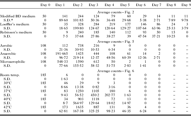
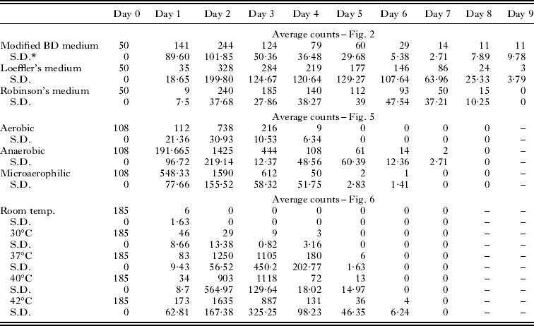
Dientamoeba trophozoites were added to Loeffler's medium at a total concentration of 185 cells/μl. Two of these quantified cultures were grown at each of the different temperatures, including room temperature, 30°C, 37°C, 40°C and 42°C (a total of 10 bottles of medium). All cultures were grown under microaerophilic conditions as already described. This was repeated for each of the 4 clinical isolates of D. fragilis. Cultures were then incubated under microaerophilic conditions and cell counts were made each day until trophozoites were no longer observed. Numerical data obtained for counting experiments relating to various media formulations, atmospheric conditions and temperatures are shown in Table 4.
DNA extraction
DNA was extracted from a 2 mg portion of the sediment of modified BD cultures containing D. fragilis trophozoites. Extractions were performed using a QIAamp™ DNA stool minikit (Qiagen, Hilden, Germany) using the stool extraction protocol described by the manufacturer. This was performed for each of the 4 D. fragilis isolates obtained in this study.
PCR and sequencing
A conventional PCR assay was employed as described previously (Stark et al. Reference Stark, Beebe, Marriott, Ellis and Harkness2005a) for the amplification of the SSU rDNA gene of D. fragilis from DNA extracted from modified BD culture sediments. Following PCR, all PCR products were subjected to agarose gel electrophoresis in a 1% gel. PCR products were visualized under UV light and excised from the gel using a fine scalpel. The PCR products were then extracted from the gel slice using a QIAquick gel extraction kit (QIAGEN) according to the manufacturer's instructions. Samples for sequencing were prepared to contain 10 pmoles of the primers DF 400 or DF 1250 (Stark et al. Reference Stark, Beebe, Marriott, Ellis and Harkness2005a) and approximately 25 ng of PCR product in a total volume of 16 μl. Sequencing was performed at least once in both forward and reverse directions. All sequencing was performed by the service provider SUPAMAC. Sequences obtained from all isolates obtained in this study were aligned with other SSU rDNA genes of D. fragilis available on GenBank (Accession nos U37461 and AY730405) using a Clustal W program.
Identification of microbial flora in Dientamoeba cultures
After several passages in modified BD medium, under microaerophilic conditions at 37°C, bacterial cultures were performed on the sediments of culture vessels from each isolate of Dientamoeba. Cultures were inoculated onto Columbia horse blood agar plates (Oxoid Cat. No. PP2001) and incubated under aerobic conditions at 37°C, and anaerobic media plates (Oxoid cat. No. PP2039) incubated under anaerobic conditions at 37°C. After 24–48 h incubation the microbial flora was identified using standard phenotypic laboratory techniques.
Cryopreservation method 1
This method is a modification of that described by Sawangjaroen et al. (Reference Sawangjaroen, Luke and Prociv1993) with the addition of glucose as suggested by Dwyer and Honigberg (Reference Dwyer and Honigberg1971) for a concentration of 5% DMSO. A solution of 5% (w/v) D-glucose (Chem-Supply Pty Ltd) in single-strength PBS was prepared. Solutions of 5%, 7·5%, 10%, 12·5% and 15% (v/v) DMSO (Sigma-Aldrich Cat. No. 154938-100 ml) in single-strength PBS were also prepared. Dientamoeba fragilis trophozoites from each isolate were grown in 4 vials of Loeffler's slope medium. The contents of these vials were pooled by removing the entire liquid portion (including sediments) and placing into 10 ml vol. Falcon tubes. Tubes were centrifuged at 500 g for 5 min. The supernatant was discarded. One ml of 5% glucose (w/v) in PBS was added to the pellet and tubes were inverted several times to obtain an even cell suspension. Cell counts were then performed as described previously. The quantified cell suspensions were then diluted with 5% glucose (w/v) in PBS to a final concentration of 180 trophozoites/μl.
Five hundred μl of each of the 5 different DMSO solutions were placed into a single Microbank™ vial (Pro-Lab Diagnostics, USA) for cryofreezing (Microbank™ vials for this experiment were first prepared by pouring off the medium and beads within the tubes followed by rinsing several times with PBS and blotting dry on a paper towel). Five hundred μl of quantified cell suspension (180 trophozoites/μl) from a single isolate of D. fragilis were then added to each of the 5 tubes containing different concentrations of DMSO. This resulted in solutions of 2·5%, 3·75%, 5%, 6·25% and 7·5% DMSO (v/v) in a 2·5% glucose (w/v) solution in PBS and a final trophozoite concentration of 90 trophozoites/μl. This was repeated for each of 4 isolates of D. fragilis. These 20 tubes were then placed overnight in a −80°C freezer. The following morning the tubes were then removed from the –80°C freezer and placed in a liquid nitrogen freezer (CHART(R), Model MVE TEC 3000) for 4 days. On the morning of the fifth day, each tube was removed from liquid nitrogen and placed directly in a 37°C water bath to thaw. Once thawed, the total contents of each tube were used to inoculate vials of fresh Loeffler's medium. Each of these vials was then incubated at 37°C for 48 h. After this time, a drop of sediment from each of these cultures was examined at 400×magnification under phase-contrast microscopy for the presence of motile trophozoites of D. fragilis (Fig. 1C and D). When motile trophozoites were observed, cell counts were performed as described previously.
Cryopreservation method 2
This method is a modification of that described by Dwyer and Honigberg (Reference Dwyer and Honigberg1971). Solutions of 2%, 2·25%, 2·5%, 2·75%, 3% and 3·25% (v/v) DMSO in single-strength PBS were prepared. Dientamoeba fragilis trophozoites from each isolate were grown in 4 vials of Loeffler's medium. The 4 culture vials from each isolate were pooled in 10 ml vol. Falcon tubes. Tubes were centrifuged at 500 g for 5 min and the supernatant was discarded. One ml of PBS was added to the pellet and tubes were inverted several times to obtain an even cell suspension. Cell counts were then performed as described previously. The quantified cell suspensions were then diluted in PBS to a final concentration of 106 trophozoites/μl.
Five hundred μl of each of the 6 different DMSO solutions were placed into a single Microbank™ vial (Microbank™ vials were first prepared by removal of the beads and media as described above). Five hundred μl of quantified cell suspension (106 trophozoites/μl) from a single isolate of D. fragilis was then added to each of the 6 tubes containing different concentrations of DMSO. This resulted in solutions of 1%, 1·125%, 1·25%, 1·375%, 1·5% and 1·625% (v/v) DMSO in PBS and a final trophozoite concentration of 53 trophozoites/μl. This was repeated for each D. fragilis isolate. Each Microbank™ vial was then frozen as described above. Each vial was thawed on the morning of day 5 post-freezing as described previously and inoculated into fresh Loeffler's medium. After 48 h incubation, a drop of sediment from each of these cultures was examined at 400×magnification under phase-contrast microscopy for the presence of motile trophozoites of D. fragilis. Where motile trophozoites were observed cell counts were performed as described previously.
Statistical analysis of growth data from all experiments
In order to statistically compare the differences in cell density between the growth plots obtained in all experiments, cumulative cell counts were calculated for each day and analysed using a paired t-test.
RESULTS
Comparison of all media
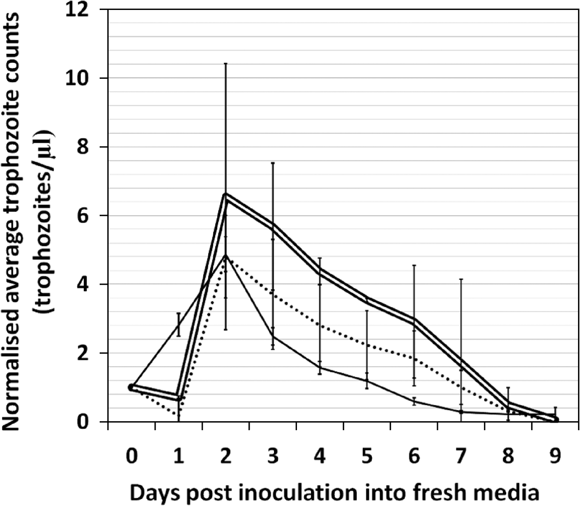
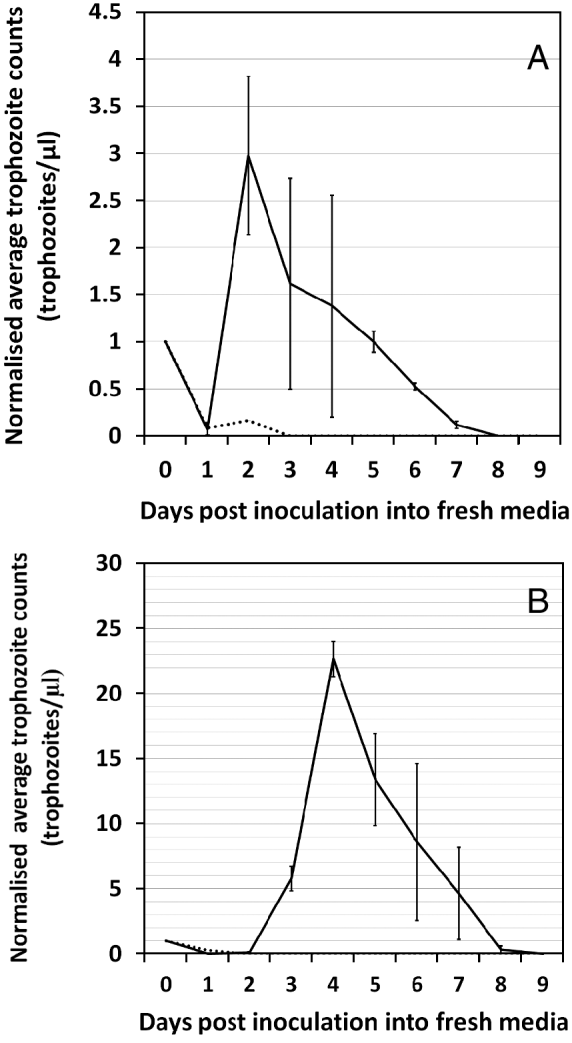
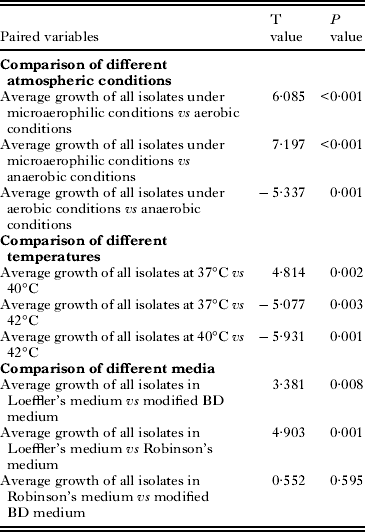
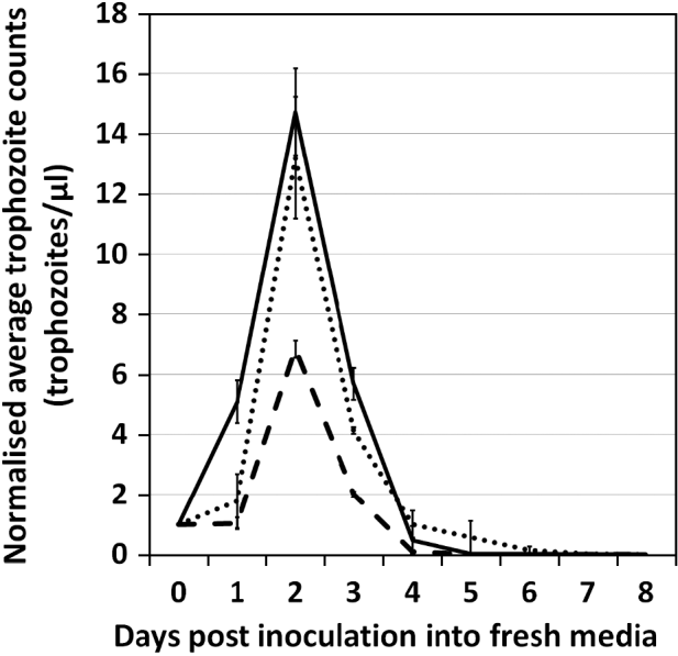
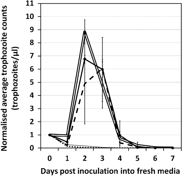
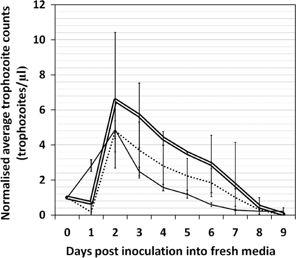
Figure 2 shows the average growth pattern of all 4 clinical isolates of D. fragilis in Loeffler's medium, modified BD medium and Robinsons’ medium at 37°C under microaerophilic conditions. Tritrichomonas foetus medium and Trichosel, failed to support the growth of D. fragilis entirely and so were excluded from further study. Loeffler's medium supported significantly higher cell densities of all clinical isolates when compared to all other media (Table 1). While the average growth of all Dientamoeba isolates in Robinson's medium was slightly higher than that of modified BD medium (T value 0·552), the cell densities achieved for these media were not significantly different from each other (P value 0·595). TYGM-9 and M199 supported very poor growth of 3 of 4 isolates. Isolate 4 demonstrated visibly better growth in TYGM-9 and in particular, M199 when compared to Isolates 1, 2 and 3 and, as such the results for these two media were excluded from Fig. 2. Figure 3 shows the average growth of Isolate 4 in TYGM-9 and M199 compared to the average growth of Isolates 1, 2 and 3. This figure demonstrates the obvious phenotypic difference between the growth of Isolate 4 compared to Isolates 1, 2 and 3 in TYGM-9 and M199.
Growth plots showing the average growth of Dientamoeba fragilis Isolates 1, 2, 3 and 4 in Loeffler's medium ( ), Robinson's medium (• • • •) and modified BD medium (
 ) under microaerophilic conditions at 37°C. Initial cell counts on Day 0 were 50 trophozoites/μl for all culture tubes. To simplify these results and to enable an easier comparison between the different figures, all raw values were normalized by dividing the averages of all raw trophozoite counts by 50 in order to obtain a value of 1 trophozoite/μl for Day 0. As the error bars may be difficult to discern, the value and standard deviation of each point plotted in Figs 2, 5 and 6 is presented in numerical form in Table 4.
Growth plots comparing the average growth of Dientamoeba fragilis Isolates 1, 2 and 3 (• • • •) with the growth of Isolate 4 () in TYGM-9 (A) and M199 (B) under microaerophilic conditions at 37°C. Initial cell counts on Day 0 were 50 trophozoites/μl for all culture tubes. All raw values were normalized by dividing the averages of all raw trophozoite counts by 50 in order to obtain a value of 1 trophozoite/μl for Day 0.

P and T values obtained using a paired t-test to compare the growth of Dientamoeba under different conditions
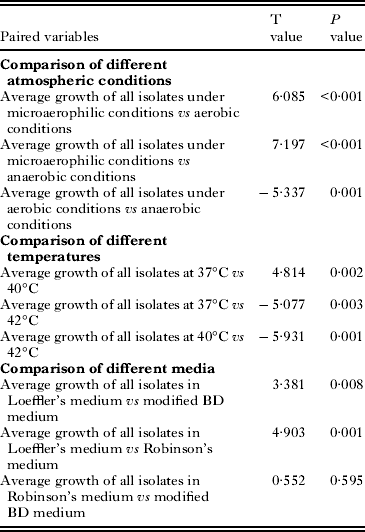
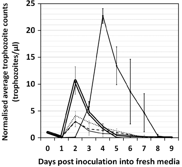
Figure 4 compares the growth of Isolate 4 in MBD medium, Loeffler's slope medium, Robinsons’ medium, TYGM-9 and M199. This figure shows that M199 is a suitable medium for the growth of Isolate 4 only. Despite this observation, Loeffler's medium followed by the modified BD medium and Robinson's medium support (on average) the most efficient growth of D. fragilis.
Growth plots comparing the growth of Dientamoeba fragilis Isolate 4 in Loeffler's slope medium (), M199 ( ), modified BD medium (
 ), Robinson's medium (• • • •) and TYGM-9 medium (— –) under microaerophilic conditions at 37°C. Initial cell counts on Day 0 were 50 trophozoites/μl for all culture tubes. All raw values were normalized by dividing the averages of all raw trophozoite counts by 50 in order to obtain a value of 1 trophozoite/μl for Day 0.

Effect of atmospheric composition on in vitro growth
The results shown in Fig. 5 indicate that concentrations of oxygen found in the terrestrial atmosphere are not favourable for the growth of D. fragilis. Concentrations of oxygen equal to or below 6% are conducive for the growth of D. fragilis. Interestingly, D. fragilis appears to prefer oxygen concentrations higher than those observable in anaerobic environments, demonstrating significantly better growth under microaerophilic conditions when compared to anaerobic conditions (P value 0·000).
Growth plots comparing the growth of Dientamoeba fragilis under aerobic (— –), anaerobic (• • • •) and microaerophilic () conditions at 37°C in Loeffler's slope medium. Initial cell counts on Day 0 were 108 trophozoites/μl for all culture tubes. All raw values were normalized by dividing the averages of all raw trophozoite counts by 108 in order to obtain a value of 1 trophozoite/μl for Day 0.

Effect of temperature on in vitro growth
The data in Fig. 6 show high Dientamoeba trophozoite numbers in cultures grown between 37 and 42°C. Temperatures between that of ambient room temperature and 30°C failed to support any growth of the organism. Growth of Dientamoeba at 42°C was significantly better when compared to the growth of Dientamoeba at lower temperatures (Table 1). Growth at 37°C was found to be significantly better than growth at 40°C although the calculated standard error was quite large for values obtained at 40°C (Fig. 6).
Growth plots comparing the growth of Dientamoeba fragilis at various temperatures [room temperature (• • • •), 30°C (), 37°C ( ), 40°C (— –) and 42°C ()] in Loeffler's slope medium under microaerophilic conditions. Initial cell counts on Day 0 were 185 trophozoites/μl for all culture tubes. All raw values were normalized by dividing the averages of all raw trophozoite counts by 185 in order to obtain a value of 1 trophozoite/μl for Day 0.

PCR and sequencing
Despite the observable differences between the growth of some isolates of D. fragilis in certain media, no genetic differences were observed in a section of the SSU rDNA gene sequenced for all 4 isolates in this study. Clinical isolates from this study are of a similar SSU rDNA genotype to the more common genotype 1 (GenBank Accession no. AY730405), previously identified in patients from Sydney, Australia and dissimilar to SSU rDNA genotype 2 (the Bi/pa genotype (GenBank Accession no. U37461)).
Identification of microbial flora in Dientamoeba cultures
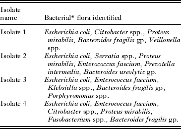
Several species of bacteria were identified in the cultures from each isolate of D. fragilis (Table 2). No eukaryotic species other than D. fragilis was identified in these cultures. The predominant bacterial species in cultures from all isolates was Escherichia coli.
Microbial flora identified in the cultures of each isolate of Dientamoeba fragilis
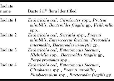
* No eukaryotic organisms other than D. fragilis were identified in cultures.
Cryopreservation
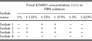
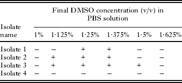
With respect to cryopreservation method 1, all DMSO/glucose solutions failed to successfully cryopreserve D. fragilis trophozoites. Regarding cryopreservation method 2, final concentrations of DMSO between 1·125 and 1·5% allowed for the successful cryopreservation of some isolates of D. fragilis (not Isolate 4) as motile trophozoites were observed after thawing and culture (Table 3). A final DMSO concentration of 1·375% allowed for excellent recovery and growth of trophozoites from Isolates 1, 2 and 3 after thawing and 3 days of in vitro growth, compared to other DMSO concentrations. For example, using a DMSO concentration of 1·375% after thawing and 3 days of in vitro growth, trophozoite numbers of Isolate 3 increased to 259% of the original number of trophozoites cryopreserved. For all DMSO concentrations that allowed for successful cryopreservation, in vitro growth could still be observed after several subsequent passages in Loeffler's medium. Cryopreservation of Isolate 4 was unsuccessful at all concentrations of DMSO used in these experiments.
The presence (+) or absence (−) of in vitro growth for each isolate of Dientamoeba fragilis after cryopreservation using different concentrations of DMSO followed by cultivation in Loeffler's medium
DISCUSSION
Dientamoeba fragilis is a protozoan parasite of the gastrointestinal tract of humans which has recently gained recognition as a significant human pathogen (Johnson et al. Reference Johnson, Windsor and Clark2004; Stark et al. Reference Stark, Beebe, Marriott, Ellis and Harkness2006, Reference Stark, Barratt, Ellis, Harkness and Marriott2009, 2010). Despite this, comparatively little information is available about the biology of this organism including genome and proteome studies. Also, the most effective drug treatment for dientamoebiasis is yet to be defined, although progress has been made (Stark et al. Reference Stark, Barratt, Roberts, Marriott, Harkness and Ellis2010). The ability to cryogenically preserve this organism and to culture it in vitro at high cell densities will facilitate the development of new diagnostics, the characterization of novel antigens, the study of this organism's life cycle and biology and the development of in vitro methods to assess drug treatments.
Four of 7 different types of media were able to support the growth of all 4 clinical isolates of D. fragilis. These 4 media were TYGM-9 broth, Robinson's medium, modified BD medium and Loeffler's medium. Tritrichomonas foetus medium and Trichosel failed to support the growth of D. fragilis. On average, the growth of D. fragilis was significantly better in Loeffler's slope medium compared to Robinson's medium and modified BD medium. This is in contrast to the results of Sawangjaroen et al. (Reference Sawangjaroen, Luke and Prociv1993) who found that the modified BD medium was the only medium which supported the good long-term growth of Dientamoeba when they compared its growth in monophasic TYSGM-9 (TYGM-9 medium with the addition of serum), Cleveland and Collier's medium and a modified BD medium. Sawangjaroen et al. (Reference Sawangjaroen, Luke and Prociv1993) found that the modified BD medium was the only medium which supported the good long-term growth of the organism (Sawangjaroen et al. Reference Sawangjaroen, Luke and Prociv1993).
Dientamoeba was found to grow well under both microaerophilic (6% O2, 7·2% CO2, 3·6% H2, 83·3% N2) and anaerobic (0·2% O2, 9·9% CO2, 5% H2, 84·9% N2) conditions. However, D. fragilis does not appear to grow as well in the presence of atmospheric levels of oxygen. The trophozoite densities obtained under microaerophilic conditions were significantly greater when compared to the trophozoite densities obtained under both aerobic and anaerobic conditions. The comparatively poor growth of Dientamoeba at higher oxygen concentrations reflects the fragile nature of D. fragilis once it is passed from the body and comes into contact with the atmosphere. The results suggest that Dientamoeba is best grown at 42°C. However, it appears that temperatures between 37°C and 42°C are acceptable for D. fragilis growth. Significantly higher cell densities are obtained at 42°C compared to 40°C and 37°C. Dobell previously reported optimum growth of D. fragilis at 41°C (Dobell, Reference Dobell1940) and the results of the current study support Dobells’ observations. The growth of D. fragilis at 42°C is an interesting observation as one would expect an optimum D. fragilis growth temperature closer to the core body temperature of humans (∼37°C) rather than that of birds (∼42°C). All isolates demonstrated similar temperature affinities although it was often observed that the trophozoite densities from 2 culture vials of the same isolate, grown at the same temperature were quite different from each other. This phenomenon was particularly apparent when isolates were grown at 40°C and is the reason for the large error bars observable in Fig. 6. Trophozoite densities obtained at 37°C were significantly greater than those obtained at 40°C. This seems paradoxical as the highest cell densities were achieved at 42°C and one would expect greater cell densities at temperatures approaching 42°C. All temperature experiments were repeated to exclude human error, though with a similar paradoxical outcome. The reason behind this phenomenon is unclear although we recommend that for cultivation of Dientamoeba, multiple culture vessels of a given isolate should always be maintained, and that subcultures should be made from the ‘healthiest’ of these cultures only in order to select protozoa that grow vigorously. However, it should also be taken into account that selecting certain subpopulations of trophozoites introduces bias and has the potential to reduce the genetic variability of the original clinical isolate. As such, this kind of selection will reduce the similarity between the original wild-type organisms and the cultured organisms. These points should be considered when undertaking this kind of selection.
During the course of these experiments, it also became apparent that 1 of the clinical isolates of D. fragilis was quite different from the other 3 in terms of its ability to grow comparatively well in 2 monophasic liquid media (TYGM-9 broth and M199) while all other isolates could not. The growth of this isolate (Isolate 4) in Robinson's medium, modified BD medium and Loeffler's medium followed a very similar growth pattern to all other isolates and so raw counts made for Isolate 4 were included when calculating the average cell densities shown in Fig. 1. However, due to the visibly different patterns of growth observed for Isolate 4 in TYGM-9 broth and M-199, a separate figure was created in order to compare the growth of Isolate 4 to Isolates 1–3 in these monophasic media. Fig. 3 clearly demonstrates the ability of Isolate 4 to grow well in TYGM-9 broth and M-199 when compared to all other isolates.
Sequencing of the small subunit gene of all 4 isolates demonstrated that they were all greater than 99% similar in the sequenced region of the SSU rDNA. All 4 isolates demonstrated almost identical sequences to 7 other sequences derived from a previous PCR study of D. fragilis performed in Sydney (GenBank Accession no. AY730405). Undoubtedly, some genetic differences must exist between Isolate 4 and all other isolates and future work may investigate these differences. Interestingly while M199 failed to support 3 isolates of D. fragilis, it seems that M199 was able to support high cell densities for Isolate 4 at 4 days of growth (higher cell densities than were achieved for this isolate in any other media). While this may be the case we do not consider M199 a good medium for the cultivation of D. fragilis for the obvious reason that growth in this media is isolate/strain dependent.
Given the obvious phenotypic differences observable between Isolate 4 and all other isolates, and the similarity between the SSU rDNA sequences obtained from all isolates, we conclude that the SSU rDNA gene is well conserved, even amongst very different isolates of D. fragilis, suggesting that the SSU rDNA gene is a poor genetic marker for distinguishing amongst different strains of D. fragilis. This point is supported by the findings of other studies (Stark et al. Reference Stark, Beebe, Marriott, Ellis and Harkness2005a; Bart et al. Reference Bart, van der Heijden, Greve, Speijer, Landman and van Gool2008). Only 2 SSU rDNA genotypes of D. fragilis are known to exist (genotypes 1 and 2) and all isolates in this study are similar to SSU rDNA genotype 1, which is the first (and only) SSU rDNA genotype identified in Sydney to date (Stark et al. Reference Stark, Beebe, Marriott, Ellis and Harkness2005a). Genotype 1 was also found to be more common than genotype 2 (the Bi/pa genotype) in previous studies (Johnson and Clark, Reference Johnson and Clark2000; Windsor et al. Reference Windsor, Clark and Macfarlane2004).
In order to loosely explore the possibility that D. fragilis may exhibit better growth in the presence of certain bacterial species, the bacterial flora accompanying the cultures of each isolate of Dientamoeba was defined. As mentioned previously, the growth of all Dientamoeba isolates was similar in Loeffler's medium, Robinson's medium and modified BD medium. This was in spite of the slight variation in the bacterial flora accompanying each isolate in culture. Escherichia coli was the predominant bacterial species within all cultures. As such, we can conclude that D. fragilis will grow happily with a support flora consisting mostly of E. coli. We also suggest that slight variations in the species of prokaryotic support flora present within D. fragilis cultures, are unlikely to exhibit any significant effect on growth. It is possible that D. fragilis could exhibit better (or worse) growth if the predominant bacterial species within a culture is changed, although no conclusions regarding this hypothesis can be drawn from the present study. Differences in growth patterns exhibited by Isolate 4 compared to other isolates of Dientamoeba are unlikely to be attributable to the bacterial flora. Once again, E. coli was the predominant bacterium within cultures of Isolate 4 and nearly all other bacterial species present within Isolate 4 cultures were also present in cultures of other isolates. However, the presence of Fusobacterium spp. was a feature unique to Isolate 4. Despite this, Fusobacterium spp. were not a major constituent of the flora within cultures of Isolate 4 and we feel that the differences observable for Isolate 4 are inherent within the isolate itself rather than the support flora.
It is possible that the temperature dependence observed for all Dientamoeba isolates could be directly related to the effect of changes in temperature on the microbial support flora. It is possible that an increase in temperature may reduce the doubling time for bacteria and have little/less effect on Dientamoeba, thus increasing the availability of certain nutrients to Dientamoeba. It is also possible that an increase in temperature directly increases the doubling time of Dientamoeba. However, it should also be noted that some species of enteric bacteria (such as E. faecium) have an optimal growth temperature of 42–45°C (Zanoni et al. Reference Zanoni, Garzaroli, Anselmi and Rondinini1993). Clearly, these hypotheses require further investigation. The development of cultures with defined bacterial species’ (monoxenic or dixenic cultures) or preferably, axenic cultures, would be useful to explore the effects of temperature on Dientamoeba growth.
No monoxenic, dixenic or axenic cultures of D. fragilis exist to our knowledge (at the time of writing). According to studies performed by Brug (Reference Brug1938) and Jacobs (Reference Jacobs1953), Dientamoeba trophozoites are heavily dependent on the presence of live bacteria as a food source, with dead bacteria being insufficient for Dientamoeba growth (Jacobs, Reference Jacobs1953). Brug (Reference Brug1938) observed that if bacteria become scarce in cultures, Dientamoeba trophozoites eventually die off. This suggests that an axenic culture of Dientamoeba would be difficult to achieve (if this is achievable at all). However, the creation of monoxenic or dixenic cultures of Dientamoeba seems more obtainable. According to Jacobs (Reference Jacobs1953), a monoxenic culture of Dientamoeba with Clostridium perfringens was obtained through the use of various antibiotics. Jacobs (Reference Jacobs1953) also noted that Dientamoeba was quite tolerant to fairly high concentrations of the drugs penicillin, streptomycin and sulfadiazine. As such, the use of various antibiotics may represent a simple means of obtaining monoxenic or dixenic cultures of Dientamoeba. Monoxenic or dixenic cultures of Dientamoeba would prove useful in genetic and proteomic studies as the defined support flora could then be used as a negative control. Experiments which test the in vitro susceptibility of Dientamoeba to various anti-microbial drugs would also benefit from the existence of such cultures. Experiments which aim to achieve monoxenic, dixenic and/or axenic cultures of Dientamoeba are currently underway.
A final concentration of 1·375% DMSO (as described in cryopreservation method 2) resulted in excellent recovery and growth of trophozoites when compared to other concentrations of DMSO used. This is in agreement with the study by Dwyer and Honigberg (Reference Dwyer and Honigberg1971) who found the highest percentage recoveries at a final DMSO concentration of 1·375%. Cryopreservation method 1 failed to preserve viable D. fragilis trophozoites entirely. Interestingly, Isolate 4 was the only isolate that could not be cryogenically preserved using method 2. We speculate that this is probably the result of genetic differences between isolates. Future efforts will be made to identify these genetic differences and to develop a method for cryopreservation of Isolate 4. Dwyer and Honigberg (Reference Dwyer and Honigberg1971) utilized a liquid nitrogen freezer with controlled cooling capabilities in their D. fragilis cryopreservation experiments. Phillips et al. (Reference Phillips, Boreham and Shepherd1984) noted higher recoveries of Giardia trophozoites when they were frozen using a liquid nitrogen freezer with controlled cooling capabilities compared to simply placing trophozoites into a −70°C freezer. This equipment was not available to us although we believe that the cryopreservation of D. fragilis trophozoites may be improved using a controlled cooling freezer. Final DMSO concentrations >1·5% failed to preserve viable D. fragilis trophozoites entirely. This is in contrast to the findings of Sawangjaroen et al. (Reference Sawangjaroen, Luke and Prociv1993), who reported the successful cryopreservation of viable D. fragilis trophozoites in a final concentration of 7·5% DMSO.
To conclude, our studies indicate that the best conditions for the growth of D. fragilis in the media tested in this study are as follows; microaerophilic conditions, at 42°C in Loeffler's slope medium (a modified Cleveland and Collier's medium). However, D. fragilis will also grow quite well in modified BD medium or Robinson's medium under microaerophilic conditions at 37°C and 40°C. It should also be noted that while M199 was able to support good growth of 1 isolate of Dientamoeba only, Loefflers’ medium, modified BD medium and Robinson's medium were able to support good growth of all isolates in these experiments. Therefore these 3 media are recommended for those attempting to cultivate clinical isolates of D. fragilis from fresh stool specimens. Furthermore, effective cryopreservation of D. fragilis trophozoites is best achieved using method 2 of cryopreservation described herein at a final DMSO concentration of 1·375% (v/v) in single-strength PBS.
ACKNOWLEDGMENTS
We acknowledge the help of the staff at St Vincent's Hospital Microbiology Department in the collection and processing of stool samples positive for D. fragilis. We especially acknowledge the assistance of Tamalee Roberts in typing the bacterial species in our D. fragilis cultures.
FINANCIAL SUPPORT
This work was supported by the Australian Research Council Linkage. Project Number LP0775326 entitled ‘Gastrointestinal Parasites and their Diagnosis’.