Introduction
External surgical approaches to medial orbital tumours such as transcaruncular or medial transconjunctival orbitotomy have been well described.Reference Karaki, Kobayashi and Mori1–Reference Castelnuovo, Dallan, Locatelli, Battaglia, Farneti and Tomazic9 Nevertheless, these tumours are difficult to access and several authors have described the endoscopic transnasal approach as a feasible, less-invasive alternative. We present our experience with endoscopic transnasal management of medial intraconal and extraconal lesions.
Case report
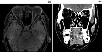
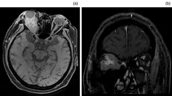
An endoscopic transnasal approach to intra-orbital lesions was used for nine patients. The main clinical features are shown in Table I. Pre-operative computed tomography and magnetic resonance imaging were used to evaluate the lesions in all patients (Figures 1 and 2).
Patient characteristics

Pt no = patient number; y = years; M = male; F = female
Pre-operative axial magnetic resonance imaging (a) and coronal computed tomography (b) scans showing a left extraconal pseudotumour in patient 5.

Pre-operative axial (a) and coronal (b) magnetic resonance imaging scans showing a right intraconal non-Hodgkin's lymphoma in patient 7.

Surgical techniques
All patients were placed supine under general anaesthesia and the nasal cavity was decongested. The middle turbinate was then gently medialised, followed by uncinectomy and wide middle meatal antrostomy. Next, anterior and posterior ethmoidectomy were performed; sphenoidectomy was then carried out for visualising the optic canal.
The lamina papyracea was removed to expose the periorbita. An incision was then made in the periorbita with a sickle knife in a posterior to anterior direction, starting from the inferior part so that the surgical view was not obstructed by the prolapsing orbital contents. To achieve adequate orbital decompression, the medial third of the orbital floor was removed by downward fracture with a Freer elevator, and three to four parallel, horizontal incisions were made through the periorbita.
For intraconal lesions, the extraconal fat was reduced by bipolar cautery, where necessary, to enable identification of the rectus muscles. The medial rectus muscle was elevated to allow access into the intraconal orbital space. The tumour was then located, excised and sent for histopathological analysis.
Meticulous haemostasis was secured using adrenaline-soaked gauze, bipolar cauterisation and Surgicel® haemostat. The orbital fat was pulled over the muscles to prevent scarring; no bony reconstruction was performed. Both nasal cavities were packed with Gelfoam® and Merocel® nasal tampons.
Post-operative course
A total of nine patients underwent an endoscopic transnasal procedure: four patients had intraconal lesions, and five had lesions in the extraconal space. Post-operatively, seven patients reported improved visual acuity and two had stable vision. There were no complications of cerebrospinal (CSF) leakage or diplopia. Patients 2, 4, 7 and 8 underwent further chemotherapy and radiotherapy, and patient 5 and 6 each received a tapering dose of corticosteroids. Unfortunately, patient 1 died as a result of disease progression. Patient 3 had no recurrence and good visual acuity at follow up; there was no encroachment on the optic nerve and the lesion was completely excised.
Discussion
The surgical approach to the orbital content can be daunting. The factors involved in determining the most suitable approach are tumour location, extent and pathology.Reference Karaki, Kobayashi and Mori1, Reference Castelnuovo, Dallan, Locatelli, Battaglia, Farneti and Tomazic9 Although the surgeon will endeavour to maintain ocular structure and function, the aim is to achieve adequate decompression and resection. Thus, the endoscopic transnasal approach is the least invasive option for managing medial orbital tumours.
The endoscopic transnasal route provides a safe, direct approach to the medial orbital space. This is particularly important because the orbital anatomy is complex, with several important structures contained within a small volume. Procedures involving the orbit can cause a significant rise in orbital pressure because of the limited size of the cone-shaped space. Removing the lamina papyracea in transnasal procedures lessens this risk by enabling the orbital content to be displaced to some extent.Reference Castelnuovo, Dallan, Locatelli, Battaglia, Farneti and Tomazic9
The endoscopic transnasal approach is minimally invasive, thus causing less tissue damage and better cosmetic outcomes. The recovery period is therefore significantly reduced, enabling shorter hospital stays and allowing adjuvant therapy to commence without delay. The endoscope also provides excellent illumination and visualisation through a narrow surgical corridor.
External approaches to the orbit have several limitations. Although transconjunctival orbitotomy has shown favourable results for anterior orbital lesions, it is extremely challenging to access those lesions located in the posterior half of the orbit.Reference Castelnuovo, Dallan, Locatelli, Battaglia, Farneti and Tomazic9 Additionally, extra-ocular muscle detachment may be necessary for resecting intraconal lesions; this can affect extra-ocular movement, causing strabismus.Reference Murchison, Rosen, Evans and Bilyk6, Reference Dallan, Seccia, Lenzi, Castelnuovo, Bignami and Battaglia10 External approaches also entail significant displacement of orbital structures, including the globe.Reference Castelnuovo, Dallan, Locatelli, Battaglia, Farneti and Tomazic9
In our experience, the endoscopic transnasal method is effective for resecting both extraconal and intraconal tumours. The main anatomical difference between these lesions is their location relative to extra-ocular muscle boundaries. In this case series, the first step was to identify the medial and inferior rectus muscles by establishing an inferomedial periorbital window. Following that, the extraconal tumours could be located and resected.
Intraconal lesions present a greater challenge: it is imperative that the dissection is performed between the medial and inferior rectus muscles, and not through the muscle fibres.Reference McKinney, Snyderman, Carrau, Germanwala, Prevedello and Stefko4 In this case series, bipolar cautery was used to reduce the amount of extraconal fat in the vicinity of the medial rectus muscle. We consider bipolar cautery to be very precise and safe, with a minimal risk of fibrosis and resulting diplopia. The medial rectus muscle is essential for identifying the medial orbit. Some authors advocate stiffening the extra-ocular muscles by methods such as subconjunctival looping of the medial and inferior rectus muscles to isolate them or anchoring the medial rectus muscle to the septum with a suture.Reference McKinney, Snyderman, Carrau, Germanwala, Prevedello and Stefko4, Reference Castelnuovo, Dallan, Locatelli, Battaglia, Farneti and Tomazic9 We found that stiffening the extra-ocular muscles was unnecessary because elevating the medial rectus muscle provided adequate exposure and access to the medial orbit.
To minimise the risk of irreversible damage, it is important to avoid crossing the optic nerve.Reference McKinney, Snyderman, Carrau, Germanwala, Prevedello and Stefko4 For this reason, superolateral lesions are contraindicated; instead, superior or lateral orbitotomy should be considered.Reference McKinney, Snyderman, Carrau, Germanwala, Prevedello and Stefko4 In addition, injury to the ethmoid arteries can cause retrobulbar haemorrhage and vision loss. Hence, it is important to ensure that the lamina papyracea is accessed below the ethmoidal foramina.
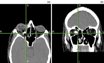
Although some authors suggest that the narrow surgical field available in endoscopic procedures may result in limited instrumentation access, inadequate visualisation and problems in achieving haemostasis, no such difficulties were encountered in this case series.Reference Karaki, Kobayashi and Mori1, Reference Miller, Agrawal, Sciubba and Lane2, Reference Yoshimura, Kubo, Yoneda, Hasegawa, Tominaga and Yoshimine5 Moreover, these drawbacks can be overcome by enlarging the orbital window and using neuronavigation.Reference Castelnuovo, Dallan, Locatelli, Battaglia, Farneti and Tomazic9 Neuronavigation should be considered for complex cases or when a difficult dissection is anticipated, as used for patient 9 (Figure 3). This patient had acquired a penetrating orbital injury at work, resulting in an intraconal metallic foreign body. As a retained foreign body can lead to chronic orbital inflammation, osteomyelitis, thrombotic vasculitis and septicopyaemia, surgical removal is essential. Neuronavigation is therefore a valuable tool for localising small objects, especially in traumatised anatomical structures, and may improve intra-operative safety.
Axial (a) and coronal (b) intra-operative neuronavigation images showing a right intraconal foreign body in patient 9. A = anterior; P = posterior; S = superior; I = inferior

Karaki et al. suggested reconstructing the bony medial orbital wall with nasal mucosa or bone depending on the defect size.Reference Karaki, Kobayashi and Mori1 Other authors, however, maintain that reconstruction of the medial orbital wall or periorbita is unnecessary because fibrotic healing is sufficient.Reference Castelnuovo, Dallan, Locatelli, Battaglia, Farneti and Tomazic9, Reference Dallan, Seccia, Lenzi, Castelnuovo, Bignami and Battaglia10 Nevertheless, reconstruction is advised when resection is very wide, and different methods involving the fascia lata or a septal flap have been used.Reference Castelnuovo, Dallan, Locatelli, Battaglia, Farneti and Tomazic9, Reference Dallan, Seccia, Lenzi, Castelnuovo, Bignami and Battaglia10
• The endoscopic transnasal approach is a minimally invasive technique for medial orbital lesion
• It causes less tissue damage, with shorter hospital stays and earlier commencement of adjuvant therapy
• Neuronavigation should be considered for complex cases or potentially difficult dissections
Damage to the neural, muscular or vascular structures of the orbit can have grave outcomes. Optic nerve injury and haemorrhage may lead to irreversible compressive optic neuropathy or central retinal occlusion.Reference Miller, Agrawal, Sciubba and Lane2 Other risks are CSF leakage and persistent diplopia due to damage to extra-ocular muscles or their nerves.Reference Miller, Agrawal, Sciubba and Lane2 However, only a few complications have been reported: these include decreased visual acuity in two patients, diplopia in three patients and CSF leakage in one patient.Reference Murchison, Rosen, Evans and Bilyk6, Reference Castelnuovo, Dallan, Locatelli, Battaglia, Farneti and Tomazic9 There were no complications in our case series.
Conclusion
This case series has demonstrated the safety and effectiveness of the endoscopic transnasal approach in managing intraconal and extraconal lesions. This minimally invasive technique should be considered a valid alternative for accessing orbital tumours, particularly those located in the medial compartment.