After birth, ductus arteriosus closes spontaneously. An open ductus arteriosus after the first 72 hours of life is defined as patent ductus arteriosus. The incidence of patent ductus arteriosus in term infants is 57 per 100,000 live births. The incidence of patent ductus arteriosus is 60–70% in premature younger than 28 weeks and 20% in premature older than 32 weeks. Reference Köksal, Aygün and Uras1 Indomethacin, ibuprofen, and paracetamol can be used for medical patent ductus arteriosus closure. Percutaneous or surgical closure of patent ductus arteriosus must be the treatment option for the cases that medical closure failed. Surgery is a preferred method to close the patent ductus arteriosus in the neonatal period. Some surgical complications are bleeding, recanalization, aneurysm, and vocal cord paralysis. Other extremely rare surgical complications are ligation or clipping of left pulmonary artery, left main bronchus, and aorta. Reference Russell, Leblanc and Potts2 We report a newborn case whose left pulmonary artery ligated accidentally during patent ductus arteriosus surgery and surgical correction of this complication in the early post-operative period.
Case report
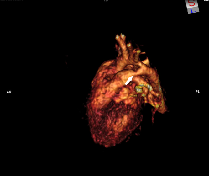
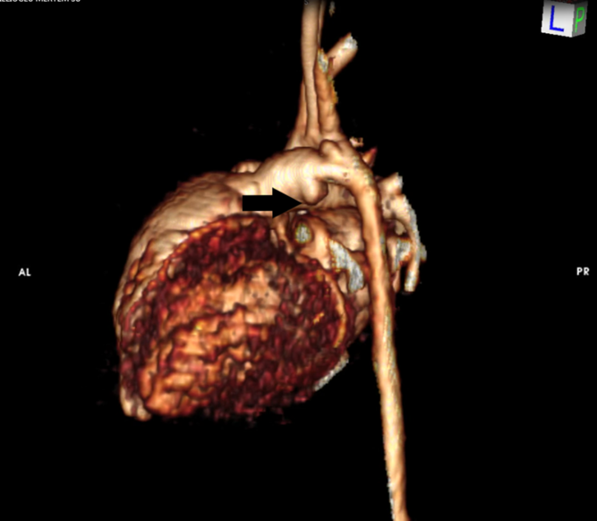
A 17-day-old girl was admitted to our hospital with the diagnosis of pneumonia. The patient was born via elective caesarean section at 37 weeks gestation, weighing 2200 g. Parenteral antibiotic treatments were initiated. The patient was intubated. On physical examination, the continuous murmur was detected. Echocardiography revealed a large patent ductus arteriosus. Computed tomography angiography was done to patient in order to investigate aortic coarctation (Fig 1). On echocardiographic examination, aortic coarctation was also suspected. Medical treatment was started to close the patent ductus arteriosus. Computed tomography was performed to clearly view aortic coarctation. Because if patent ductus arteriosus could be closed medically, balloon angioplasty would be planned for the treatment of aortic coarctation. Despite three cures of ibuprofen treatment, patent ductus arteriosus could not be closed. Due to clinical findings of haemodynamically significant patent ductus arteriosus like persisting need for assisted ventilation, it was decided to close patent ductus arteriosus surgically. The patient was taken to the operating table at the age of 32 days. The left posterolateral thoracotomy incision was performed. Patent ductus arteriosus ligated uneventfully. In our clinic, we perform temporary occlusion test routinely for patent ductus arteriosus ligation operation. We waited approximately a minute during temporary occlusion and then it is observed whether there is hypoxia, bradycardia, and hypotension. If these findings are not observed, the patent ductus arteriosus is ligated. In the first operation of our patient, temporary occlusion test was performed and there were no signs of hypoxia, bradycardia, or hypotension. No improvement in the patient’s pulmonary functions was observed on post-operative day 2. Echocardiography determined that patent ductus arteriosus was still open and there was no flow through the left pulmonary artery. At the end of the first operation, we thought that the patent ductus arteriosus was closed uneventfully. Echocardiography revealed that patent ductus arteriosus flow continued on post-operative day 2. Control computed tomography was done to confirm the diagnosis of this complication by an objective imaging method. On the computed tomography angiography, it was determined that patent ductus arteriosus flow was still continuing, and there was no flow in the left pulmonary artery and its branches (Fig 2). The patient was reoperated on the 3rd post-operative day, and patent ductus arteriosus was ligated. Two ligation silk sutures on the left pulmonary artery were removed. On the intraoperative echocardiography, it was confirmed that patent ductus arteriosus was closed, and there was blood flow in the left pulmonary artery. The patient’s pulmonary functions were gradually improved. The patient was removed from the ventilator on the 6th post-operative day. The patient was discharged on the 14th post-operative day. Control echocardiography in the 1st post-operative month revealed minimal gradient (3 mmHg) of blood flow in the left pulmonary artery, which is not significant.
Preoperative three dimensional CTA image. Large PDA (white arrow).

Postoperative three dimensional CTA image. Ligated left pulmonary artery (black arrow).

Discussion
In the embryological period, the 6th left aortic artery – one of the symmetrical aortic arteries arising from the dorsal aorta – remains unchanged and functions as a “ductus arteriosus”. Reference İrdem, Kervancıoğlu and Başpınar3 Prostaglandins, nitric oxide, and relative hypoxic environment prevent closure of ductus arteriosus during the intrauterine period. After birth, ductus arteriosus closes due to increase in blood oxygen saturation and decrease in prostaglandin levels (since the placenta is disabled). Prostaglandin synthetase inhibitors such as indomethacin, ibuprofen, and paracetamol may be used with the aim of medical closure of haemodynamically significant patent ductus arteriosus, which did not close spontaneously. Reference Bardanzellu, Neroni and Dessì4 In case of medical closure failure, percutaneous or surgical closure of patent ductus arteriosus are the treatment options. Closure of patent ductus arteriosus with percutaneous technique involves some technical difficulties (vascular injury and embolism) in the neonatal period. Surgical treatment is often the preferred method for closure of patent ductus arteriosus in the neonatal period. The most common surgical complications are bleeding, recanalization, aneurysm, wound infection, pneumothorax, vocal cord paralysis, phrenic nerve injury, and chylothorax. Other extremely rare surgical complications are ligation or clipping of left pulmonary artery, left main bronchus, and aorta. The incidence of inadvertent left pulmonary artery ligation is estimated to be almost about 1 in 1000 cases. Reference Panagopoulos, Tatooles and Aberdeen5 However, the fact that there is no post-operative routine echocardiographic examination for these patients in many centres suggests that this rate may be much higher than expected. In the early post-operative period, patients with the following findings should be suspected to ligate the left pulmonary artery instead of patent ductus arteriosus: decreased pulmonary vascular markings of left lung on chest X-ray, continuous murmur and persisting necessity to mechanical ventilation without improvement of pulmonary function. Echocardiography should be done immediately for these patients. If echocardiography determines that patent ductus arteriosus flow continuing and there is no flow in the left pulmonary artery, computed tomography angiography should be done to the patient immediately. Kim et al published a case report that left pulmonary artery was ligated instead of patent ductus arteriosus. In this publication, they emphasised that performing control echocardiography and computed tomography scan is important for early detection of this complication in patients whose expected signs of improvement in the early post-operative period are not observed. They treated this complication successfully by performing emergency reoperation to their patient on the 2nd post-operative day. Reference Kim, Kim and Shin6 Orzel et al published a case report in which they used pulmonary perfusion scintigraphy to diagnose this complication. The authors emphasised that this diagnostic method is non-invasive and portable. Reference Orzel and Monaco7 The patient should be reoperated as soon as possible after the ligation of the left pulmonary artery and continuing patent ductus arteriosus are clearly defined. Delay of the reoperation leads to further impairment of the patient’s pulmonary functions. In addition, in a delayed reoperation, the possibility of recanalisation of the left pulmonary artery decreases and the risk of mortality increases. Because irreversible wall damage occurs at the ligated segment of the left pulmonary artery, hypoplasia and dysplasia develop in the patient’s left lung. There are some important issues to be kept in mind during the operation in order to avoid this complication in the neonatal period: In patients with patent ductus arteriosus larger than 4–5 mm, the aortic arch and other vascular structures must be carefully distinguished. It should be avoided to retract the left lung inferiorly during the operation. Because, this manoeuvre can cause confusion of the patent ductus arteriosus and left pulmonary artery. Reference Pontius, Danielson and Noonan8,Reference Tefera, Bermudez-Cañete and van Doorn9 In patients whose vascular anatomy is suspected to be abnormal, intraoperative echocardiography should be planned and performed to avoid this complication. The right lateral decubitus positioning during the surgical procedure allows echocardiography examination on the right side of the patient under the sterile surgical area.
Conclusion
Patent ductus arteriosus closure surgery has some complications, such as left pulmonary artery ligation. All necessary attention should be paid to prevent this complication. This surgical complication rate is higher in neonatal patients with large patent ductus arteriosus. In the early post-operative period, physical examination and chest X-ray evaluation should be carefully performed. Echocardiography should be used routinely. Reference Terlemez, Işık and Şahin10 In order to perform successful surgical treatment, neonatal specialist, paediatric cardiologist, and cardiovascular surgeon must work as a team.
Acknowledgements
None.
Author Contribution
All authors have major contribution to manuscript preparation and approval of the final version.
Financial Support
This research received no specific grant from any funding agency or from commercial or not for-profit sectors.
Conflicts of Interest
The authors declare that they have no conflict of interest.