Case report
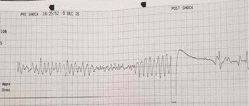
We report a 9-year-old African-American girl who presented with Brugada electrocardiographic (EKG) pattern, polymorphic ventricular tachycardia (VT), and pulseless arrest following adenotonsillectomy earlier that day (at noon). Sevoflurane, propofol (2 mg/kg intravenous bolus), fentanyl, morphine, dexamethasone, ondansetron, and naloxone were administered during the procedure for anaesthetic and pain management. The operation and the post-operative recovery periods were both uneventful. She was transferred over to post-anaesthesia care unit after the operation at approximately 13:00 hours and was discharged to home at 16:30 hours with a prescription for extended release oxycodone for pain management. The patient was connected to a continuous cardiac monitor upon admission earlier that day, and the monitoring was continued during the operative and the post-operative recovery periods. No arrhythmias or ectopy was noted on the cardiac monitor, and the patient was afebrile during the entire duration of her hospital stay. On her way home, the patient was found to be unresponsive by the mother in the backseat of her car at approximately 5:40 PM and was immediately brought to our Emergency room (ER) (at 5:52 PM) where she was found to be pulseless. The patient was promptly connected to a cardiac monitor which showed polymorphic VT (Fig. 1). Cardiopulmonary resuscitation (CPR) was initiated and she required six defibrillation shocks before reverting back to a stable atrial rhythm. The defibrillation attempts were successful in terminating polymorphic VT transiently, but the tachycardia restarted immediately. Three doses of epinephrine, 2 g of intravenous magnesium, and a single intravenous amiodarone bolus (300 mg) were administered during CPR.
Polymorphic ventricular tachycardia observed in this patient upon presentation which terminates in response to a defibrillation shock.

The patient had otherwise been completely asymptomatic from a cardiovascular standpoint without any past history of syncope, dizziness, palpitations, or febrile seizures. Her family history was negative for any known cardiac diseases, unexplained syncope, sudden cardiac death, single person motor vehicle accident, unexplained drowning, and defibrillator or pacemaker implantation at a young age.
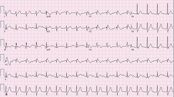
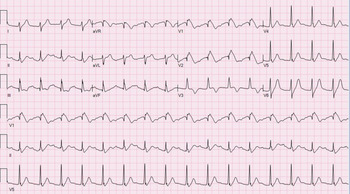
Evaluation of 12-lead EKG in sinus rhythm showed type 1 Brugada pattern in leads V1 and V2 (Fig. 2). This was further clarified by repeating the EKG with leads V1 and V2 in the second intercostal space (Fig. 3), which confirmed sinus rhythm with first-degree AV block and type 1 Brugada pattern (right bundle branch block and coved-type ST-segment elevation).
Type 1 Brugada pattern and first-degree atrioventricular block are noted during sinus rhythm.

Brugada pattern is brought out by repositioning of leads V1 and V2 in the second intercostal space.

A limited echocardiogram was performed in the ER during atrial rhythm which showed normal biventricular size, low normal biventricular function, and no evidence of pericardial effusion. A head CT scan and CT angiogram were also obtained and did not show any clinically significant abnormality.
She was subsequently transferred to paediatric ICU where she was noted to have fixed, dilated pupils, and her coagulation profile was indicative of disseminated intravascular coagulation. The patient started to bleed profusely from her venous and arterial puncture sites and became progressively bradycardic. CPR was restarted and continued for 30 minutes without any clinical improvement.
Blood sample for genetic testing was obtained post-mortem and an arrhythmia panel which included Brugada genes (Rhythm First, Ambry Genetics, Aliso Viejo, CA, USA) was sent. The panel was positive for a variant of unknown significance in Desmoplakin (DSP) gene (c.4527_4599del33 variant also known as p.R1509_S1519del). An autopsy could not be performed as the family did not consent for the same.
Discussion
First described in 1992, Brugada syndrome (BrS) is an autosomal dominant disease which is characterised by EKG abnormalities (right bundle branch block pattern and coved-type ST-segment elevation in the precordial leads V1–V3) and predisposition to life-threatening ventricular arrhythmias.Reference Brugada and Brugada1 Despite the underlying genetic basis, mutations are only identified in only a minority of patients (11–30%) with BrS.Reference Behere and Weindling2 Diagnosis of BrS rests on demonstrating either spontaneous or class I antiarrhythmic-induced type 1 EKG morphology (coved-type ST-segment elevation with ≥2 mm elevation in ≥1 lead among the right precordial leads V1, V2, positioned in the 2nd, 3rd, or 4th intercostal space) or conversion of type 2 and 3 EKG morphology (ST-segment elevation in ≥1 lead among the right precordial leads V1, V2 positioned in the 2nd, 3rd, or 4th intercostal space) to type 1 upon provocative drug testing with intravenous administration of Class I antiarrhythmic drugs.Reference Priori, Wilde and Horie3 Two-thirds of patients with BrS are asymptomatic at presentation, whereas a third are diagnosed after symptoms (syncope or aborted SCD) most of which occur at rest or during sleep when the vagal tone is high.Reference Gourraud, Barc, Thollet, Le Marec and Probst4 Fever and several medications, particularly sodium channel blockers, unmask the EKG findings of BrS in asymptomatic patients and can precipitate ventricular arrhythmias.Reference Andorin, Behr and Denjoy5 Patients with BrS are at increased risk of malignant ventricular arrhythmias during perioperative period due to medications, electrolyte imbalance, fever, and changed in autonomic nervous system response. Symptomatic BrS-related polymorphic VT, as seen in our patient, has been shown to have a high mortality.Reference Junttila, Gonzalez and Lizotte6
A common anaesthetic, propofol, has been associated with BrS-like EKG findings and malignant ventricular arrhythmias in adults. Prolonged infusion of propofol produces a distinct syndrome known as propofol infusion syndrome which is characterised by metabolic acidosis, rhabdomyolysis, hyperkalemia, and most importantly, BrS-like EKG changes and malignant ventricular arrhythmias.Reference Inamura, Okamoto, Kuroiwa and Hoka7–Reference Weiner, Haddad and Raj10 The exact incidence of propofol infusion syndrome and the dose of propofol required to produce these symptoms are not known. It is also unclear if genetic predisposition underlies the pathogenesis of propofol infusion syndrome. Autopsy studies have shown skeletal and cardiac myocytolysis and widespread fat accumulation in myocardium of patients with propofol infusion syndrome.Reference Jorens and Van den Eynden11 A high mortality (30%) has been reported in these patients, particularly in young males and in those with cardiac manifestations, rhabdomyolysis, hypotension, renal failure, metabolic acidosis, and dyslipidaemia.Reference Fong, Sylvia, Ruthazer, Schumaker, Kcomt and Devlin12 Recent studies have found lower incidence of ventricular arrhythmias following administration of a single dose of propofol in an unselected cohort as well as adults with BrS.Reference Wutzler, De Asmundis and Matsuda13–Reference Kloesel, Ackerman, Sprung, Narr and Weingarten17 Interestingly, a bolus dose of propofol has been shown to attenuate BrS-associated arrhythmogenic risk in high-risk adults with BrS patients in a recent study.Reference Ciconte, Santinelli and Brugada18 In contrast to adults in these studiesReference Wutzler, De Asmundis and Matsuda13–Reference Ciconte, Santinelli and Brugada18 who did not have clinically significant symptoms following propofol administration (mean dose 2.2–3.3 mg/kg), those with propofol infusion syndrome were administered a higher dose of propofol (4.5–7.3 mg/kg/hour).Reference Vernooy, Delhaas and Cremer8 EKG changes in patients with BrS phenocopy due to propofol infusion syndrome have been shown to normalise as early as 24 hours after withdrawal of propofol, and provocative testing with sodium channel blockers typically does not reveal an underlying predisposition to BrS.Reference Weiner, Haddad and Raj10 The effects of propofol on arrhythmogenesis in children have however not been systematically evaluated. The dose of propofol (2 mg/kg) used in our patient is comparable to the dose utilised in adults who were administered a single propofol bolus (mean dose 2.2–3.3 mg/kg).Reference Wutzler, De Asmundis and Matsuda13,Reference Kwon, Kim and Park14,Reference Ciconte, Santinelli and Brugada18 However, long-term use of antiarrhythmics such as amiodarone, β-blockers, calcium-channel blockers, and sodium channel blockers, which could have a bearing on the EKG changes and the risk of arrhythmogenesis during the procedure, was reported in one of the larger studies that found a low incidence of propofol infusion syndrome during and after short-term infusion of propofol.Reference Wutzler, De Asmundis and Matsuda13 Moreover, drug-induced BrS EKG changes can persist beyond the expected period of complete elimination of the offending drug from the body.Reference Weiner, Haddad and Raj10 Taking into consideration all the available evidence, it is quite likely that our patient had drug-induced BrS rather than propofol infusion-related BrS phenocopy.
Autonomic nervous system activity has a major bearing on the risk of arrhythmogenesis in patients with BrS. Therefore, it is possible that altered autonomic tone in the post-operative period may have contributed to enhanced ventricular arrhythmogenesis in this patient.
Repolarisation abnormalities such as prolonged QTc interval, ischaemia-like EKG changes, inverted T waves with a wide base (cerebral T waves), and morphological end-repolarisation abnormalities have usually been reported in patients with acute brain injury in the setting of a significant cerebral nervous system (CNS) insult such as stroke, subarachnoid haemorrhage, cerebral aneurysm rupture, head trauma, or subdural haemorrhage.Reference Katsanos, Krantzopoulos and Tsivgoulis19 Since the patient was found unresponsive, a head CT scan was promptly obtained but did not show any significant abnormality. Moreover, while ischaemia-like ST elevation has been reported with acute neurological injury, BrS-like EKG changes following acute neurological injury have only been reported in the setting of propofol infusion.Reference Otterspoor, Kalkman and Cremer20 Thus, the abnormal EKG findings in our patient are unlikely to be due to cardiac arrest-related acute neurological insult alone.
Preoperative work-up often includes an EKG in older adults; however, as per the current American Academy of Pediatrics recommendations for evaluation and preparation of children undergoing anaesthesia, a 12-lead EKG is not recommended.21 There is an ongoing debate in paediatric electrophysiology community regarding the utility of universal EKG screening in prevention of sudden cardiac death events in children. The benefits of universal EKG screening include higher sensitivity than the current standard (history and physical examination), lower costs with higher cost-effectiveness than other methods, ease of training a wide assortment of healthcare professionals, and technological assistance with computer-generated algorithms. However, EKG screening is associated with a number of important limitations which include more false positives when compared to the current standard (history and physical examination), additional cost of testing, additional testing required for further evaluation, errors in computer-generated readings, and lack of recognition of differences by age, gender, race/ethnicity.Reference Vetter22 In addition, EKG screening for BrS has lower sensitivity and specificity than EKG screening for other cardiac disorders as younger patients with BrS often do not have spontaneous BrS EKG pattern even with lead placement in the second intercostal space or the pattern is intermittent and therefore can be missed on a one time screening EKG. No consensus has been reached, and universal EKG screening is therefore not recommended at this time.Reference Vetter22
The complexity in clinically diagnosing BrS has led to increased utilisation of genetic testing. The largest proportion of identified genetic mutations have been demonstrated in SCN5A gene (20–25%); however, other genes have recently been identified, but their clinical significance remains uncertain.Reference Hosseini, Kim and Udupa23 Our patientʼs genetic testing was remarkable for a variant of uncertain significance in DSP gene which codes for DSP, a critical component of desmosomes in cardiac muscle cells. Our understanding of the role of DSP gene is etiopathogenesis of arrhythmogenic disorders is evolving and in addition to BrS,Reference Zhao, Chen and Peng24 DSP gene mutations have also been associated with arrhythmogenic right ventricular dysplasia,Reference Zhao, Chen and Peng24 and progressive cardiac conduction disease.Reference Kiselev, Mikhaylov and Pamon25 However, based on the current evidence, it is unlikely that DSP gene plays a causative role in the pathogenesis of BrS.Reference Hosseini, Kim and Udupa23
To conclude, there is a small risk of fatal ventricular arrhythmias in the post-operative period in patients who receive propofol anaesthesia; however, the precise determinants of this risk and the factors that differentiate drug-induced BrS from propofol infusion-related BrS phenocopy remain poorly defined. Though universal EKG screening in children remains controversial and is not recommended at this time, BrS-like EKG changes are lead specific (best seen in V1 and V2) and can therefore easily be missed on cardiac monitors. Pre- and post-operative 12-lead EKG is a low cost, low risk, non-invasive intervention which might have some utility in a small subset of children who are deemed to be at a higher risk of an inherited arrhythmia syndrome based on personal or family history. However, given the fact that BrS/BrS phenocopy events are overall quite rare in paediatric population (to the best of our knowledge, this is the only reported post-operative BrS-related death in a child), recommending the same universally might be unnecessarily alarmist and runs the risk of generating a large number of false positives.
Acknowledgements
None.
Financial Support
This research received no specific grant from any funding agency, commercial, or not-for-profit sectors.
Conflict of Interest
None.
Ethical Standards
This report does not involve any human subjects research or animal experimentation.