Case description
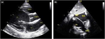
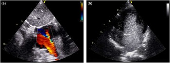
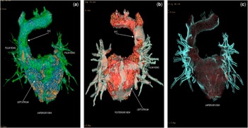
A 2-year-old girl presented with a history of cyanosis during the past 6 months. The child had undergone echocardiographic evaluation twice previously and was reported as normal. The child had clubbing and was cyanosed at a room air saturation of 85%. The praecordial examination was unremarkable. Further, two-dimensional echocardiography showed a tubular (vascular) structure connected to the left atrium in the modified parasternal long-axis view (Fig 1a). The subcostal view clearly showed the superior caval vein draining into the left atrium (Fig 1b). Color Doppler imaging confirmed draining of the superior caval vein into the left atrium (Fig 2a). In addition, there was a small atrial septal defect of the ostium secundum type with left-to-right shunting. Saline contrast injection through either upper limb showed immediate opacification of the left atrium (Fig 2b). The structure of the coronary sinus was normal. The inferior caval vein was normally connected to the right atrium. Cardiac multidetector CT with three-dimensional reconstruction showed direct drainage of the right superior caval vein into the left atrium and anomalous drainage of the right upper pulmonary vein to the superior caval vein (Fig 3a–c). The child underwent surgical correction. A vertical incision was made from the superior caval vein across the atriocaval junction, through the atrial septum, creating an atrial septal defect, which was extended onto the anterolateral wall of the right atrium. A pericardial patch was used to create an intra-atrial baffle diverting the flow from the superior caval vein to the right atrium. The superior margin of the patch was sutured to the posterior wall of the superior caval vein, and the inferior margin was sutured to the caudal edge of the created atrial septal defect. The right upper pulmonary vein was routed to the left atrium. The periprocedural period was uneventful.
( a ) Echocardiographic parasternal long-axis view showing a vascular structure (superior caval vein (SVC)) attached to the left atrium (LA). ( b ) Echocardiographic subcostal view showing the anomalous direct connection of the superior caval vein to the left atrium. LV=left ventricle; RA=right atrium.

( a ) Color Doppler echocardiographic image showing anomalous connection of the right superior caval vein to the left atrium. ( b ) Saline-contrast echocardiography showing opacification of the left-sided chambers with minimal contrast in right atrium secondary to the atrial septal defect.

( a – c ) Multidetector cardiac CT showing the anomalous drainage of the right superior caval vein (SVC) to the left atrium. In addition, anomalous connection of the right upper pulmonary vein to the SVC is also seen.

The embryological basis of the condition is believed to be a sinus venosus defect of the superior caval vein type in association with atresia of the right superior caval vein orifice. Cyanosis is usually the dominant clinical feature. The risks of polycythemia, shortness of breath, decreased exercise tolerance, systemic emboli, brain abscess, and other cerebrovascular complications increase with age. Since the first case reported by Wood in 1957, a little more than a dozen cases have been published: 10 of these patients underwent surgery, and in nine of them the right upper lobe pulmonary veins drained into the right superior caval vein. Alternative surgical procedures include the Warden procedure, in which the superior caval vein is divided, and the cephalic superior caval vein is anastomosed to the right atrial appendage, whereas the caudal segment serves as a pulmonary drainage conduit. In patients with borderline pulmonary ventricles requiring complex repairs, a bidirectional Glenn procedure can be performed without superior caval vein hypertension and without the potential risks of atrial arrhythmias and baffle complications. Complete repair has also been performed using a veno-atrial shunt and without the use of cardiopulmonary bypass.Reference Ahmad and Sell 1 The present case emphasises the importance of suspecting extracardiac right-to-left shunt in any case of unexplained cyanosis.
Acknowledgement
None.
Financial Support
The evaluation of the case described received no specific grant from any funding agency or from commercial or not-for-profit sectors.
Conflicts of Interest
None.