Atrial septal aneurysm is a localised “saccular” deformity of the interatrial septum, generally at the level of the fossa ovalis, which bulges into the right or left atrium or both.Reference Olivares-Reyes, Chan, Lazar, Bandlamudi, Narla and Ong 1 It was initially thought to be a rare congenital abnormality; however, with the advent of two-dimensional echocardiography and, more recently, the widespread use of trans-oesophageal echocardiography, it has become more easily and more frequently identified in patients. The first autopsy of such a case was reported over 80 years ago in 1934.Reference Lang and Posselt 2 There are few papers in the literature regarding the prevalence of atrial septal aneurysms in the paediatric population, which by means of transthoracic echocardiography ranges from 0.9% to 1.7% in children and almost 7.6% in neonates.Reference Olivares-Reyes, Al-Kamme and Gonzalez 3 – Reference Ozcelik, Atalay, Tutar and Ekici 5 On the contrary, many papers have shed light on their prevalence in adulthood, which ranges from 2.2% up to 10% in trans-oesophageal series.Reference Schneider, Hanrath, Vogel and Meinertz 6 – Reference Mattioli, Aquilina, Oldani, Longhini and Mattioli 10
Our study aims to enlighten the prevalence of atrial septal aneurysms and concurrent abnormalities in children, their natural course, and the clinical implications to children's life.
Materials and methods
From March, 2005 to February, 2010, we studied 4522 healthy children and patients aged more than 12 months, referred for transthoracic echocardiography either for an audible murmur or within scheduled follow-up for their medical history or just for an ordinary check-up. This prospective study was performed according to the provisions of the Declaration of Helsinki and written informed consent was obtained from the parents of the children. The study was also reviewed and approved by the AHEPA University Hospital's committee on human experimentation. Neonates and infants younger than 12 months were excluded as most atrial septal aneurysms diagnosed at this age range subside within months.Reference Ozcelik, Atalay, Tutar and Ekici 5 , Reference Hung, Lu and Hung 11 Once an aneurysm was found, a second transthoracic echocardiography was performed after 1 year so as to re-evaluate its size.
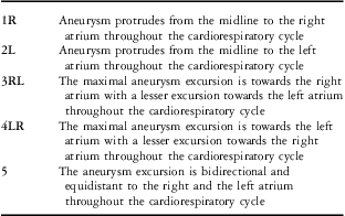
Atrial septal aneurysm was defined according to criteria previously published by Hanley et alReference Hanley, Tajik and Hynes 12 diameter of the base of the aneurysmatic portion of the septum of at least 15 mm and either protrusion of the septum or part of it of at least 15 mm beyond its plane or phasic excursion during the cardiorespiratory cycle of at least 15 mm in total amplitude. Although any cut-off in the definition is arbitrary, the 15 mm cut-off adopted in the present study provides relatively high specificity for atrial septal aneurysm diagnosis. Therefore, the parameters evaluated were: length of the aneurysm in the plane of the atrial septum, maximal excursion or protrusion of the aneurysm beyond the plane of the septum, direction of maximal protrusion into the right or left atrium, spontaneous oscillation of it during normal cardiorespiratory cycle, maximal extent of protrusion during oscillation, and other structural abnormalities associated with the atrial septum as the presence of a patent foramen ovale or an atrial septal defect. Further analysis was based on the classification proposed by Olivares-Reyes et al in 1997.Reference Olivares-Reyes, Chan, Lazar, Bandlamudi, Narla and Ong 1 According to this classification, there are five different types of atrial septal aneurysms. More specifically, an aneurysm is labelled as being type 1R when it protrudes from the midline to the right atrium throughout the cardiorespiratory cycle. It is labelled as being type 2L when it protrudes from the midline to the left atrium throughout the cardiorespiratory cycle. It belongs to the type 3RL when the maximal excursion is towards the right atrium with a lesser excursion towards the left atrium. It is named type 4LR when the maximal excursion of the aneurysm is towards the left atrium with a lesser excursion towards the right atrium. It is labelled type 5 when the movement is bidirectional and equidistant to the right and the left atrium during the cardiorespiratory cycle. Atrial septal aneurysm classification is summarised in Table 1. Transthoracic studies were obtained with the use of a Vivid 3 Pro ultrasonography system (General Electric Medical Systems, Milwaukee, Wisconsin, USA) with a 5 MHz phased array imaging transducer, and all recordings were analysed by the same paediatric cardiologist. Patients underwent standard views including parasternal long-axis, short-axis, apical four- and five-chamber views, as well as suprasternal and subcostal views. The studies were performed with the patient in supine and left lateral decubitus positions – for the older and greater body habitus ones – during quiet respiration. The most valuable were the subcostal and apical four-chamber views, with appropriate transducer angulation to visualise the atrial septum completely. Quiet respiration and sustained inspiration were conducive to getting the best images, which allowed precise measurements of morphological characteristics of atrial septal aneurysms. All procedures followed were in full accordance with the recommendations of the American Society of Echocardiography.Reference Lai, Geva and Shirali 13 Bubble studies were not performed.
Atrial septal aneurysm classification.

Results
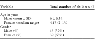
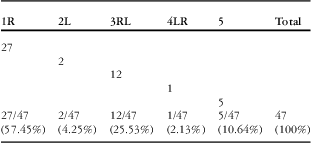
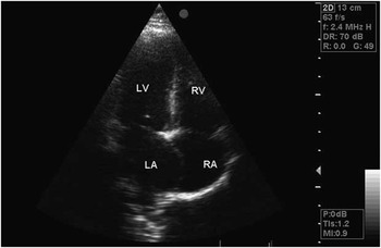
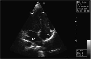
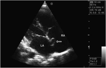
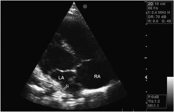
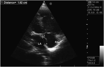
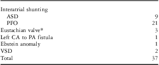
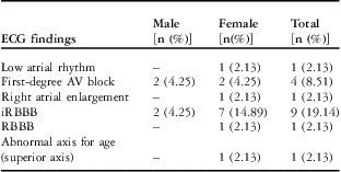
A total of 4522 children were enrolled in the study. Of them, 47 fulfilled the echocardiographic criteria for atrial septal aneurysm (1.04%). The demographical characteristics of these children are reported in Table 2. Overall, atrial septal aneurysms bulged towards the right atrium in 27 patients (57.78%) and towards the left atrium in two (4.44%) patients. They bulged predominantly towards the right atrium and less to the left in 12 patients (24.44%) and predominantly towards the left atrium and less to the right in one patient (2.22%). In five patients (11.11%), the septum showed the same oscillation between the two atria during a normal cardiorespiratory cycle. Table 3 summarises the incidence of each type of atrial septal aneurysm as determined by transthoracic echocardiography. Figures 1–6 show some types of aneurysms recorded. The aneurysm involved almost the entire septum in 14 patients (28.89%) and was limited to the fossa ovalis in 33 (71.11%). None was detected to be limited only to the septum primum. Aneurysms were often detected along with other cardiac abnormalities (n = 37), in particular atrial septal defect and patent foramen ovale. In 10 patients (35.56%), it was an isolated structural abnormality – called “lone” atrial septal aneurysm. The concurrent cardiac abnormalities are listed in Table 4. The electrocardiographic findings of children with aneurysms are listed in Table 5. None of these children had non-invasive evidence of increased pulmonary pressures and the estimated right ventricular pressure from the tricuspid regurgitation jet – when a full envelope enabled us to estimate such pressure – never exceeded one-third of systemic. Pulmonary pressure was also low in both children with a ventricular septal defect, as the peak gradient across it was 62 and 72 mmHg, respectively with normal systemic pressures. All the 47 patients with an atrial septal aneurysm underwent a second transthoracic study after a year, and in no participant was any difference recorded, that is, neither enlargement of the aneurysm nor subsidence of it.
Demographical characteristics of children with an atrial septal aneurysm.

Incidence of each type of atrial septal aneurysm as determined by transthoracic echo.

Atrial septal aneurysm type 1R involving the entire septum. LA = left atrium; LV = left ventricle; RA = right atrium; RV = right ventricle.

Atrial septal aneurysm type 2L involving the entire septum. LA = left atrium; LV = left ventricle; RA = right atrium; RV = right ventricle.

Atrial septal aneurysm type 3RL involving the fossa ovalis. The aneurysm bulges into the right atrium. LA = left atrium; RA = right atrium.

Atrial septal aneurysm type 3RL involving the fossa ovalis. The same aneurysm as in Fig 3 bulges into the left atrium. LA = left atrium; RA = right atrium.

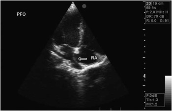
Atrial septal aneurysm type 5 involving the entire septum. The aneurysm bulges into the right atrium. Note the equal protrusion as in Fig 6. PFO = patent foramen ovale; RA = right atrium.

Atrial septal aneurysm type 5 involving the entire septum. The same aneurysm as in figure 5 bulges into the left atrium. Note the equal protrusion as in figure 5. LA = left atrium; RA = right atrium.

Concurrent cardiac abnormalities associated with an atrial septal aneurysm.

ASD = atrial septal defect; CA = coronary artery; PA = pulmonary artery; PFO = patent foramen ovale; VSD = ventricular septal defect
*Although a Eustachian valve is a normal finding, in these particular patients they were abnormally large and mobile
ECG findings of children with an atrial septal aneurysm.

AV = atrioventricular; ECG = electrocardiogram; iRBBB = incomplete right bundle branch block; RBBB = right bundle branch block
Discussion
From March, 2005 to February, 2010, we studied 4522 healthy children and patients aged more than 12 months by the means of transthoracic echocardiography. We believe that the big number of participants is well reflective of the general paediatric population. The atrial septal aneurysm is a congenital cardiac abnormality that is characterised by “saccular” formation of the interatrial septum. Redundant septal tissue results in bulging of the septum into either or both atria during the cardiac cycle. Aneurysm formation can also be secondary to interatrial pressure differences.Reference Silver and Dorsey 14 That could also explain the much higher incidence in adult population as thin tissue under chronic pressure becomes more aneurysmal over time. In 1997, Olivares proposed a new classification of atrial septal aneurysms,Reference Olivares-Reyes, Chan, Lazar, Bandlamudi, Narla and Ong 1 which replaced the existing one that had been proposed by Hanley in 1985.Reference Hanley, Tajik and Hynes 12 According to the new classification, there are five different types and this is the classification used in our study.
An atrial septal aneurysm is clinically significant in adulthood because of its association with cerebral ischaemia and stroke, especially when there is a concurrent patent foramen ovale.Reference Cabanes, Mas and Cohen 15 – Reference Ueno, Shimada and Tanaka 23 Of the 47 patients with an aneurysm, 21 (44.68%) had at the same time a patent foramen ovale. The parents of these children were informed thoroughly about the possible association with cerebral ischaemia and stroke in adulthood, and this fact was recorded in the child's file. However, life shows that these patients make it so far into adult life without an embolic event, and thus it was highlighted that the risks are quite small in otherwise healthy children.
Other reports in adults have also noted that atrial septal aneurysms are also associated with supraventricular tachyarrhythmias, mitral valve prolapse, and migraines with aura.Reference Morelli, Voci and Morabito 24 – Reference Carerj, Narborne and Zito 27 The aforementioned conditions may be found sporadically in childhood as well. In our study, none of the children diagnosed to have an aneurysm had a medical history of palpitations or supraventricular tachyarrhythmia, one had a mitral valve prolapse, and one had migraines with aura and was on medication by a paediatric neurologist.
It has also been reported that non-specific right atrial electrocardiographic signs such as peaked and moderately enlarged P-waves in leads II, III, and aVF and rotation of the frontal P-wave axis may be indicative of asymptomatic “lone” aneurysm.Reference Scholz, Zitron, Katus, Karle and Mereles 28 In our study, all electrocardiographic recordings were within normal for childhood apart from that of the child with the Ebstein form of the tricuspid valve, which showed signs of right atrial enlargement and right bundle branch block.
In every case, it should be stated clearly that no action is to be taken during childhood, as a child with an atrial septal aneurysm is not at increased risk compared with a child without one. Some propose that once a child is diagnosed to have such an aneurysm he/she be checked once again after a year to detect any differences. However, no consensus has been reached regarding the follow-up, which is then scheduled on an individual basis. In our study, all the 47 children underwent a second transthoracic study a year after the first one and no difference in aneurysm size was recorded. This finding is reassuring of the benign course of such aneurysms in childhood. However, all these 47 patients will be invited to undergo a transthoracic echo 5 years after the diagnosis, so as to establish a much more convincing evidence of their natural history in childhood. In the literature, there are also data regarding the familial occurrence of atrial septal aneurysms.Reference Paraskevaidis, Tsiapras, Kyrzopoulos and Kremastinos 29 , Reference Werren and Alberti 30 In the near future – in collaboration with adult cardiologists – we plan to invite all the first relatives of these children to undergo a transthoracic echo to further record the prevalence of the familial occurrence. We also aim to further develop our study by using bubble studies.
Acknowledgement
The authors would like to thank Mrs Dimitra Papazisi for the secretarial support throughout the study period. They have no conflicts of interest to disclose.