Implications
The increasing growth rate and body size of modern hybrid birds recently caused the appearance of breast muscle abnormalities such as white striping (WS) and wooden breast. Recently, it has been observed a new abnormality termed ‘spaghetti meat (SM)’ which exhibits an altered structural integrity of the breast fillet. Because of impaired appearance, poultry plants tend to downgrade abnormal breasts and potentially divert the meat into processed products with considerable economic losses. Therefore, poultry industry reveals a growing interest to understand how this new muscle abnormalities can affect meat quality and, more importantly, how to deal with this significant problem.
Introduction
In the past few years, several studies evidenced that the genetic selection in broilers has led to muscle fibers hypertrophy and altered their structural, functional and metabolic characteristics (Petracci et al., Reference Petracci, Mudalal, Soglia and Cavani2015; Velleman and Clark, Reference Velleman and Clark2015). Indeed, if compared with their unselected counterpart, fibers composing the muscle tissues of fast-growing birds exhibited larger diameter, lower capillary-to-fiber ratio, increased intercapillary distance and lower protein degradation rate (Branciari et al., Reference Branciari, Mugnai, Mammoli, Miraglia, Ranucci, Dal Bosco and Castellini2009; Velleman and Clark, Reference Velleman and Clark2015). Therefore, the remarkable increase in muscle size and growth rate have inevitably led to the appearance of several muscle myopathies (Petracci et al., Reference Petracci, Mudalal, Soglia and Cavani2015; Kuttappan et al., Reference Kuttappan, Shivaprasad, Shaw, Valentine, Hargis, Clark, McKee and Owens2016). In detail, the main myopathies affecting the Pectoralis major muscles are WS, wooden breast condition and, recently, the SM abnormality. Considering that both wooden and WS cases exhibited similar histological lesions (including myodegeneration and regeneration, fibrosis and lipidosis) (Sihvo et al., Reference Sihvo, Lindén, Airas, Immonen, Valaja and Puolanne2016; Soglia et al., Reference Soglia, Laghi, Canonico, Cavani and Petracci2016a; Radaelli et al., Reference Radaelli, Piccirillo, Birolo, Bertotto, Gratta, Ballarin, Vascellari, Xiccato and Trocino2017) and they often occur within the same fillet, a common underlying mechanism might be hypothesized (Petracci et al., Reference Petracci, Mudalal, Soglia and Cavani2015; Kuttappan et al., Reference Kuttappan, Shivaprasad, Shaw, Valentine, Hargis, Clark, McKee and Owens2016).
Recently, it has been observed that the WS defect is also associated with another muscle abnormality termed ‘SM’ and exhibiting an altered structural integrity of the cranial surface of the P. major muscle (Bilgili, Reference Bilgili2015; Sirri et al., Reference Sirri, Maiorano, Tavaniello, Chen, Petracci and Meluzzi2016). Some years ago, also Swatland (Reference Swatland1990) observed an analogous abnormality affecting turkey breast muscles exhibiting a loose structure in which muscle fiber bundles could be pull away by fingers. Thus, as an increased cross-sectional area was observed in muscle fibers from disintegrated meat, Swatland (Reference Swatland1990) suggested that these fibers have outgrown their connective tissue. As a result, SM defect seemed to mainly affect the connective tissue within the perymisial compartments leading to the formation of large intracellular spaces. Thus, the fluid lost by myofibrils during postmortem period might result in muscle disintegration. In addition, more recently Ahn et al. (Reference Ahn, Zheng, Li, Zeng, Qu, Xu and Yang2010) observed that the perymisial septa of breast muscles in fast-growing birds was thinner than the slow-growing counterpart.
As a consequence of their impaired appearance and quality traits, these abnormal fillets are normally downgraded by the poultry meat industry and used for further processing. Thus, there is serious interest in the meat industry to understand the effect exerted by the occurrence of these abnormalities on meat quality traits. Within this context, although both quality and histological traits of WS and wooden breast fillets have been extensively studied (Petracci et al., Reference Petracci, Mudalal, Soglia and Cavani2015; Velleman and Clark, Reference Velleman and Clark2015; Kuttappan et al., Reference Kuttappan, Shivaprasad, Shaw, Valentine, Hargis, Clark, McKee and Owens2016), no information are available concerning the SM abnormality. In addition, as the SM defect often appears in association with WS, this study aimed at evaluating the effect exerted by WS and SM abnormalities (occurring alone or combined within the same P. major muscle) on muscle histology and meat quality traits.
Material and methods
Sample collection
The study was conducted on two flocks of broiler chickens reared and slaughtered under commercial conditions into 2 consecutive weeks. The birds were homogeneous in genotype (Ross 308 strain), gender (males), age and weight at slaughter (47 days and 2.8 kg) as well as feeding plan (ad libitum access to a wheat/sorghum-soybean multiphase diet). Before slaughter, broilers were subjected to a total feed withdrawal of 10 h, including a 3 h lairage time at the processing plant.
Subsequently, birds were processed under commercial conditions: exposed to carbon dioxide for stunning, bled for 180 s and then after being conveyed through scalding tanks (52°C for 220 s), plucked by rotating rubber fingers. After evisceration, carcasses were air-chilled passing through a cold-air flow tunnel (−6°C for 150 min) until reaching 2°C to 3°C at the core. Then, a total of 96 P. major muscles (48 samples/each replication) were selected at 3 h postmortem in the deboning area of the same commercial processing plant. The samples were classified by two experienced people according to the presence of WS and SM abnormalities (see figure provided in supplementary material). In detail, the P. major muscles were classified into four experimental groups as follow (by excluding those having signs of wooden breast condition):
-
∙ normal (N): fillets exhibiting neither white striations nor tendency toward separation of the muscle fiber bundles composing the P. major;
-
∙ white striping: exhibiting superficial medium-to-thick white striations in the cranial part of the fillet;
-
∙ spaghetti meat: exhibiting an overall impaired integrity and tendency toward separation of the muscle fiber bundles composing the P. major muscle especially within the cranial part of the fillet; and
-
∙ white striping/spaghetti meat: affected by both WS and SM abnormalities.
Sample preparation and measurements of weight and dimension
After being collected, the samples were bagged by group and brought to the laboratory under refrigerating conditions. Then, muscles were trimmed from superficial fat, visible cartilage and connective tissues and subsequently stored at 2°C to 4°C until 24 h postmortem. The whole fillet was weighed and morphometric measurements (length, width and height) were assessed with an electronic calliper as previously described by Mudalal et al. (Reference Mudalal, Lorenzi, Soglia, Cavani and Petracci2015). Then, according to the same procedure adopted in our previous study (Soglia et al., Reference Soglia, Gao, Mazzoni, Puolanne, Cavani and Petracci2017) each fillet was cut in order to separate the superficial layer from the deep one and used to assess proximate composition and histological features, functional properties and color as well as to evaluate both myofibrillar and sarcoplasmic proteins profile.
Histology
For histological investigations five samples of P. major muscle belonging from each experimental group (N, WS, SM and WS/SM for a total of 20 samples) were considered. Each sample was cut in order to separate the superficial from the deep layer according to the sampling protocol previously described by Soglia et al. (Reference Soglia, Gao, Mazzoni, Puolanne, Cavani and Petracci2017). Subsequently, the samples were immediately fixed in a 10% buffered formalin solution for 24 h at room temperature. After that, specimens were oriented for transverse and longitudinal fiber sectioning, dehydrated in a graded series of ethanol and embedded in paraffin. From each sample, serial transverse and longitudinal sections (6 μm thick) were obtained, mounted on polylysine-coated slides and stained with Masson’s trichrome.
Proximate composition
Proximate composition (moisture, protein, fat, ash and collagen) of N, WS, SM and WS/SM samples was determined on both the superficial and the deep layer of each P. major muscle following standard methods. Moisture content was assessed as the percentage of weight lost after drying 5 g of sample in oven (105°C for 16 h) using standard Association of Official Analytical Chemists (AOAC) method (Washington, DC, USA). Crude protein was determined according to Kjeldahl method by using copper sulphate as catalyst using standard AOAC method, whereas lipids were extracted following the Folch chloroform–methanol extraction procedure. Similarly, the colorimetric method by Kolar (Reference Kolar1990) was applied in order to quantify the exact amount of hydroxyproline in the sample and calculate the total collagen (considering 7.5 as conversion factor).
Functional properties and color
Meat color (Commission Internationale de l‘Éclairage, L*=lightness, a*=redness and b*=yellowness) was measured in triplicate on the bone-side surface of both the superficial and the deep layer of each fillet using a Chroma Meter CR-400 (Minolta Corp., Milan, Italy). Similarly, ultimate pH (pHµ;) of N, WS, SM and WS/SM samples was assessed at 24 h postmortem following the procedure described by Jeacocke (Reference Jeacocke1977). Total protein solubility was measured following the procedure described by Bowker and Zhuang (Reference Bowker and Zhuang2015). In brief, triplicate 1 g muscle samples were homogenized (30 s at 13 500 r.p.m., in ice) in 1.1 M KI+0.1 M potassium phosphate buffer (pH 7.2). Samples were stored at 4±1°C for 20 h and then centrifuged at 2600×g for 30 min at 4°C. Then, protein concentration was measured by Bradford assay by using bovine serum albumin as a standard. Myofibril Fragmentation Index (MFI) was assessed in duplicates on both the superficial and the deep layer of each P. major muscle. The method is based on that described by Culler et al. (Reference Culler, Parrish, Smith and Cross1978) with slight modifications (Hopkins et al. Reference Hopkins, Littlefield and Thompson2000). Finally, protein oxidation was assessed following the novel DNPH-based method proposed by Soglia et al. (Reference Soglia, Mudalal, Babini, Di Nunzio, Mazzoni, Sirri and Petracci2016b).
Sodium dodecyl sulphate-polyacrylamide gel electrophoresis analysis
Myofibrillar and sarcoplasmic protein extracts were prepared according to Liu et al. (Reference Liu, Ruusunen, Puolanne and Ertbjerg2014) by homogenizing (30 s at 13 500 r.p.m., in ice) two grams of frozen P. major muscle in 40 ml of cold Rigor Buffer (75 mM KCl, 10 mM KH2PO4, 2 mM MgCl2, 2 mM Ethylene glycol-bis(2-aminoethylether)-N,N,N′,N′-tetraacetic acid; pH 7.0). Each sample was run in duplicates. The homogenate was centrifuged for 10 min at 10 000×g at 4°C and the supernatant collected as the sarcoplasmic protein fraction. The procedure was repeated twice and the resultant pellet, composed by myofibrillar proteins, was re-suspended by homogenization in 20 ml of cold Rigor Buffer. Protein concentration of each extract was adjusted to 1.0 mg/ml and each sample was mixed 1 : 1 (v/v) with Sample Buffer (50 mM Tris-HCl, 8 M Urea, 2 M Thiourea, 75 mM dithiothreitol, 3% (v/v) SDS) (Fritz et al., Reference Fritz, Swartz and Greaser1989). Sodium dodecyl sulphate-polyacrylamide gel electrophoresis analysis was run, in duplicates, on 15 μg of proteins according to the procedure described by Laemmli (Reference Laemmli1970) by using 7.5% polyacrylamide hand-cast gels. A molecular-weight marker (Precision Plus Standard Proteins, All Blue Prestained; Bio-Rad, Hercules, CA) was loaded into each gel to assess the molecular weight of the protein bands. Gels were run on a Bio-Rad Mini Protean II electrophoresis apparatus at 110 V constant voltage for about 1 h. Gels were stained with Coomassie Brilliant Blue R-250 (Sigma Aldrich, San Louis, Missouri, USA) (1 g/l) containing 40% (v/v) methanol and 10% (v/v) acetic acid in distilled water and destained in distilled water. Gel images were acquired by using a GS-800™ Calibrated Densitometer (Bio-Rad) to quantify the relative abundance of each protein band.
Nuclear Magnetic Resonance (NMR) relaxation measurements
Proton transverse relaxation decay curves in breast meat samples were recorded, at the operating frequency of 20 MHz, with a Bruker (Milan, Italy) Minispec PC/20 spectrometer using a Carr-Purcell-Meiboon-Gill (CPMG) pulse sequence, set as described by Petracci et al. (Reference Petracci, Mudalal, Bonfiglio and Cavani2014). The samples had a weight of about 600 mg, with a height not exceeding the active region of the radio frequency coil. All the measurements were performed at a constant temperature of 24°C. The CPMG decays were normalized to the sample weight and transformed into relaxograms (i.e. continuous distributions of relaxation times) through the program UPEN. Each relaxogram was interpreted in terms of bound water, intra-myofibrillar and extra-myofibrillar proton pools in agreement with previous studies (Petracci et al., Reference Petracci, Mudalal, Bonfiglio and Cavani2014; Soglia et al., Reference Soglia, Petracci and Ertbjerg2016c). To separately observe such protons populations, the relaxograms were fit to the sum of four exponential curves and the two with intermediate relaxation times (T2), describing intra-myofibrillar protons behaviors, were merged.
Statistical analysis
Data were analyzed using the ANOVA option of the GLM procedure present in SAS software (SAS Institute Inc., Cary, NC, USA) and testing the main effects for type of meat quality abnormality (N, WS, SM and WS/SM), position (superficial and deep) and replication, as well as the interaction terms on meat quality traits. Means were separated using Tukey’s honestly test multiple range test of the GLM procedure.
Results
Histology
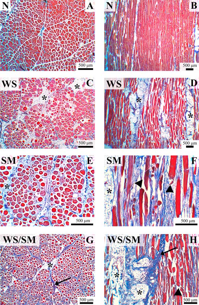
The findings of the histological observations performed on SM and/or WS fillets revealed that, if compared with their unaffected counterpart (N), all samples affected by muscle abnormalities exhibited degenerative processes affecting the P. major muscles. Notwithstanding, the microscopic histological features were consistent with the macroscopic grading (classification) of the fillets. Indeed, myodegenerative fibers were not observed within the N samples whose fibers exhibited a regular polygonal profile, normal cross-striated architecture as well as a structured endo- and peri-mysial connective tissue (Figure 1a and b).
Chicken Pectoralis major muscle, Masson’s trichrome. Representative images of transversal (a, c, e and g) and longitudinal (b, d, f and h) histological sections of normal (N), white striping (WS), spaghetti meat (SM) and fillets affected by both abnormalities (WS/SM) (Supplementary Figure S1). In N samples skeletal muscle fibers show a regular polygonal profile, compact and normal shape and size: also pery- and endo-mysial spaces are normal (a and b). In samples affected by WS is peculiar abundant endomysial fatty tissue infiltrated and, above all, perimysial (c and d, asterisks): fatty infiltration in some cases matches the fibrillar part. Even in this case, there were observed rounded fibers of different dimensions. In the SM samples muscle fibers were reduced in number and spaced apart (sometimes almost isolated) from each other; such fibers have a rounded profile and very variable dimensions. The peri- and endo-mysial connective tissue is compromised (particularly rarefied around some fibers) (e and f), whereas adipose tissue infiltrates the space between the muscle fibers (e and f, asterisks). In the longitudinal section were observed numerous split fibers immersed in the rarefied endomysial connective tissue and infiltrated by inflammatory cells (f, arrowheads). White striping/spaghetti meat breasts muscle show histopathological features related to those previously described in the SM and WS. In this case, it is observed in some parts of the section an abundant proliferation of endomysial and perimysial connective (g and h, arrows). In addition, it is possible to observe split fibers (h, arrowhead) and adipose infiltration (g and h, asterisks).

With regard to the WS, the main histological features associated with the occurrence of this defect were nuclear internalization, loss of cross striations, vacuolar and hyaline degeneration, necrosis and lysis of the fibers, inflammatory cells infiltration, variable cross-sectional area (degenerating and regenerating fibers), lipidosis and fibrosis. A distinctive features observed within the WS samples were the increased deposition of adipocytes within the connective tissue (lipidosis) (Figure 1e) and fat infiltrations attaining the amount of fibrillar components (Figure 1f, longitudinal section). On the other hand, when palpated after chilling, the P. major muscles affected by SM defect were extremely soft and stringy (especially in the ventro-cranial portion). In general, this myopathy exhibited histological features reported for associated myopathies (e.g. WS, wooden breast) including: extensive fibers degeneration and regeneration, hyalinization, poor fiber uniformity, compromised connective and fat tissue deposition. However, a progressive rarefaction of the endo- and peri-mysial connective tissue leading to muscle fibers detachment from each other (as showed in Figure 1c) was observed in SM muscles. In addition, thin and split fibers surrounded by loose (immature) connective tissue and abundant inflammatory cells infiltrations were found by examining the longitudinal sections (Figure 1d). Interestingly, the samples affected by both WS and SM defect exhibited an alternation of the pathological traits previously described. In detail, some areas exhibited the typical features of the SM defect whereas others evidenced the pathological changes largely associated with WS (Figure 1g and h). Overall, the most severe histopathological lesions frequently affected only the superficial section of the fillet whereas the deep one often exhibited normal traits.
Weight and dimension of raw fillets
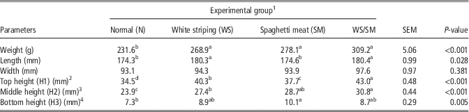
The results for weight and dimensions of the breast fillets are shown in Table 1. Overall, if compared with N, all abnormal samples exhibited significantly higher weight (P<0.001) and thickness (P<0.05) measured at the top (H1), middle (H2) and bottom (H3) positions. As for length, WS and WS/SM samples revealed significantly higher (P<0.005) values in comparison with both N and SM fillets, which did not differ between each other. None of the abnormalities significantly affects the width.
Effect of breast abnormalities on weight and dimension of raw chicken fillets

SEM=standard error of means.
a,b,c,dMean values within the same parameter followed by different superscript letters significantly differ (P<0.05).
1 n=24/group.
2 H1 was measured at the thickest point in the cranial part.
3 H2 was measured at the half distance of the breast length.
4 H3 was measured far from the end of the caudal part by 1 cm toward a dorsal direction.
Proximate composition
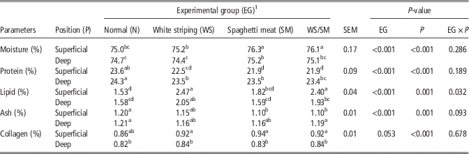
The results for proximate composition are shown in Table 2. If compared with the deep section, a significantly higher (P<0.001) moisture content was found within the superficial layer of the P. major muscles belonging from all the experimental groups. In addition, SM and WS/SM meat exhibited significantly higher (P<0.001) moisture values in both the superficial and the deep sampling position. With regard to protein, a significantly higher (P<0.001) content was found in both the superficial and deep section of N fillets. In addition, if compared with the deep counterpart, a significantly lower (P<0.001) protein content was observed within the superficial layer of all the abnormal samples with the major differences being measured in SM and WS/SM fillets (WS: 22.5% v. 23.5%; SM: 21.9% v. 23.5%; WS/SM: 21.9% v. 23.4%). As for lipid content, both the superficial and deep sections of WS and WS/SM samples exhibited significantly higher (P<0.001) values, the last displaying a higher lipid content on the surface layer rather than in the deep (2.40% v. 1.93%; P<0.05). Although no significant differences among all the experimental groups were found within the deep section, a significantly higher (P<0.001) ash content was found in the superficial portion of N samples. In addition, if the effect of the sampling position is considered, a lower ash content was found within the superficial layer of WS/SM samples (1.10% v. 1.19%; P<0.05). At last, although no effect of the sampling position was found for N, an increase (P<0.05) in collagen content was observed when comparing the deep and superficial sections of all abnormal fillets.
Effect of breast abnormalities on proximate composition of chicken meat
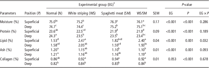
SEM=standard error of means.
a,b,c,dMean values within each parameter considering both the experimental groups (same row) and the sampling position (same column) followed by different superscript letters significantly differ (P<0.05).
1 n=24/group.
Functional properties and color
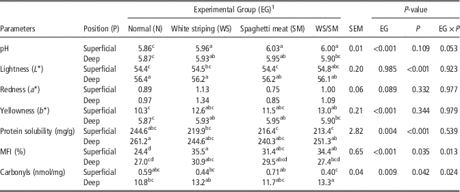
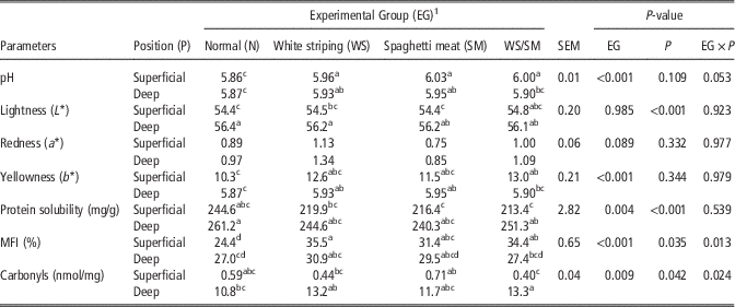
The results for functional properties and color of breast fillets are shown in Table 3. Overall, if compared with N, all abnormal fillets exhibited significantly higher ultimate pH on both the superficial and the deep sections, with the first exhibiting the highest values. As for raw meat color, no significant differences were found among the samples with the only exception of both the superficial and the deep layers of the WS/SM fillets which exhibited a significantly higher yellowness in comparison with N. In addition, although redness was not affected by sampling position, all the deep sections were lighter than their superficial counterparts.
Effect of breast abnormalities on color and functional properties of chicken meat
SEM=standard error of means; MFI=Myofibril Fragmentation Index.
a,b,c,dMean values within each parameter considering both the experimental groups (same row) and the sampling position (same column) followed by different superscript letters significantly differ (P<0.05).
1 n=24/group
With regard to total protein solubility, even if lower values were found in fillets affected by muscle abnormalities, significant differences were found only when the deep layer of N fillets was compared with superficial section of samples affected by SM abnormality (SM and WS/SM groups). In addition, a significant increase in total protein solubility was observed moving from the superficial toward the deep layer of WS/SM samples. As for MFI, if compared with N, significantly higher (P<0.001) values were found in the superficial section of all the abnormal groups. In addition, even if according to the statistical analysis, both the main effects and the interaction term were significant, no clear trends were found for carbonyls.
Electrophoretic analysis
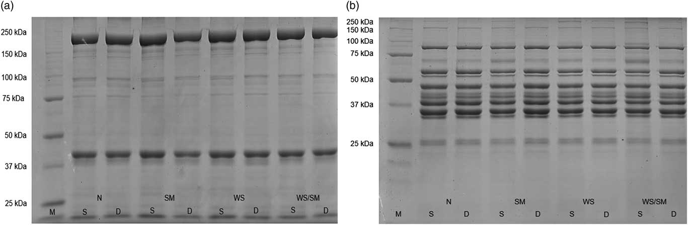
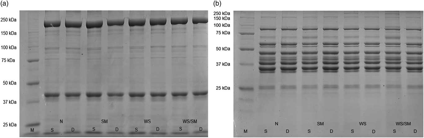
Figures 2a and b, respectively, show the electrophoretic patterns of myofibrillar and sarcoplasmic proteins for both the superficial and deep sections of N, WS, SM and WS/SM samples. Overall, no significant differences were found by comparing the deep sections of the four experimental groups. On the other hand, an increased number of high molecular-weight bands (ranging in size from 100 to 250 kDa) ascribed to both the myofibrillar and the sarcoplasmic protein fractions was found within the superficial layer of all abnormal samples.
Polyacrylamide gel (7.5%) electrophoresis pattern of myofibrillar (a) and sarcoplasmatic (b) proteins obtained from superficial (S) and deep (D) sections of chicken breast fillets belonging from different experimental groups (normal (N), spaghetti meat (SM), white striping (WS) and WS/SM). M=molecular-weight marker.
NMR relaxation measurements
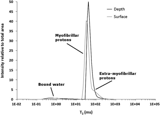
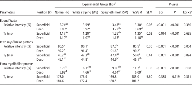
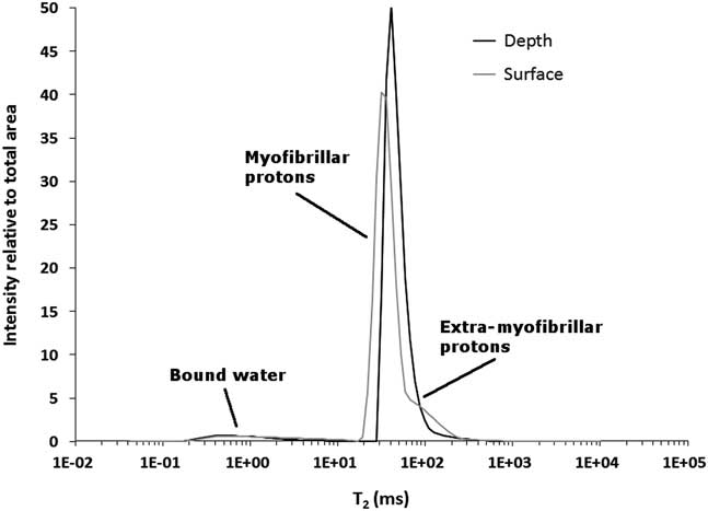
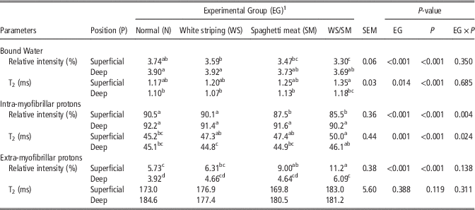
Figure 3 shows, for one of the samples pertaining to the N group, T2 spectra from a portion collected in depth and from a portion collected on the surface. Relative intensity and T2 of the three protons populations observed in the present work are reported in Table 4. Water partition throughout meat compartments was influenced both by abnormality and position. Superficial samples having SM abnormality (SM and WS/SM groups) were characterized by higher percentages of extra-myofibrillar water, at the expenses of water located in the myofibrils spaces (intra-myofibrillar). In addition, superficial WS/SM samples had a lower percentage of bound water if compared with N and WS group, whereas SM showed intermediate values. These differences mostly disappeared in depth, with the exception of extra-myofibrillar water which was higher in WS/SM group if compared with controls. The reduction of water in a single compartment was accompanied, with few exceptions, by an inversely proportional change in T2. Overall, the consequence of the different levels of distribution of water between intra- and extra-myofibrillar compartments was that, compared with N samples, WS/SM samples showed higher differences in water intensities between samples collected on the surface and in depth. In details, even if extra-myofibrillar T2 was not modified by experimental factors, WS/SM group exhibited increased intra-myofibrillar T2 in respect to N samples (50.0 v. 45.2 ms; P<0.05). Moreover, white striped samples (WS and WS/SM) showed an increase of bound water T2 when superficial and deep position are compared. WS samples also had a concomitant increase of intra-myofibrillar T2 (47.3 v. 44.8 ms; P<0.05)
T2 spectra obtained on the samples collected in depth (black solid line) and surface (gray solid line) on a breast from normal (N) group. To allow for a direct comparison, the intensities are scaled so that the total area equals 100.
Effect of breast abnormalities on relative intensity (%) and T2 relaxation time (ms) of the protons populations identified through nuclear magnetic resonance
SEM=standard error of means.
a,b,c,dMean values within each parameter considering both the experimental groups (same row) and the sampling position (same column) followed by different superscript letters significantly differ (P<0.05).
1 n=24/group.
Discussion
The remarkable increase in growth rate and breast yield achieved in the past years have recently been associated with the higher incidence of several muscle abnormalities affecting chicken pectoral muscles (Petracci et al., Reference Petracci, Mudalal, Soglia and Cavani2015; Velleman and Clark, Reference Velleman and Clark2015; Kuttappan et al., Reference Kuttappan, Shivaprasad, Shaw, Valentine, Hargis, Clark, McKee and Owens2016). The eventual alterations in muscle growth were tested by measuring fillets weight and dimensions. In general, the findings of the present study revealed that, if compared with normal, fillets affected by WS and SM abnormalities exhibited higher weights and increased thickness and length. Therefore, as previously observed for WS (Kuttappan et al., Reference Kuttappan, Hargis and Owens2012 and Reference Kuttappan, Brewer, Mauromoustakos, McKee, Emmert, Meullenet and Owens2013b; Mudalal et al., Reference Mudalal, Babini, Cavani and Petracci2014) and wooden breast (Mudalal et al., Reference Mudalal, Lorenzi, Soglia, Cavani and Petracci2015; Tasoniero et al., Reference Tasoniero, Cullere, Cecchinato, Puolanne and Dalle Zotte2016; Soglia et al., Reference Soglia, Laghi, Canonico, Cavani and Petracci2016a), birds displaying higher breast-size seem to be more prone to develop the SM abnormality.
In this study, the effect of the sampling position (superficial v. deep) on the main quality parameters of the chicken breast meat have been considered. With regard to proximate composition, the findings revealed that SM occurrence is associated with a remarkable decrease in protein content coupled with a relevant increase in moisture level, which were previously observed also for the wooden breast condition (Soglia et al., Reference Soglia, Laghi, Canonico, Cavani and Petracci2016a). On the other hand, in agreement with previous studies (Kuttappan et al., Reference Kuttappan, Hargis and Owens2012; Soglia et al., Reference Soglia, Laghi, Canonico, Cavani and Petracci2016a) fat content was affected only by the concurrent occurrence of WS. Notwithstanding, the occurrence of these muscle abnormalities seems to mainly affect the superficial portion of the P. major muscles rather than the deep one. In addition, as collagen was barely affected by the occurrence of WS and SM abnormalities, an increase in its content might be associated with the wooden breast condition (Tasoniero et al., Reference Tasoniero, Cullere, Cecchinato, Puolanne and Dalle Zotte2016; Soglia et al., Reference Soglia, Laghi, Canonico, Cavani and Petracci2016a).
The different modern myopathies described so far share some microscopic aspects. We described a range of microscopic lesions such as internalization of nuclei, loss of cross striation, vacuolar and hyaline degeneration, necrosis and lysis of fibers, inflammatory cells infiltration, degenerating and regenerating fibers of variable size, lipidosis, proliferating connective tissue and fibrosis. In this regard, Kuttappan et al. (Reference Kuttappan, Shivaprasad, Shaw, Valentine, Hargis, Clark, McKee and Owens2016) reported that hereditary muscular dystrophies in the chicken, WS and wooden breast closely share some gross and/or histological lesions. Whether WS and wooden breast are different expressions of the same myopathy or not is yet to be demonstrated, as stated by Velleman and Clark (Reference Velleman and Clark2015), but the histological lesions described are very similar. In a recent publication, Radaelli et al. (Reference Radaelli, Piccirillo, Birolo, Bertotto, Gratta, Ballarin, Vascellari, Xiccato and Trocino2017) in chicken breast muscles affected by WS or wooden breast at 46 days of age did not observe any difference regarding the histological characteristics.
The histopathological features associated with the occurrence of SM abnormality are in agreement with those previously reported by Bilgili (Reference Bilgili2015). In addition, the same author states that SM histologically shows morphological changes similar to those reported for other myopathies (e.g. WS, woody breast): extensive fiber degeneration and regeneration, hyalinization, poor fiber uniformity, increased fat and connective tissue deposition. A particular characteristic observed in the SM sections were the progressive rarefaction of the endo- and peri-mysium connective tissue. It is likely that the architecture and structural integrity is affected by the immaturity of the newly deposited collagen as previously described by Bilgili (Reference Bilgili2015). With regard to WS meat, the microscopic lesions include vacuolar and hyaline degeneration, lysis, mild mineralization, fibers scattered in an abundant collagen-rich connective tissue and exhibited a high variability in size (degeneration and regenerating fibers) and interstitial inflammation along with fibrosis. However, the most severe histological lesions were observed within the samples concurrently affected by both WS and SM abnormality with a high connection existing between the occurrence of histopathological lesions, proximate composition and an increased proportion of both intra- and extra-myofibrillar water. Abundant adipose tissue deposition infiltrating the endo- and peri-mysial spaces were observed in agreement with Kuttappan et al. (Reference Kuttappan, Brewer, Apple, Waldroup and Owens2013a) and Radaelli et al. (Reference Radaelli, Piccirillo, Birolo, Bertotto, Gratta, Ballarin, Vascellari, Xiccato and Trocino2017). These results are also consistent with the findings of proximate composition. Indeed, the white striped samples exhibited the highest lipid content.
With regard to ultimate pH, if compared with the normal, all abnormal fillets (apart from the type of abnormality) exhibited significantly higher values within the superficial portion of the P. major muscle. Thus, it seems that ultimate pH is altered to the same extent by the occurrence of both SM and WS conditions. Besides, previous studies evidenced that ultimate pH was remarkably altered following the occurrence of wooden breast (Mudalal et al., Reference Mudalal, Lorenzi, Soglia, Cavani and Petracci2015).
In a recent study, a moderate positive genetic correlation was found to exist between pHu and WS condition (Alnahhas et al., Reference Alnahhas, Le Bihan-Duval, Baéza, Chabault, Chartrin, Bordeau, Cailleau-Audouin, Méteau and Berri2016). Hence, a link between the occurrence of the WS abnormality and the energetic status of the P. major muscle during life was hypothesized (Alnahhas et al., Reference Alnahhas, Le Bihan-Duval, Baéza, Chabault, Chartrin, Bordeau, Cailleau-Audouin, Méteau and Berri2016) and confirmed by Zambonelli et al. (Reference Zambonelli, Zappaterra, Soglia, Petracci, Sirri, Cavani and Davoli2016). Indeed, the reduced muscular vascularization and glycogen reserves observed in modern heavy broilers may impair the energy supply to muscle fibers (Alnahhas et al., Reference Alnahhas, Le Bihan-Duval, Baéza, Chabault, Chartrin, Bordeau, Cailleau-Audouin, Méteau and Berri2016). In addition, selection for increased pectoral muscle pH can result in reduced capillary density leading to an inadequate oxygen supply and metabolic waste products displacement (Alnahhas et al., Reference Alnahhas, Berri, Chabault, Chartrin, Boulay, Bourin and Le Bihan-Duval2015). An overall reduction in glycogen content within the wooden breast fillets was recently associated with a decreased amount of some glycolytic intermediates (i.e. glucose 6-phosphate and fructose 6-phosphate) (Abasht et al., Reference Abasht, Mutryn, Michalek and Lee2016). On the contrary, although an increased pHu was found in wooden samples as a result of in vivo reduction in muscular glycogen reserves, an intensified glycolytic activity was evidenced (Zambonelli et al., Reference Zambonelli, Zappaterra, Soglia, Petracci, Sirri, Cavani and Davoli2016). Thus, the findings of these studies suggested that changes in glycolysis may arise from a different glucose utilization rather than from its availability (Abasht et al., Reference Abasht, Mutryn, Michalek and Lee2016; Zambonelli et al., Reference Zambonelli, Zappaterra, Soglia, Petracci, Sirri, Cavani and Davoli2016).
The occurrence of muscle abnormalities did not exert any relevant effect on meat color with the only exception of the yellowness which displayed significantly higher values within the fillets affected by muscle abnormalities. In detail, in agreement with our previous study (Petracci et al., Reference Petracci, Laghi, Rimini, Rocculi, Capozzi and Cavani2013), WS samples exhibited the highest values, as a consequence of an increased deposition of intramuscular fat (Bianchi et al., Reference Bianchi, Petracci, Sirri, Folegatti, Franchini and Meluzzi2007).
As for the functional properties, if compared with their abnormal counterpart, normal fillets exhibited a higher protein solubility measured within the superficial portion. Overall, these findings are both consistent with the protein content observed in the present study and in agreement with previous studies performed on WS (Mudalal et al., Reference Mudalal, Babini, Cavani and Petracci2014; Bowker and Zhuang, Reference Bowker and Zhuang2016). Indeed, even if no relevant effect was observed within the dorsal portion, an overall reduction in sarcoplasmic protein solubility was found to affect the ventral surface of the cranial end in moderate and severe WS fillets (Bowker and Zhuang, Reference Bowker and Zhuang2016). On the other hand, myobrillar protein solubility was not affected in both the superficial and the deep sampling position. Protein solubility is normally used as an index to evaluate protein functionality, denaturation and their impact on water holding capacity of meat (Mudalal et al., Reference Mudalal, Babini, Cavani and Petracci2014). According to the findings of our previous study, the occurrence of muscle abnormalities (either alone or combined within the same P. major muscle) significantly affect the water holding capacity of meat (Mudalal et al., Reference Mudalal, Lorenzi, Soglia, Cavani and Petracci2015). As the T2 relaxation times were successfully used to study the interaction existing between water molecules and muscle structure (Bertram and Andersen, Reference Bertram and Andersen2006), this study aimed at evaluating the impact of WS and SM abnormalities onto water partition through NMR. According to our findings, the superficial section of all the samples affected by muscle abnormalities exhibited a higher proportion of extra-myofibrillar water, with the SM samples displaying the highest values. In addition, the lower proportion of bound and intra-myofibrillar water coupled with the increased intra-myofibrillar T2 relaxation time observed within the abnormal samples revealed a greater mobility of water within the meat structure. Similarly, an increased proportion of extra-myofibrillar water was previously found in WS and wooden breast meat (Soglia et al., Reference Soglia, Petracci and Ertbjerg2016c). This might be the result of the degenerative processes taking place within the muscle fibers and leading to an increased moisture within the extra-cellular spaces and, consequently, to an overall reduction in water holding capacity of meat (Mudalal et al., Reference Mudalal, Lorenzi, Soglia, Cavani and Petracci2015), in agreement with our histological observations. Indeed, if compared with the deep portion, several degenerating and necrotic fibers were found within the superficial layer of the affected fillets. Similarly, a clear gradual decrease in the histopathological lesions was previously found moving toward the deep portion (about 1 cm deep) of the wooden breast muscles (Soglia et al., Reference Soglia, Laghi, Canonico, Cavani and Petracci2016a; Tasoniero et al., Reference Tasoniero, Cullere, Cecchinato, Puolanne and Dalle Zotte2016).
As for carbonylation level as an index for protein oxidation, no clear trends were observed. In agreement with a previous study performed by Soglia et al. (Reference Soglia, Petracci and Ertbjerg2016c), protein carbonylation level was mainly affected by the occurrence of wooden breast rather than WS abnormality. However, the amount of carbonyls measured within the present study is slightly lower than those reported in our previous works (Soglia et al., Reference Soglia, Mudalal, Babini, Di Nunzio, Mazzoni, Sirri and Petracci2016b and 2016Reference Soglia, Petracci and Ertbjergc). If compared with their normal counterpart, the superficial section of the fillets affected by muscle abnormalities exhibited a higher MFI. Thus, it seems reasonable to associate the occurrence of muscle abnormalities with a more intense proteolytic degradation of muscle tissue. Accordingly, the electrophoretic separation of the myofibrillar protein fraction belonging from the superficial section of the abnormal muscles evidenced an increased number of high molecular-weight bands. This is likely due to the proteolytic processes affecting the structural proteins (such as titin, nebulin and vinculin) composing the sarcomere. Similarly, an increased number of high molecular-weight bands was found within the water soluble fraction ascribed to sarcoplasmic proteins. Although unexpected, this result might be explained considering that, as a consequence of proteolysis, the fragments resulting from the high molecular-weight myofibrillar proteins can exhibit a different solubility and thus being extracted together with the water soluble fraction. Clearly these bands cannot be ascribed to the protein fragments which typically result from the proteolytic processes occurring during the postmortem period, but might be attributed to the degenerative processes associated with the occurrence of muscle abnormalities. Indeed, in a previous study performed by Lee et al. (Reference Lee, Santè-Lhoutellier, Vigorux, Briand and Briand2008) proteolysis generated fragments exhibiting a molecular weight ranging from 30 to 110 kDa.
Conclusion
This study reveals that both WS and SM abnormalities have adverse effects on quality traits and histological features of broiler meat. From histological comparison, WS abnormality shows a greater adipocyte deposition and fat infiltration, otherwise SM defect displays a progressive rarefaction of the endo- and pery-misial connective tissue, which likely is involved in an altered structural integrity of the P. major surface. Overall, proximate composition of abnormal samples was found to be significantly modified according the type of abnormality, especially within the superficial layer of the fillets. In fact, WS fillets have a higher lipid content, whereas in SM samples was observed a remarkable decrease in total protein content coupled with an increased moisture level. From NMR investigation, SM fillets show a relevant increase of extra-myofibrillar water portion at the expenses of the intra-myofibrillar one and, as a consequence, display a reduced water holding capacity even in respect to WS fillets. Moreover, the occurrence of muscle abnormalities are associated with a more intense proteolytic degradation of muscle tissue which leads to the formation of high molecular-weight protein fragments.
Overall results reveal that WS and SM myopathies primarily affect the superficial layer of P. major muscle and just mildly the deep section. Furthermore, the occurrence of SM abnormality generally leads to a more pronounced modification of the meat quality traits rather than the mere presence of WS myopathy.
Acknowledgment
The authors thank the Gesco Consorzio Cooperativo (Cesena, Italy), for supplying samples for this research.
Supplementary material
To view supplementary material for this article, please visit https://doi.org/10.1017/S1751731117001069