Abnormalities of the heart and major head and neck vessels are seen in almost 50% of women with Turner syndrome.Reference Dawson-Falk, Wright, Bakker, Pitlick, Wilson and Rosenfeld1, Reference Ho, Bakalov and Cooley2 The abnormalities include bicuspid aortic valve (10–30%),Reference Dawson-Falk, Wright, Bakker, Pitlick, Wilson and Rosenfeld1, Reference Mazzanti and Cacciari3–Reference Volkl, Degenhardt, Koch, Simm, Dorr and Singer7 coarctation of the aorta (7–18%)Reference Dawson-Falk, Wright, Bakker, Pitlick, Wilson and Rosenfeld1, Reference Gotzsche, Krag-Olsen, Nielsen, Sorensen and Kristensen6 and elongated transverse aortic arch (49%)Reference Ho, Bakalov and Cooley2 while abnormalities such as aberrant right subclavian artery are less frequent. Interestingly, the vascular defects observed in Turner syndrome involve not only the aorta but also the carotid and brachial arteries.Reference Ostberg, Donald, Halcox, Storry, McCarthy and Conway8, Reference Baguet, Douchin, Pierre, Rossignol, Bost and Mallion9 The vascular abnormalities often coexist and interact to increase the overall burden of risk to the patient. The abnormalities thereby play a key role to the increased early-life morbidity and the more than fourfold increased risk of premature death in adulthood, with important contributions from dissection of a gradually dilating ascending aortopathy.Reference Bondy10–Reference Schoemaker, Swerdlow, Higgins, Wright and Jacobs14 Previously the risk of aortic dilatation or dissection has been related to the presence of a bicuspid aortic valve and aortic coarctation, irrespective of previous repair. No similar association has been established with the elongated transverse aortic arch, the most frequently encountered abnormality in Turner syndrome, or with dilation of the aortic branch arteries. In contrast, the presence of the classic 45,X monosomy karyotype is proposed to be of particular importance not only for congenital cardiovascular abnormalities but also for the development of aortopathy.Reference Ho, Bakalov and Cooley2, Reference Sachdev, Matura and Sidenko4, Reference Prandstraller, Mazzanti and Giardini15
In this study, we set out to define the prevalence of congenital arterial abnormalities and dilatation of the entire thoracic aorta and the proximal segments of the head and neck branch arteries in Turner syndrome. In addition, we aimed to further elucidate the associations between these abnormalities and karyotype.
Material and Methods
Patients
As part of a prospective observational study of cardiovascular morbidity in Turner syndrome, technically successful magnetic resonance imaging scans and echocardiograms were performed in 99 of 102 non-selected women with Turner syndrome. These patients were recruited through The National Society of Turner Syndrome Contact Groups in Denmark and an endocrine outpatient clinic. The mean age was 37 plus or minus 10 years, ranging from 18 to 62 years. Inclusion required confirmation of either 45,X, 57 of 99 patients (57.6%), or other karyotypes specific for Turner syndrome, mosaic or isochromosomal as seen in 42 of 99 patients (42.4%). Inclusion required successful examination with both echocardiography and magnetic resonance imaging in the setting of the current study. Exclusion criteria included malignancy, significant liver disease, contraindications to magnetic resonance imaging scans, and the presence of mechanical aortic valve prosthesis. Eight patients (8%) had undergone either surgical or catheter-based treatment of aortic coarctation. One patient was retrospectively excluded with a previously undiagnosed chronic type A dissection detected by magnetic resonance imaging. This patient had no history of heart disease, and had never undergone echocardiography or magnetic resonance imaging; her aortic valve was bicuspid, the branch arteries originated normally, and the extent of the dissection did not allow for diagnosis of coarctation of the aorta. A total of 33 healthy women of a mean age of 42 plus or minus 10 years, ranging from 25 to 63 years, were recruited as a control group by advertisement. All were asked for details of their medical history and had a brief cardiovascular examination to exclude symptomatic cardiovascular disease, including hypertension.
The Aarhus County Ethical Scientific Committee approved the protocol (no. 20010248). All participants provided written and oral consent.
Magnetic resonance imaging
Non-contrast aortic magnetic resonance imaging scans were performed on a 1.5 tesla whole body scanner, ACS-NT, Philips Medical Systems, Best, The Netherlands; maximum gradient performance 30 millitesla/metre amplitude; slew rate 150 tesla/metre/second using a five-element cardiac coil. Initial scouts were performed. From these scout images a three-dimensional data stack was acquired, measuring 27 centimetres (anterior-posterior) × 15 centimetres (foot-head) × 36 centimetres (left-right) and covering the entire thoracic aorta. A nearly isotropic three-dimensional steady-state free precession navigator with an electrocardiogram-triggered sequence acquired in diastole (250 milliseconds acquisition window) was used.Reference Sorensen, Korperich and Greil16 The voxel size was 1.4 millimetres × 1.4 millimetres × 1.5 millimetres.
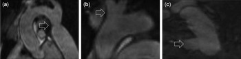
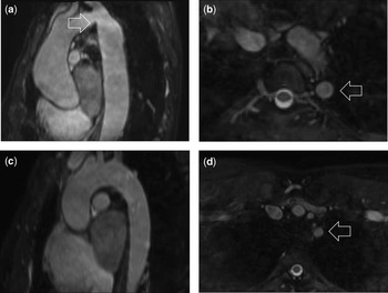
The complete data set was systematically reviewed. The encountered morphologic abnormalities were classified as aortic coarctation: concentric narrowing of the aortic lumen with a posterior shelf-like structure; aberrant right subclavian artery: origin of the right subclavian artery distally to the left subclavian artery; and bovine aortic arch: common aortic origin of the innominate and left subclavian arteries (Figs 1 and 2).Reference Layton, Kallmes, Cloft, Lindell and Cox17 A review for the presence of an elongated transverse aortic arch was also performed; this was carried out according to the previously introduced definition: an origin of the left subclavian artery from the aortic arch posterior to the trachea, with kinking or inward indentation of the lesser curvature at the aortic isthmus.Reference Ho, Bakalov and Cooley2 In an attempt to further delineate this aortic arch phenotype, the distances from the origin of the left subclavian artery to both the innominate and left common carotid arteries were measured, from anterior margin to anterior margin of the branch arteries.
Aortic coarctation (a), bovine aortic arch (b), and aberrant right subclavian artery (c) as seen by magnetic resonance imaging in Turner syndrome.

The normal and elongated transverse aortic arch as seen by magnetic resonance imaging in Turner syndrome. The abnormality is defined by the coexistence of kinking of the inferior aortic contour at the aortic isthmus (a) and a left subclavian artery origin from a position posterior to the trachea in the horizontal plane (b). The normal transverse aortic arch (c) and normal origin of the left subclavian artery (d) are illustrated for comparison; kinking is absent and the left subclavian artery departs anterior to the posterior aspect of the trachea.

Measurements were obtained with dedicated software, Systematic Software Engineering, Aarhus, Denmark, that allowed real-time, all-plane reconstruction of the full three-dimensional data set. Measurement planes were interactively positioned perpendicularly to the aorta at the eight positions: sinotubular junction (pos1), ascending aorta at the level of the right pulmonary artery (pos2), ascending aorta proximal to the innominate artery (pos3), aortic arch midway between the innominate and the left common carotid arteries (pos4), aortic arch just proximal to the left subclavian artery (pos5), the isthmus distally to the left subclavian artery (pos6), descending aorta at level between the left pulmonary artery and the top of the left atrium (pos7), and descending aorta at the most caudal border of the left atrium (pos8). The diameters of the innominate, left common carotid and left subclavian arteries were measured 5 millimetres distal to their origin. In cases with a bovine aortic arch, neither the innominate artery nor the left common carotid artery was measured. Bland–Altman plots evaluated the reliability of measurements. The average inter-observer and intra-observer variability was small, varying from −0.28 to 0.66 millimetres and from −0.12 to 0.18 millimetres, respectively, depending on the position. There was no systematic variation, neither at specific levels nor with aortic dimensions. Normal arterial dimensions were defined from the data acquired in the healthy controls. Dilatation was defined as arterial diameters 1.96 standard deviations above the normal mean. Aneurism was defined arterial diameters larger than 1.5 times the normal mean.
Echocardiography
Echocardiography was performed by a single observer on a GE Vivid Seven, GE Healthcare, Horten, Norway, with a 2.5 megahertz transducer using second harmonic imaging. Aortic valve morphology and function were noted. Stenosis was diagnosed from Doppler tracings as mild with peak gradient 16–36 millimetres of mercury, moderate with peak gradient 37–50 millimetres of mercury, and severe with peak gradient above 50 millimetres of mercury.Reference Oh, Taliercio and Holmes18 Aortic regurgitation was defined from colour Doppler and from the width of the vena contracta in the parasternal long axis view classified as mild: above 0.3 centimetres, moderate: 0.3–0.6 centimetres, and severe:above 0.6 centimetres.Reference Bonow, Carabello and Kanu19
Statistics
Statistical computations were performed using SPSS 15.0. Normal distribution of data was tested by the Shapiro–Wilk test. Data are expressed as means with plus or minus standard deviation and compared by independent t-test following testing for equal variances by Levene’s test. Associations were tested by Pearson’s coefficient of correlation or by binomial distributions, and expressed as percentages and relative risks expressed with 95% confidence intervals. They were tested for significance by χ2 tests, if the expected distribution was above five in all cells, or Fisher’s exact test, if the expected distribution was less than six in one or more cells; p < 0.05 was considered statistically significant.
Results
Aortic valve morphology and function
Aortic valve morphology was normal in all controls. Three controls (9.1%) had trivial aortic regurgitation; none had aortic stenosis.
Aortic valve morphology could not be precisely determined in 4 of 99 (4%) patients with Turner syndrome. Bicuspid aortic valve was seen in 26 of 95 (27.4%) patients, Turner syndrome versus controls; p = 0.001. Aortic valve function was abnormal in 28 of 99 (28.2%) patients, Turner syndrome versus controls; p = 0.001. Regurgitation was present in 18 of 99 patients (18.1%) and classified as mild in 13 (72.2%) and moderate in five (27.8%). Stenosis was observed in 16 of 99 patients (16.2%) and rated mild in 11, moderate in four, and severe in one. Moderate-to-severe stenosis or regurgitation was only present in patients with bicuspid aortic valves. Stenosis and regurgitation coexisted in six patients. The association between valve morphology and function is shown in Table 1.
The prevalence of bicuspid and tricuspid aortic valve morphology in Turner syndrome (N = 95) and relative risks of aortic regurgitation (n = 18) and stenosis (n = 16).

*Any degree of stenosis and regurgitation
**χ2 test
Arterial abnormalities
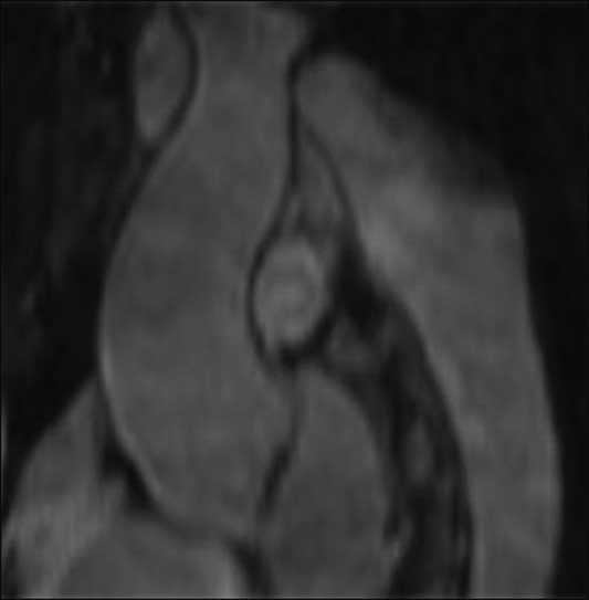
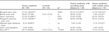
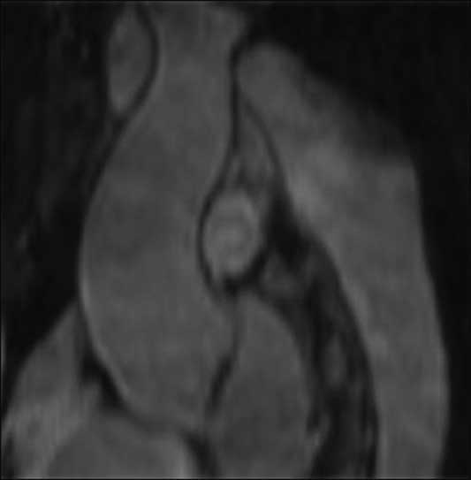
A bovine aortic arch was the only abnormality seen in the controls (4 of 33, 12.1%). Two patients with Turner syndrome had a structural aortic abnormality that did not readily match the four predefined groups. Both had severe narrowing and shortening of the aortic arch (retrospectively classified as “aortic arch hypoplasia”); one of these patients also had a bicuspid aortic valve with dilation of the ascending aorta (Fig 3). The majority of the patients (69 of 99, 69.7%) had at least one structural abnormality. The proximal branch arteries were also more commonly dilated, relative risk: 7.7 (3.2; 29.1), Turner syndrome versus controls; p < 0.001. Not only bicuspid aortic valve but also elongated transverse aortic arch (p < 0.001) and aortic coarctation (p = 0.03) were overrepresented in Turner syndrome, whereas the prevalence of an aberrant right subclavian artery, aortic arch hypoplasia, and bovine aortic was statistically similar (Table 2).
Aortic arch hypoplasia in a 36-year old patient with Turner syndrome with bicuspid aortic valve and ascending aortic dilatation.
The prevalence of arterial abnormalities in Turner syndrome and healthy female controls.

*χ2 test or Fishers exact test comparing the specific prevalence in all patients with Turner syndrome and controls
**The total number refers to the number of magnetic resonance imaging scans where analysis for the abnormality in question could be technically successfully conducted
***Overall equals one or more of the detected arterial abnormalities
ns, non-significant
Aorta and the major head and neck arteries
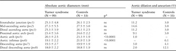
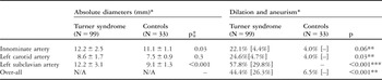
Mean aortic dimensions were similar at all points, except at pos5–6, the classical coarctation site, where aortic diameters were smaller in Turner syndrome (Table 3). Dilation or aneurism of the ascending aorta (pos1–3) was seen in 20 of 99 (20.2%) patients and in one control (3.0%). This conferred with a 6.7 fold increased risk of aortic dilatation or aneurism in Turner syndrome; p = 0.01; such predilection to ectatic aortopathy was also seen by the wide range of diameters in Turner syndrome. Aneurysm was only encountered in Turner syndrome (2 of 99, 2.0%), and the innominate and left subclavian arteries were also significantly larger in these women (Table 4).
Absolute aortic dimensions and the prevalence of aortic dilation and aneurism in Turner syndrome and healthy female controls.

*Independents t-test, equal variances assumed
ns, non-significant
Dimensions of the innominate, the left carotid and left subclavian arteries measured just after their aortic origin in Turner syndrome and in healthy female controls.

[%]The percentage of all patients with Turner syndrome or controls with aneurism of the artery in question
*Diameters and dilation quantified in subjects with normal departure of the arteries in question
**Fishers exact test (dilation and aneurism compared as one)
***χ2 test (dilation and aneurism compared as one)
‡Independents t-test, equal variances assumed
Delineation of the vascular phenotype
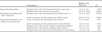
In Turner syndrome, the presence of a bicuspid aortic valve was associated with elongated transverse aortic arch and aortic coarctation (Table 5), whereas no statistically significant associations were found among elongated transverse aortic arch, aberrant right subclavian artery, bicuspid aortic valve, bovine aortic arch, and aortic coarctation.
Statistically significant relatives risks of the coexistence of congenital arterial abnormalities and the relation of these to dilation of the major branch arteries and the aorta in Turner syndrome.

*χ2 test or Fishers exact test
Dilation of the ascending aorta was more frequent in patients with bicuspid aortic valves; aneurisms were restricted to the same bicuspid subgroup. Dilation of the descending aorta (pos8 and pos7) was only encountered in patients with aortic coarctation (p = 0.002). Neither the presence of aberrant right subclavian artery nor bovine aortic arch was associated with aortic dilation. Elongated transverse aortic arch was associated with a borderline significantly higher prevalence of dilation at pos7 (p = 0.06) that corresponded to a general increase of aortic diameters, elongated transverse aortic arch versus non-elongated transverse aortic arch at pos7: 19.9 plus or minus 3.0 versus 18.8 plus or minus 2.5 millimetres; p = 0.03. The distances between the left subclavian artery and both the left common carotid and the innominate artery were longer in Turner syndrome when compared with healthy female controls; this was also the case when comparing patients with Turner syndrome designated as having an elongated transverse aortic arch to those without this morphological arch abnormality (Table 6). In patients with Turner syndrome, but not controls, the distance from the innominate to the left subclavian artery correlated with ascending and descending aortic diameter, Pearson’s coefficient of correlation: 0.3–0.4 (all p < 0.005) in all ascending and the two most distal descending aortic positions. No such correlation was seen in the aortic arch or at the aortic isthmus (pos4–6). The innominate to left subclavian arch distance also predicted branch artery diameter in patients with Turner syndrome, Pearson’s coefficient of correlation: 0.3–0.5, all p < 0.006. Age did not predict the length of the aortic arch, but it was longer in the presence of bicuspid aortic valve, aortic coarctation and 45,X (all p < 0.003).
Aortic arch length in Turner syndrome measured as the distance from the origin of the left subclavian artery to both the innominate and left subclavian arteries.

ETA, elongated transverse aortic arch
*Two-tailed p-test
**Independent t-test, equal variances assumed comparing Turner syndrome patients with elongated transverse aortic arch with patients with no such morphological abnormality
A positive correlation between aortic dimensions and the diameters of the innominate artery, in all positions, the left common carotid artery, in most positions, and the left subclavian artery, in the descending aorta (Table 7) was found irrespective of the coexistence of bicuspid aortic valve, elongated transverse aortic arch, or aortic coarctation. Coarctation of the aortic arch was associated with dilation of the innominate and the left common carotid arteries, while a bicuspid aortic valve was more frequent in patients with dilation of the left common carotid and left subclavian arteries. Patients with aortic dilation (at one or more positions) more often had dilation or aneurism of the innominate artery, relative risk 3.4 (1.4; 7.2); p = 0.009, but no such correlation was present with the other branch arteries. Dilation of the left subclavian artery was more often observed in patients with an elongated transverse aortic arch. No similar associations were present in the control group.
Pearsons bivariate coefficient of correlation (r) between aortic dimensions and dimensions of innominate, left carotid and left subclavian arteries in Turner syndrome.

ns, non-significant
*Independent t-test, equal variances assumed
Karyotype
Elongated transverse aortic arch and aortic coarctation were predominantly found in patients with 45,X when compared with other karyotypes; elongated transverse aortic arch: relative risk 1.8 (1.2; 3.1), p = 0.01; aortic coarctation: relative risk 3.9 (1.1; 24.6), p = 0.04). A similar association was seen between 45,X and dilation of the ascending aorta; the relative risk at pos1 was 6.6 (1.3; 119.0), p = 0.04 and at pos2 it was 4.1 (1.1; 25.2). Karyotype and bicuspid aortic valve did not associate at a statistically significant level, 45,X versus other karyotypes: 18/54 versus 8/41; p = 0.2. In the major branch arteries, only dilation of the left subclavian artery was associated with the 45,X karyotype, relative risk 2.2 (1.3; 3.6); p < 0.001.
Discussion
The study demonstrates that almost 70% of adult patients with Turner syndrome have at least one congenital abnormality of the thoracic aorta and the proximal parts of the head and neck vessels. Compared with healthy females, this corresponds to a nearly eightfold higher prevalence of morphological abnormalities in the supra-diaphragmatic aorta, which is significantly higher than previously reported in patients with asymptomatic Turner syndrome recruited by similar means but examined with a different magnetic resonance technique.Reference Ho, Bakalov and Cooley2 The data confirms that serious cardiovascular abnormalities appear irrespective of karyotype, although 45,X associates predominantly with important phenotypic features such as elongated transverse aortic arch, aortic coarctation and dilatation of the ascending aorta.Reference Prandstraller, Mazzanti and Giardini15
The high prevalence of aortic coarctation and bicuspid aortic valve seen in this study corroborates earlier reports and reiterates the finding that women with Turner syndrome generally present with cardiovascular risk profiles known to increase the risk for not only aortic dilation but also dissection.Reference Dawson-Falk, Wright, Bakker, Pitlick, Wilson and Rosenfeld1, Reference Ho, Bakalov and Cooley2, Reference Sachdev, Matura and Sidenko4–Reference Volkl, Degenhardt, Koch, Simm, Dorr and Singer7, Reference Bonamico, Pasquino and Mariani20 Our study, accounting for dimensions along the entire thoracic aorta, also indicates that elongated transverse aortic arch should be added to the growing list of risk factors for aortic dilation in Turner syndrome. The predictive capacity of elongated transverse arch to aortic size is a novel finding. Whether it can predict aortic dissection in afflicted individuals will of course need validation in other populations. The correlation of this arch abnormality with aortic caliber together with the confirmation of a true association with longer transverse aortic arch indicates that this arch phenotype is a true separate and morphologically classifiable entity. This abnormality may represent the morphologically classifiable end of a spectrum of aortic arch elongations in Turner syndrome. Interestingly, such elongation did appear to be specific component of the thoracic aortopathy, as longer arches associated with larger aortic size as well as some of the other adverse features encountered in the thoracic aorta in Turner syndrome. It is beyond the scope of this study to determine a cause or effect relationship of this widespread elongation, but interestingly there was no correlation between age and arch length. Our findings underline the importance of imaging not only the ascending but also the descending aorta; considerable pathology is found throughout the thoracic aorta that could contribute to the increased risk of both ascending and descending aortic dissection as reported in case studies.Reference Carlson and Silberbach11
The relatively common finding of an aberrant right subclavian artery reinforced the results of previous studies in Turner syndrome,Reference Ho, Bakalov and Cooley2 whereas aortic arch hypoplasia, defined by severe shortening and narrowing of the aortic arch, is a novel finding. Although the relatively small population size precludes any firm statement to conclude that a hypoplastic arch associates with ascending aortic dilation, it is noted that both aortic arch hypoplasia cases had significant ascending aortopathy. In contrast to these abnormalities, the bovine aortic arch was a frequent finding in both healthy individualsReference Layton, Kallmes, Cloft, Lindell and Cox17 and Turner syndrome, but it did not associate with aortic size. A bicuspid aortic valve, aortic coarctation, and the elongated transverse aortic arch were more commonly, but not exclusively, seen in patients with the 45,X karyotype. Our data clearly indicates that the dilated aorta and other presumed markers of an increased risk of aortic dissection occurred with some, but far from total concordance in Turner syndrome.
The majority of aortic dissections in Turner syndrome occur in patients with bicuspid aortic valve, coarctation of the aorta, pre-existing aortic dilation, or the 45,X karyotype. However, at least 10% of all dissections occur in patients with no apparent risk factors and apparently normal-sized ascending aortas.Reference Carlson and Silberbach11 This suggests that the currently used risk markers fail to provide optimal identification of women with Turner syndrome at risk of aortic dissection. It is therefore of key importance to consider that the major body of studies on Turner syndrome have the limitation of investigating the risk of aortic dissection by the use of aortic dimensions as the primary surrogate marker of increased risk in a population where prospective data and clinical follow-up is limited. In addition, the lack of accurate definitions of “normal” aortic dimensions in the syndrome remains a limitation when attempting to put our observations into clinical perspective. That is, milder forms of aortopathy could easily be overlooked if we uncritically compare aortic measurements in Turner syndrome to those acquired in taller healthy women who are unaffected by hypertension, metabolic syndrome, or other well-known risk factors reported to be associated with the syndrome.Reference Hjerrild, Mortensen and Gravholt21 Unfortunately, adjustment for body surface area or the use of an aortic ratio in order to avoid the issue of normality in Turner syndrome has not been successful in this cohort.Reference Hjerrild, Mortensen and Sørensen22 With our current knowledge, we should therefore view aortic size as an additional surrogate marker of the risk of future dissection only on a backdrop of congenital malformations and hypertension. Prospective data is needed to determine the impact of relative aortic size on the individual risk of aortic dissection in Turner syndrome.
Dilation of the proximal parts of major branch arteries was more prevalent in Turner syndrome. This supports the hypothesis that vascular involvement is widespread and that dilation of these vessels potentially serves as an additional surrogate marker for an increased risk of cardiovascular complications. Despite a significant correlation between the absolute dimensions of the aorta and the branch arteries, dilation or aneurism formation did not uniformly coexist. Rather a state of dilatory vasculopathy was more commonly present in the conduit arteries, but our findings suggest a common underlying pathology since both aortic and branch artery diameters associated with bicuspid aortic valve morphology, elongated transverse aortic arch, aortic coarctation and 45,X. The discrepancy between the presence of aortic vasculopathy and dilation of the branch arteries reflects the important issue of defining normality in Turner syndrome since, as noted earlier, aortopathy may be more widespread than suggested by the current definitions of a dilatory aortopathy. In previous studies of the branch arteries in Turner syndrome, the arterial dimensions were determined more peripherally and by ultrasound, which may explain the conflicting data available to date.Reference Ostberg, Donald, Halcox, Storry, McCarthy and Conway8, Reference Baguet, Douchin, Pierre, Rossignol, Bost and Mallion9 Interestingly, the widespread branch artery dilation not only raises the possibility of a more universal vasculopathy but also suggests that these arteries could be susceptible to dissections with entry in the left common carotid artery, as previously reported.Reference Fuentes, Silveira and Papamitsakis23 The clinical impact on cardiovascular morbidity as well as the potential for dissection and propagation into the aorta remain to be investigated.
Limitations
Selection bias could play a potential role in studies of any Turner syndrome population, because of the well-known delay in diagnosis or even widespread non-diagnosis,Reference Stochholm, Juul, Juel, Naeraa and Gravholt24 making it nearly impossible to select a random sample in Turner syndrome. In this study, patients were recruited through the national patient organisation or the hospital’s endocrine clinic. This could skew patient inclusion towards those more severely affected by the stigma of the syndrome. However, supportive of the absence of significant selection into this specific study of more adversely affected Turner syndrome phenotypes, the prevalence of the congenital arterial abnormalities (bicuspid aortic valve, elongated transverse aortic arch, and aortic coarctation) and coarctation repair was comparable to previous studies (coarctation repair in 8% versus previously 5%).Reference Ho, Bakalov and Cooley2 Furthermore, patient recruitment from a non-cardiology ward minimized the risk of skewing the cohort towards an unrepresentative and more severe cardiovascular phenotype than that of the general adult Turner syndrome population.
Conclusion
This magnetic resonance imaging study shows that morphologic abnormalities in aorta are more common than previously reported in women with Turner syndrome. It further documents that the multiple cardiovascular defects do not uniformly relate to specific karyotypes. Cardiovascular follow-up is therefore of key importance in all subgroups. This standpoint is supported by the fact that more than 10% of aortic dissections occur in patients with Turner syndrome considered at low risk of the aortic event when using the current indices of risk. However, patients with Turner syndrome with bicuspid aortic valve and aortic coarctation clearly comprise a subpopulation with a particularly high risk of developing aortic dilation or dissection, and require particular clinical attention.
Acknowledgements
The authors thank Bente Mortensen, Lone Svendsen, Merete Møller, Joan Hansen, Karen Mathiasen, and Inga Bisgaard for their expert technical help and Line Gether for valuable secretarial assistance. The study was supported by unrestricted grants from the Danish Agency for Science Technology and Innovation, the Danish Heart Foundation, Novo Nordisk, Korning Fonden, Hede Nielsens Fond, Eva og Henry Fraenkels Minde Fond, and Snedkermester Sophus Jacobsen og hustru Astrid Jacobsens Fond.