Introduction
Bilateral vocal fold paralysis (BVFP) is a rare cause of respiratory distress in the newborn age group and is frequently managed by tracheostomy.Reference Lesnik, Thierry, Blanchard, Glynn, Denoyelle and Couloigner1 BVFP is typically characterised based on aetiology, including traumatic, iatrogenic, neurologic and idiopathic. BVFP is exceedingly rare after cardiac surgery. One case series identified only two cases of BVFP requiring tracheostomy out of nearly 4,000 cardiac surgeries in children under one year of age.Reference Wynne, Kong and Berkowitz2 In both of their cases, the patient was suspected of having a left recurrent laryngeal nerve (RLN) injury from the cardiac surgery itself, with injury to the right RLN from cannulation of the internal jugular vein for intra-operative monitoring.
The surgical management of these patients varies based on clinical presentation, degree of airway obstruction, co-morbidities and surgeon’s experience. Tracheostomy has historically been the mainstay surgical intervention in these patients, with tracheostomy rates previously reaching as high as 90 per cent.Reference Thorpe and Kanotra3 Anterior–posterior cricoid split (APCS) is an option both in the setting of previous tracheostomy to maximise the chance of successful decannulation and as a primary surgical intervention to avoid tracheostomy.Reference Rutter, Hart, de Alarcon, Daniel, Parikh and Balakrishnan4 Other described surgical interventions include cordotomy/cordectomy, arytenoidectomy, laryngotracheal reconstruction (LTR) with cartilage grafting, laryngeal reinnervation and injection of botulinum toxin into the cricothyroid muscle.Reference Thorpe and Kanotra3, Reference Hoey, Hall, Butler, Frauenfelder and Wyatt5 Here we present the case of a patient with BVFP following extensive surgical repair of congenital cardiac abnormalities managed with endoscopic APCS.
Case Report
The patient was a newborn female born 39 weeks gestational age admitted to the neonatal intensive care unit for multiple cardiac abnormalities, including hypoplastic transverse aortic arch, coarctation of the aorta, hypoplastic bicuspid aortic valve, atrial septal defect (ASD), large ventricular septal defect (VSD) and large bidirectional patent ductus arteriosus (PDA). On days of life (DOL) 1-6, the patient had no noisy or laboured breathing, dysphagia or dysphonia. The patient was experiencing intermittent self-resolving bradycardia that was not associated with laboured or obstructed breathing.
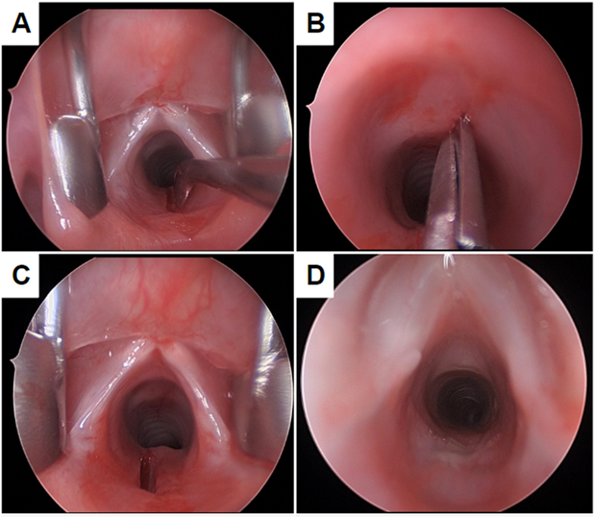
On DOL 7, the patient underwent patch ASD closure, PDA ligation and division and patch aortoplasty with inter-digitated extended end-to-end coarctation repair under deep hypothermic circulatory arrest. The patient was extubated the following day to non-invasive ventilation (NIV). ENT was consulted on DOL 12 for stridor and inability to wean NIV. Flexible fiberoptic laryngoscopy (FFL) demonstrated immobile bilateral vocal folds in the paramedian position with minimal posterior glottic gap, which was unchanged on repeat FFL 48 hours later. The patient’s respiratory distress subsequently worsened, and she was placed on full-face bilevel positive airway pressure (BiPAP) with intubation imminent. The patient was brought to the operating room (OR) on DOL 21 for direct laryngoscopy, bronchoscopy (DLB) and endoscopic APCS using a previously described technique (Figure 1).Reference Rutter, Hart, de Alarcon, Daniel, Parikh and Balakrishnan4 At the end of the procedure, the patient was nasally intubated with a 4.0 cuffless endotracheal tube and brought back to the intensive care unit (ICU) and kept sedated and ventilated for 13 days.
Intra-operative endoscopic laryngeal photographs. (A) Posterior cricoid split using a laryngeal D-knife. (B) Anterior cricoid split using an upwards-curved laryngeal scissor. (C) Subglottis following dilation with an airway balloon. (D) Subglottis on post-operative day 13.

On DOL 34 (day 13 post APCS) repeat DLB found good distraction of the airway with adequate mucosalisation. The patient was extubated to nasal cannula the following day and subsequently weaned to room air. Modified barium swallow study (MBSS) on DOL 41 revealed silent aspiration; after conservative measures and continued work with speech therapy, repeat MBSS on DOL 65 was normal.
At two months of age, the patient was having intermittent stridor when agitated but was feeding well, gaining weight appropriately and had no episodes of laboured breathing or cyanotic spells. FFL revealed minimal mobility of the right true vocal fold (TVF) and an immobile left TVF. At six months of age, the patient had only occasional noisy breathing and FFL demonstrated improved right TVF mobility, stable left TVF immobility and good glottic closure with phonation. Repeat FFL at 11 months of age (10 months post APCS) showed improved right TVF mobility on FFL to near-normal and unchanged left TVF immobility. At 19 months of age (18 months post APCS), the patient continued to be asymptomatic from airway, voice, sleep and swallowing perspectives. FFL at the patient’s most recent follow-up at 22 months of age (21 months post ACPS) demonstrated near complete return of function of the left TVF except at maximum abduction.
Discussion
There are two possible aetiologies for our patient’s BVFP. The first is that there was injury to both RLNs during the initial cardiac surgery. The second is that there was subclinical unilateral congenital paralysis, and injury of the contralateral RLN occurred during the cardiac procedure. Other causes to consider but thought to be less likely include: (1) intubation injury leading to immobility of one or both TVF, (2) congenital BVFP and (3) a central nervous system lesion. In our case we believe these causes to be less likely due to good mobility of the arytenoids on palpation during DLB, lack of symptoms prior to cardiac surgery and lack of concerning intra-cranial findings on brain MRI, respectively. While iatrogenic BVFP is rare in neonates, this case demonstrates the importance of FFL in the workup of respiratory distress following cardiothoracic surgery.
While there are many management options for neonatal BVFP, endoscopic APCS offers several advantages. In a select patient group, it can be used as a primary surgical intervention for BVFP to avoid tracheostomy. The endoscopic approach specifically has the benefit of improved visualisation during surgery, avoidance of neck incision and dissection and potentially decreased hospitalisation times. In patients who may have an eventual return of TVF mobility, as in our patient, APCS can be an effective option to avoid tracheostomy and to improve airway patency without limiting innate laryngeal function in the future. This procedure, however, may be difficult to perform in infants with an anterior larynx or poor pulmonary reserve. Alterations to the anatomy of the glottis are minimal but may still affect swallow function, and patients who undergo this procedure need to be assessed post-operatively for aspiration. Additionally, it may not be appropriate or successful in infants with significant co-morbidities such as bronchopulmonary dysplasia, neurologic diseases, syndromes or significant secondary airway pathology such as tracheomalacia.
What is already known:
• Bilateral vocal fold paralysis (BVFP) in paediatric patients is often managed with tracheostomy.
What this paper adds to our understanding:
• In our case, endoscopic anterior–posterior cricoid split (APCS) was effective in managing airway obstruction secondary to BVFP in a newborn following cardiac surgery.
• This surgical approach can help avoid tracheostomy in paediatric patients with BVFP.
• Bilateral vocal fold function can return to clinically normal levels in some patients, with no residual airway, voice, sleep or swallow symptoms.
Conclusion
We presented a rare case of BVFP following cardiac surgery in a neonate with no previous airway or respiratory symptoms. Endoscopic APCS was effective in managing this patient’s respiratory failure and avoiding tracheostomy, with long-term follow-up demonstrating resolution of symptoms and bilateral recovery of laryngeal mobility.
Acknowledgements
None.
Financial support
This research did not receive any specific grant from funding agencies in the public, commercial or not-for-profit sectors.
Competing interests
The authors declare none.
Ethical standards
The authors assert that all procedures contributing to this work comply with the ethical standards of the relevant national and institutional guidelines on human experimentation (West Virginia University IRB 2309853233) and with the Helsinki Declaration of 1975, as revised in 2008. Additionally, a written informed consent form was completed by the patient’s legal guardian.