




No CrossRef data available.
Article contents
Use of proteolytic enzymes as an additional tool for trypanosomatid identification
Published online by Cambridge University Press: 13 December 2004
Abstract
The expression of proteolytic activities in the Trypanosomatidae family was explored as a potential marker to discriminate between the morphologically indistinguishable flagellates isolated from insects and plants. We have comparatively analysed the proteolytic profiles of 19 monoxenous trypanosomatids (Herpetomonas anglusteri, H. samuelpessoai, H. mariadeanei, H. roitmani, H. muscarum ingenoplastis, H. muscarum muscarum, H. megaseliae, H. dendoderi, Herpetomoas sp., Crithidia oncopelti, C. deanei, C. acanthocephali, C. harmosa, C. fasciculata, C. guilhermei, C. luciliae, Blastocrithidia culicis, Leptomonas samueli and Lept. seymouri) and 4 heteroxenous flagellates (Phytomonas serpens, P. mcgheei, Trypanosoma cruzi and Leishmania amazonensis) by in situ detection of enzyme activities on sodium dodecyl sulphate-polyacrylamide gel electrophoresis (SDS–PAGE ) containing co-polymerized gelatine as substrate, in association with specific proteinase inhibitors. All 23 trypanosomatids expressed at least 1 acidic proteolytic enzyme. In addition, a characteristic and specific pattern of cell-associated metallo and/or cysteine proteinases was observed, except for the similar profiles detected in 2 Herpetomonas (H. anglusteri and H. samuelpessoai) and 3 Crithidia (C. fasciculata, C. guilhermei and C. luciliae) species. However, these flagellates released distinct secretory proteinase profiles into the extracellular medium. These findings strongly suggest that the association of cellular and secretory proteinase pattern could represent a useful marker to help trypanosomatid identification.
Keywords
Information
- Type
- Research Article
- Information
- Copyright
- © 2004 Cambridge University Press
INTRODUCTION
The Trypanosomatidae family is a cosmopolitan group of flagellates containing prominent groups with ability to parasitize a very diverse range of hosts including humans, animals, plants and other protists (Wallace, 1966; McGhee & Cosgrove, 1980). Some species are of particular interest, due to their medical importance. These are the agents of Chagas' disease (Trypanosoma cruzi), African trypanosomiasis (e.g. T. brucei complex) and species of Leishmania, which cause the various forms of leishmaniasis (Vickerman, 1994). Moreover, Phytomonas species are plant trypanosomatid parasites of phloem, latex and fruits that are responsible for important diseases in commercially important crops, such as coconut, oil palm, cassava and coffee (Camargo, Kastelein & Roitman, 1990). In addition to these digenetic parasites, several genera including Crithidia, Blastocrithidia, Herpetomonas and Leptomonas are composed wholly or largely of single host (monoxenous) parasites of the gut and associated organs of a wide range of insects (Wallace, 1966; McGhee & Cosgrove, 1980; Vickerman, 1994).
The medical and economical relevance of causative agents of dangerous diseases resulted in the preferential investigation of trypanosomatids of vertebrates and, for the same reasons, of plant parasites. On the other hand, trypanosomatids from insects remained forgotten for a long time. However, it is now becoming apparent that insect trypanosomatids play very important roles in research and are indeed probably close to the critical point of establishlishing a plausible trypanosomatid phylogeny (Podlipaev, 2001). Additionally, trypanosomatids not normally infectious to humans (e.g. Herpetomonads and Leptomonads) are also emerging as pathogens in immunosuppressed patients, mainly in HIV-infected individuals, who develop a clinical pattern very similar to visceral and/or cutaneous leishmaniasis-like syndromes (McGhee & Cosgrove, 1980; Dedet et al. 1995; Jiménez et al. 1996; Pacheco et al. 1998; Boisseau-Garsaud et al. 2000; Dedet & Pratlong, 2000; Miller, 2000). These opportunistic trypanosomatids are morphologically indistinguishable from pathogenic ones, impairing their identification. Therefore, these observations emphasize the urgent need to characterize ‘Leishmania-like’ parasites isolated from immunodeficient patients, as well as insect trypanosomatids. For example, Noyes et al. (2002) published a molecular study showing that a previously unclassified trypanosomatid responsible for human cutaneous syndrome in Martinique belongs to the most divergent member of the genus Leishmania.
Insect trypanosomatids have been traditionally allocated to a number of genera that were described based on morphological features, host and geographical origin (Wallace et al. 1983; Momen, 2001). However, for identification purposes, these criteria proved to be impractical and insufficient, because the same trypanosomatid species may be recovered from diverse species of insects and the same insect species may harbour various species of trypanosomatids. In addition, the morphology of trypanosomatid cells can be modified by environmental factors (Podlipaev, 2001; Momen, 2001, 2002). Therefore, there is a need to develop more effective means of Trypanosomatid identification. Methods such as genomic or kinetoplast DNA (k–DNA) fingerprinting and isoenzyme analysis were effective for distinguishing individual isolates, but failed as markers for generic distinction of the Trypanosomatidae family (Momen, 2001). Other biological, nutritional, immunological, biochemical and molecular criteria have been used for this purpose. Biochemistry aided in generic identification as different genera having distinct enzymes. In this sense, we focus on the proteolytic activity, since this class of enzymes is involved in a great number of physiological and pathological processes during the life-cycle of microorganisms (Rao et al. 1998). Proteases can be categorized based on their substrate specificities or mechanisms of catalysis. Enzymes cleaving within a polypeptide chain are named endopeptidases (or proteinases), while activities cleaving at the ends of polypeptides are named exopeptidases. Upon the basis of the mechanism of peptide hydrolysis, five major protease classes are known: metallo, serine, cysteine, aspartic and threonine proteases (McKerrow et al. 1993; Rao et al. 1998; Barrett, Rawlings & O'Brien, 2001). In trypanosomatids, especially in pathogenic flagellates, serine, metallo and cysteine proteinases have been extensively described.
Aiming at the detection of reliable markers for the identification of 23 distinct trypanosomatids, the cell-associated proteolytic activities were analysed by sodium dodecyl sulphate-polyacrylamide gel electrophoresis (SDS–PAGE) with gelatine incorporated into the gel as the proteinaceous substrate.
MATERIALS AND METHODS
Chemicals
The metallo (1,10-phenanthroline) and the cysteine (trans-epoxysuccinyl L-leucylamido-(4-guanidino) butane [E–64]) proteinase inhibitors as well as gelatine were purchased from Sigma Chemical Co. (St Louis, MO, USA). The other reagents used in electrophoresis and buffer components were obtained from Amersham Life Science (Little Chalfont, England).
Parasites and growth conditions
The 23 flagellate trypanosomatids used in this comparative study are listed in Table 1. For the experiments, cells were grown at 26 °C in 250 ml Erlenmeyer flasks containing 100 ml of liver infusion-tryptose (LIT) medium, except for the human pathogenic trypanosomatids (Trypanosoma cruzi and Leishmania amazonensis) that were cultured in LIT medium supplemented with 10% foetal bovine serum (FBS). Growth was estimated by determining the cell number in a Neubauer chamber. Cellular viability was assessed by motility and trypan blue dye exclusion. The viability of the parasites was not affected by the culture conditions employed in this work.

Parasite extracts
Three-day-old cultured parasites, at the log growth phase, were harvested by centrifugation at 1500 g for 10 min at 4 °C, and washed 3 times with cold PBS (150 mM NaCl, 20 mM phosphate buffer, pH 7·2). The trypanosomatids (1×10⁸ cells) were resuspended in 50 μl of PBS and lysed by the addition of 150 μl of SDS–PAGE sample buffer (125 mM Tris, pH 6·8, 4% SDS, 20% glycerol, 0·002% bromophenol blue). The cells were broken in a vortex by alternating 1 min shaking and 2 min cooling intervals, followed by a centrifugation at 5000 g for 15 min at 4 °C. The supernatants obtained after centrifugation corresponded to the whole parasite cellular extracts (Santos et al. 1999).
Culture supernatant fluids
Cell culture supernatant fluids were filtered in a 0·22–μm membrane (Millipore) and concentrated 10-fold by ultrafiltration in a 10000 molecular weight cut-off Centricon microconcentrator (AMICON, Beverly, MA, USA) at 4 °C, for 2 h. Concentrated supernatants were treated with an equal volume of SDS–PAGE sample buffer (Santos et al. 1999). To confirm that all extracellular proteolytic activities were genuine secretions, we measured in the culture supernatant fluids the lactate dehydrogenase activity, an intracellular enzyme, as previously described by Santos et al. (2002c). No lactate dehydrogenase was detected (not shown).
Gelatine–SDS–PAGE assay
The proteolytic activities were assayed and characterized by 10% SDS–PAGE with 0·1% gelatine incorporated into the gel as substrate (Heussen & Dowdle, 1980). The gels were loaded with 10 μl of each parasite extract (equivalent to 5×10⁶ cells), and alternatively with 20 μl (equivalent to 1×10⁷ cells) of concentrated supernatant fluids. Electrophoresis was performed at a constant current of 100 V at 4 °C for 2 h. After electrophoresis, SDS was removed by incubation, at room temperature under agitation, with 10 volumes of 1% Triton X-100 in the absence or in the presence of proteolytic enzyme inhibitors (10 mM 1,10-phenanthroline or 10 μM E–64) for 1 h. Then, the gels were incubated in 50 mM sodium phosphate buffer supplemented with 2 mM dithiothreitol (DTT), pH 5·5, at 37 °C, in the absence or in the presence of the same proteinase inhibitors. After incubation for 48 h, the gels were washed with 10 volumes of distilled water, stained for 2 h with 0·2% Coomassie brilliant blue R-250 in methanol-acetic acid-water (50[ratio ]10[ratio ]40 v/v/v) and destained overnight in a solution containing methanol-acetic acid-water (5[ratio ]10[ratio ]85 v/v/v), to intensify the digestion haloes. The proteolytic activities in the gels were revealed by the presence of colourless zones indicative of gelatine digestion. The gels were dried, scanned and digitally processed (Santos et al. 2002a). The molecular masses of the proteinases were calculated by comparison with the mobility of GIBCO BRL SDS-PAGE standards (Grand Island, NY, USA).
RESULTS
All the flagellate trypanosomatids examined contained at least 1 cell-associated proteolytic enzyme (Figs 1–3). This observation allowed the use of this class of enzyme as a new approach to help the identification of members of the Trypanosomatidae family. To make easier the analysis of the results obtained in the gelatine-SDS-PAGE, we separated the flagellates in 3 major groups: Herpetomonas genus (Fig. 1), Crithidia genus (Fig. 2) and the remaining flagellates (Fig. 3). When necessary, we also characterized the secretory proteinase profile (Fig. 4).

Fig. 1. Gelatine–SDS–PAGE showing the cell-associated proteolytic activity profiles detected in 9 different Herpetomonas species: (a) H. anglusteri, (b) H. samuelpessoai, (c) H. mariadeanei, (d) H. roitmani, (e) H. muscarum ingenoplastis, (f) H. megaseliae, (g) H. muscarum muscarum, (h) H. dendoderi and (i) Herpetomoas sp. The gel strips containing extracts from 5×10⁶ cells were incubated at 37 °C, for 48 h, in 50 mM sodium phosphate buffer, pH 5·5, supplemented with 2 mM DTT. Molecular masses of the proteinases, expressed in kDa, are represented on the left.

Fig. 2. Gelatine–SDS–PAGE showing the cell-associated proteolytic activity profiles detected in 7 different Crithidia species: (a) C. oncopelti, (b) C. deanei, (c) C. acanthocephali, (d) C. harmosa, (e) C. fasciculata, (f) C. guilhermei and (g) C. luciliae. The gel strips containing extracts from 5×10⁶ cells were incubated at 37 °C, for 48 h, in 50 mM sodium phosphate buffer, pH 5·5, supplemented with 2 mM DTT. Molecular masses of the proteinases, expressed in kDa, are represented on the left.

Fig. 3. Gelatine–SDS–PAGE showing the cell-associated proteolytic activity profiles detected in (a) Blastocrithidia culicis, (b) Leptomonas samueli, (c) Leptomonas seymouri, (d) Phytomonas serpens, (e) Phytomonas mcgheei, (f) Trypanosoma cruzi and (g) Leishmania amazonensis. The gel strips containing extracts from 5×10⁶ cells were incubated at 37 °C, for 48 h, in 50 mM sodium phosphate buffer, pH 5·5, supplemented with 2 mM DTT. Molecular masses of the proteinases, expressed in kDa, are represented on the left.

Fig. 4. Gelatine–SDS–PAGE showing the secretory proteolytic activity profiles detected in (a) Herpetomonas anglusteri, (b) Herpetomonas samuelpessoai, (c) Crithidia fasciculata, (d) Crithidia guilhermei, (e) Crithidia luciliae, (f) Herpetomonas sp. and (g) Phytomonas mcgheei. The gel strips containing 10-fold concentrated supernatant (equivalent to 1×10⁷ cells) were incubated at 37 °C, for 48 h, in 50 mM sodium phosphate buffer, pH 5·5, supplemented with 2 mM DTT. Molecular masses of the proteinases, expressed in kDa, are represented on the left.
In the species of Herpetomonas studied, the simplest proteolytic profile was observed in H. roitmani, an endosymbiont-bearing trypanosomatid that expressed a single faint band of 50 kDa, which was inhibited by 10 mM 1,10-phenanthroline, a potent zinc-metalloproteinase inhibitor (Fig. 1, lane d and Table 2). H. megaseliae (n=8 proteolytic bands), H. m. muscarum (n=6), H. dendoderi (n=5) and Herpetomonas sp. (n=4) showed complex and distinct proteolytic profiles containing both metallo and cysteine proteinases (Fig. 1, lanes f–i, respectively, and Table 2), as concluded from the inhibition by 1,10-phenanthroline and E–64, respectively. H. mariadeanei exhibited only cysteine proteinases with lower molecular masses (Fig. 1, lane c and Table 2), while H. m. ingenoplastis synthesised 2 metalloproteinases of 67 and 80 kDa (Fig. 1, lane e and Table 2). H. anglusteri and H. samuelpessoai expressed the same cell-associated profile, composed by a broad metalloproteinase band activity of apparent molecular mass of 60 kDa (ranging from 50 to 70 kDa) and a 45 kDa cysteine proteinase (Fig. 1, lanes a, b, respectively, and Table 2), impairing the discrimination between these 2 species. For this reason, we decided to analyse in these 2 distinct Herpetomonas species the released proteolytic pattern, in an effort to characterize specific secretory proteolytic activities. Our results demonstrated 2 distinct extracellular proteinase profiles composed of several metalloproteinases in H. anglusteri (Fig. 4, lane a) and H. samuelpessoai (Fig. 4, lane b).

In the Crithidia group analysed, heterogeneous proteolytic profiles were observed (Fig. 2). C. oncopelti (Fig. 2, lane a and Table 2), a symbiont-harbouring flagellate isolated from a plant host (Table 1), and C. deanei (Fig. 2, lane b and Table 2), a trypanosomatid isolated from an insect (Table 1) and harbouring a bacterium-like endosymbiont in its cytoplasm, expressed only 2 weak metalloproteinase activities when cultured in LIT medium. The other 5 Crithidia species studied do not possess symbionts, and expressed a more pronounced amount of proteinases (Fig. 2, lanes c–g). C. acanthocephali produced 4 cysteine proteinases of 80, 75, 70 and 50 kDa (Fig. 2, lane c and Table 2), while C. harmosa presented at least 3 metalloproteinases, 1 of them with higher activity (70–55 kDa) (Fig. 2, lane d and Table 2). C. fasciculata showed 2 metalloproteinases (90 and 70 kDa) and 1 cysteine proteinase with a broad activity range (60–40 kDa) (Fig. 2, lane e and Table 2). C. guilhermei (Fig. 2, lane f) and C. luciliae (Fig. 2, lane g) expressed the same qualitative cell-associated proteinases when compared with C. fasciculata (Fig. 2, lane e); however, quantitative differences were clearly seen in the gels. We also analysed the supernatant fluids of these 3 Crithidia species and observed a proteolytic profile resembling the released pattern of metallo-enzymes, with minor distinctive activities (Fig. 4, lanes c–e). In these experiments, at least 2 extracellular proteinases of 35 and 30 kDa were shown to be C. fasciculata species-specific (Fig. 4, lane c), while a 95 kDa band was C. guilhermei species-specific (Fig. 4, lane d).
Blastocrithidia culicis is also an endosymbiont-bearing trypanosomatid, which produced 4 major proteolytic enzymes of 102, 100 kDa (metalloproteinases), 50 and 40 kDa (cysteine proteinases) (Fig. 3, lane a and Table 2). The 2 Leptomonas species included in this study, Lept. samueli and Lept. seymouri, displayed distinct profiles composed of only metalloproteinase activities (Fig. 3, lanes b,c, respectively, and Table 2). Four digenetic trypanosomatids were also included in this comparative work: 2 Phytomonas (P. serpens and P. mcgheei), 1 Leishmania (Leish. amazonensis) and 1 Trypanosoma (T. cruzi). Only a 70 kDa cysteine proteinase was detected in P. serpens cells (Fig. 3, lane d and Table 2), while in P. mcgheei 5 distinct activities (72, 60, 50, 45 and 40 kDa) were identified (Fig. 3, lane e and Table 2). Interestingly, a closely related proteolytic profile was observed in P. mcgheei (Fig. 3, lane e) and in Herpetomonas sp. (Fig. 1, lane i) that was isolated from the salivary gland of a phytophagous insect (Table 1). This interesting result induced us to examine the secretory proteolytic enzymes in these two flagellates. Surprisingly, a similar released proteinase profile was evidenced in both trypanosomatid strains (Fig. 4, lanes f, g), differing only in 1 band with higher molecular mass (200 kDa), which was present just in P. mcgheei supernatant fluid (Fig. 4, lane g). The human pathogen T. cruzi expressed 3 well-known proteolytic enzymes (Bonaldo et al. 1991), with a prominent intracellular cysteine proteinase (cruzipain activity) of 45 kDa (Fig. 3, lane f and Table 2). Promastigote forms of Leish. amazonensis produced a great number of lower-mass cysteine proteinase activities and higher-mass metalloproteinase ones (Fig. 3, lane g and Table 2), as previously reported (Alfieri, Balanco & Pral, 1995; Soares et al. 2003).
Collectively, these results show a great variability in the cell-associated proteinase expression among parasites belonging to the Trypanosomatidae family. At least 3 distinct experiments were performed for each trypanosomatid analysed, and the proteolytic patterns were highly reproducible. This observation minimized the possible discrepancies in the zymogram analysis and the fact that some post-translational modification, such as glycosylation, could affect the number and size of proteinase bands. For some trypanosomatids, we analysed the cellular proteolytic content in more than one strain of the same species, including B. culicis (n=2), C. deanei (n=2), C. fasciculata (n=3), C. oncopelti (n=2), H. samuelpessoai (n=3), Lept. samueli (n=2), Lept. seymouri (n=2) and P. serpens (n=2). Our results demonstrated that different strains of the same parasite species showed identical proteinase profiles under the conditions employed in the present work (data not shown).
DISCUSSION
Proteases catalyse the cleavage of peptide bonds in proteins and peptides. Proteolytic enzymes execute many physiological roles and are essential factors for homeostatic control in eukaryotic cells. Their importance in conducting the essential metabolic and regulatory functions is evident from their occurrence in all forms of living organisms. In general, extracellular proteases catalyse the hydrolysis of large proteins to smaller molecules for subsequent absorption by the cell whereas intracellular proteases play a critical role in the regulation of metabolism (Rao et al. 1998). Additionally, their involvement in the life-cycle of disease-causing microorganisms has led them to become potential targets for developing therapeutic agents against fatal diseases, such as Chagas' disease and leishmaniasis (Sajid & McKerrow, 2002). Proper identification of genera and species of trypanosomatid parasites of insects still is an unsolved problem and, on the basis of these observations, we assumed that this class of enzymes has great potential in distinguishing different species, since each one possesses a distinct key metabolism.
Electrophoretic methods that permit adequate resolution and reproducibility, such as PAGE, provide a basis for microbial systematic. SDS–PAGE containing a protein as a proteolytic substrate has been widely used as a simple standard technique to assess the complexity of proteinases in vast cell systems including protozoa (Branquinha et al. 1996). For these reasons, the present study assessed the potential of gelatine–SDS–PAGE to analyse the proteinase content in several trypanosomatids in an effort to identify specific biochemical markers that allow typing of insect trypanosomatids. Gelatine was used as the proteinaceous substrate because it is easily hydrolysed by several proteinases and does not tend to migrate out of the resolving gel for electrophoretic migrations conducted at 4 °C, thus maintaining a uniform distribution of the protein substrate in the gel (Michaud et al. 1996). In addition, acidic pH (5·5) supplemented with a reducing agent (DTT) was used to evidence the proteinase activities, since the two major proteolytic classes (metallo- and cysteine proteinases) synthesized by trypanosomatids could be easily identified under the conditions employed in this comparative study.
The heterogeneity of Crithidia, Leptomonas and Herpetomonas genera has been demonstrated by different methods (Camargo et al. 1992; Fernandes et al. 1997). The genus Herpetomonas is defined as a taxon that consists of monoxenic parasites of insects that present promastigote, paramastigote and opisthomastigote forms in their life-cycle. Despite this clear-cut definition, many trypanosomatids have been erroneously placed in the genus Herpetomonas or, conversely, many Herpetomonas spp. may remain hidden in other genera. Our results showed that 7 distinct Herpetomonas species produced species-specific proteinase profiles, which can be useful in the correct identification of these parasites. The exception for this observation was seen in H. samuelpessoai and H. anglusteri, which presented a similar cell-associated enzymatic pattern. However, these two Herpetomonas species excreted distinct proteinase activities, which may be a reflection of changes in the nutritional requirements during the life-cycle of the flagellates. Therefore, we would infer that profiles of cell-associated and secretory proteinases could represent an additional criterion to be used in the identification of trypanosomatids.
The fact that H. megaseliae and H. m. muscarum produce a related cell-associated proteinase pattern is in accordance with Teixeira et al. (1997), which allocated H. megaseliae and H. m. muscarum in a same similarity cluster after examining specific k–DNA and genomic DNA markers in 14 Herpetomonas isolates. The insect trypanosomatids H. m. ingenoplastis and H. m. muscarum have been considered to be 2 subspecies of H. muscarum (Rogers & Wallace, 1971). There are, however, significant differences between these microorganisms, which suggest that they are adapted for distinctive life-styles. The growth differences in the presence of oxygen, mitochondrial structure and electron transport pathways are reflected in the fact that H. m. ingenoplastis consumes oxygen at a slower rate than does H. m. muscarum (Wallace, Wagner & Rogers, 1973). Additionally, completely distinct proteolytic enzymes were also detected in these H. muscarum subspecies, which corroborates the idea that these 2 species present great metabolic differences.
We also showed that the flagellate isolated from a phytophagous insect (Phthia picta), which was previously classified as Herpetomonas sp., demonstrated a striking similarity in the cell-associated and released proteinases to the reference strain P. mcgheei. In addition, these 2 flagellates have been used in other studies by our group, investigating the sialoglycoprotein profile (Santos, Alviano & Soares, 2002b) and the composition of cell-surface polypeptides (Santos et al., unpublished data). In those experiments, identical protein profiles were observed, suggesting that these 2 flagellates could be the same parasite, and that the phytophagous insect Phthia picta may act as natural vector of the parasite of maize. In contrast to the complex proteolytic pattern detected in P. mcgheei, the other Phytomonas species analysed, P. serpens, synthesized just an intracellular cysteine proteinase of 70 kDa.
H. roitmani, instead of typical opisthomastigotes, displays opisthomorph forms (a choanomastigote-like body shape with post-nuclear kinetoplast). These forms are present in the original drawings of H. roitmani description (Silva et al. 1991) and in some Crithidia, particularly the symbiont-harbouring species including C. deanei (Brandão et al. 2000). In the same way, our results corroborated with these morphological similarities, since the expression of proteolytic enzymes in H. roitmani and C. deanei was closely related, differing in only one 60 kDa-band detected in C. deanei. Recent studies (Brandão et al. 2000; d'Ávila-Levy et al. 2001, 2004) proposed a re-evaluation of the taxonomic status of these 2 choanomastigote-shaped trypanosomatids. In this sense, a new genus, Angomonas (Sousa & Côrte-Real, 1991) was created to allocate these 2 species in association with C. desouzai, another symbiont-containing trypanosomatid.
Our investigation showed a great diversity in the cell-associated proteolytic expression in the Crithidia genus, except for C. fasciculata, C. guilhermei and C. luciliae. Similarly, several approaches, including sialoglycoprotein expression (d'Ávila-Levy et al. 2004) and analysis of k-DNA mini-circle size (Brandão et al. 2000), have shown a high identity of these 3 Crithidia species, which could arise from a shared ancestor. Notwithstanding, distinct extracellular proteolytic profiles were generated when we assessed their supernatant culture fluids, which might reflect an adaptation to distinct invertebrate vectors (Table 1). Furthermore, our results confirm that C. oncopelti, which expressed a doublet of metalloproteinases of 70–72 kDa, is actually a singular symbiont-bearing choanomastigote-shaped trypanosomatid. This observation is in agreement with Brandão et al. (2000) who proposed the relocation of this species into another genus, whose specification could be Strigomonas, as first suggested by Lwoff & Lwoff (1931).
Promastigote forms of all Leishmania species express at the cellular surface a 63 kDa zinc-metalloproteinase called gp63. Similar proteinase activity has also been detected in non-pathogenic trypanosomatids such as Crithidia, Herpetomonas and Leptomonas (Etges, 1992; Melo et al. 2001; Jaffe & Dwyer, 2003; Santos et al. 2003), which suggests a preferential nutritional role of this proteolytic enzyme in the vector midgut, since insect colonization is the only life-cycle stage common to the monoxenous and heteroxenous flagellates. The epimastigote evolutive stage, characterised by a discrete undulating membrane, was observed in Blastocrithidia (monoxenous trypanosomatids) and Trypanosoma (heteroxenous parasites) genera. Besides their morphological similarity, the 2 genera were completely distinct in their life-cycle styles as well as in their contents of proteolytic enzymes. However, in a similar way, we conducted an experiment that demonstrated that B. culicis epimastigote forms produced a homologous cruzipain-like activity (d'Ávila-Levy et al., submitted manuscript), which is the main proteinase expressed by T. cruzi epimastigote forms. Altogether, these results pointed to the fact that similar evolutive stages, during their long evolutionary way, converged to synthesise similar molecules, some of them being conserved for a long time and probably maintaining, at least in part, their primitive physiological functions.
Even though several methods have been proposed to discriminate trypanosomatids, including protein and glycolipid profile (Branquinha et al. 1994), cell-surface polysaccharides (Abreu Filho et al. 2001), sialoglycoconjugates (Santos, Alviano & Soares, 2002b; d'Ávila-Levy et al. 2004), trans-sialidase/sialidase activities (Medina-Acosta et al. 1994), isoenzyme polymorphism (Franco et al. 1996), monoclonal antibodies (Teixeira, Campaner & Camargo, 1995), ribosomal DNA restriction analysis (Camargo et al. 1992), sequence analysis of the small subunit ribosomal RNA gene (Marché et al. 1995), k–DNA size (Brandão et al. 2000), the general consensus is that they are cumbersome and not satisfactory. However, for generic identification, species-specific characteristics capable of accommodating several species are necessary. Here, using a rapid, inexpensive and efficient way of (roughly) estimating the cell-associated and released proteinase contents by using a simple gelatine–SDS–PAGE procedure, we provided a rationale and easier diagnosis of culture forms of trypanosomatids. The results indicate the existence of a highly heterogeneity expression of metallo and cysteine cell-associated proteinases in trypanosomatids. Collectively, the results presented herein strongly suggest the value of proteolytic enzymes in distinguishing between trypanosomatid species that cannot be differentiated on structural grounds with the aid of a light microscope.
This work was supported by grants from Fundação Universitária José Bonifácio (FUJB), Financiadora de Estudos e Projetos (FINEP), Conselho Nacional de Desenvolvimento Científico e Tecnológico (CNPq), Conselho de Ensino e Pesquisa para Graduados da Universidade Federal do Rio de Janeiro (CEPG–UFRJ) and Programa de Apoio a Núcleos de Excelência (PRONEX). The authors thank Dr Maria Auxiliadora de Sousa, Márcia Attias, Elvira Saraiva and Thaís Souto-Padrón for providing the trypanosomatids. We also wish thank Dr Marta Helena Branquinha, Dr Irene von der Weid and Dr Marcio Lourenço Rodrigues for the useful critical English review as well as for the valuable suggestions on the manuscript, and Dr Cláudia Masini d'Avila-Levy for helping with the photographs.
References
REFERENCES
ABREU FILHO, B. A.,
DIAS FILHO, B. P.,
VERMELHO, A. B.,
JANKEVICIUS, S. I.,
JANKEVICIUS, J. V. &
SANTOS, R. L. (
2001).
Surface component characterization as taxonomic tools for Phytomonas spp identification.
Parasitology Research
87,
138–144.CrossRefGoogle Scholar
ALFIERI, S. C.,
BALANCO, J. M. F. &
PRAL, E. M. F. (
1995).
A radioiodinated peptidyl diazomethane detects similar cysteine proteinases in amastigotes and promastigotes of Leishmania (L.) mexicana and L. (L.) amazonensis.
Parasitology Research
81,
240–244.Google Scholar
BARRETT, A. J.,
RAWLINGS, N. D. &
O'BRIEN, E. A. (
2001).
The MEROPS database as a protease information system.
Journal of Structural Biology
134,
95–102.CrossRefGoogle Scholar
BOISSEAU-GARSAUD, A. M.,
CALES-QUIST, D.,
DESBOIS, N.,
JOUANNELLE, J.,
JOUANNELLE, A.,
PRATLONG, F. &
DEDET, J. P. (
2000).
A new case of cutaneous infections by a presumed monoxenous trypanosomatid in the island of Martinique (French West Indies).
Transactions of the Royal Society of Tropical Medicine and Hygiene
94,
51–52.CrossRefGoogle Scholar
BONALDO, M. C.,
D'ESCOFFIER, L. N.,
SALLES, J. M. &
GOLDENBERG, S. (
1991).
Characterization and expression of protease during Trypanosoma cruzi metacyclogenesis.
Experimental Parasitology
73,
44–51.CrossRefGoogle Scholar
BRANDÃO, A. A.,
MIRANDA, A.,
DEGRAVE, W. M. &
SOUSA, M. A. (
2000).
The heterogeneity of choanomastigote-shaped trypanosomatids as analyzed by their kDNA minicircle size: taxonomic implications.
Parasitology Research
86,
809–812.CrossRefGoogle Scholar
BRANQUINHA, M. H.,
BARRETO-BERGTER, E.,
MEIRELLES, M. N. L. &
VERMELHO, A. B. (
1994).
Glycolipid and protein profiles in trypanosomatids.
Parasitology Research
80,
336–341.CrossRefGoogle Scholar
BRANQUINHA, M. H.,
VERMELHO, A. B.,
GOLDENBERG, S. &
BONALDO, M. C. (
1996).
Ubiquity of cysteine- and metalloproteinase in a wide range of trypanosomatids.
Journal of Eukaryotic Microbiology
43,
131–135.CrossRefGoogle Scholar
CAMARGO, E. P.,
KASTELEIN, P. &
ROITMAN, I. (
1990).
Trypanosomatid parasites of plants (Phytomonas).
Parasitology Today
6,
22–25.CrossRefGoogle Scholar
CAMARGO, E. P.,
SBRAVATE, C.,
TEIXEIRA, M. M. G.,
ULIANA, S. R. B.,
SOARES, M. B. M.,
AFFONSO, H. T. &
FLOETER-WINTER, L. (
1992).
Ribosomal DNA restriction analysis and synthetic oligonucleotide probing in the identification of genera of lower trypanosomatids.
Journal of Parasitology
78,
40–48.CrossRefGoogle Scholar
D'ÁVILA-LEVY, C. M.,
ARAÚJO, F. M.,
VERMELHO, A. B.,
BRANQUINHA, M. H.,
ALVIANO, C. S.,
SOARES, R. M. A. &
SANTOS, A. L. S. (
2004).
Distribution of glycoproteins recognized by different lectins in seven choanomastigote-shaped trypanosomatids: taxonomic implications.
FEMS Microbiology Letters
231,
171–176.CrossRefGoogle Scholar
D'ÁVILA-LEVY, C. M.,
MELO, A. C. N.,
VERMELHO, A. B. &
BRANQUINHA, M. H. (
2001).
Differential expression of proteolytic enzymes in endosymbiont-harboring Crithidia species.
FEMS Microbiology Letters
202,
73–77.CrossRefGoogle Scholar
DEDET, J. P. &
PRATLONG, F. (
2000).
Leishmania, Trypanosoma and monoxenous trypanosomatids as emerging opportunistic agents.
Journal of Eukaryotic Microbiology
47,
37–39.CrossRefGoogle Scholar
DEDET, J. P.,
ROCHE, B.,
PRATLONG, F.,
CALES-QUIST, D.,
JOUANNELLE, J.,
BENICHOU, J. C. &
HUERRE, M. (
1995).
Diffuse cutaneous infection caused by a presumed monoxenous trypanosomatid in a patient infected with HIV.
Transactions of the Royal Society of Tropical Medicine and Hygiene
89,
644–646.CrossRefGoogle Scholar
ETGES, R. (
1992).
Identification of a surface metalloproteinase on 13 species of Leishmania isolated from humans, Crithidia fasciculata and Herpetomonas samuelpessoai.
Acta Tropica
50,
205–217.CrossRefGoogle Scholar
FERNANDES, O.,
TEIXEIRA, M. M. G.,
STURM, N. R.,
SOUSA, M. A.,
CAMARGO, E. P.,
DEGRAVE, W. M. &
CAMPBELL, D. A. (
1997).
Mini-exon gene sequences define six groups within the genus Crithidia.
Journal of Eukaryotic Microbiology
44,
535–539.CrossRefGoogle Scholar
FRANCO, A. M. R.,
MOMEN, H.,
NAIFF, R. D.,
MOREIRA, C. F. S.,
DEANE, M. P. &
GRIMALDI, G. (
1996).
Enzyme polymorphism in Endotrypanum and numerical analysis of isoenzyme data.
Parasitology
113,
39–48.CrossRefGoogle Scholar
HEUSSEN, C. &
DOWDLE, E. B. (
1980).
Electrophoretic analysis of plasminogen activators in polyacrilamide gels containing sodium dodecyl sulphate and copolymerized substrates.
Analytical Biochemistry
102,
196–202.CrossRefGoogle Scholar
JAFFE, C. L. &
DWYER, D. M. (
2003).
Extracellular release of the surface metalloprotease, gp63, from Leishmania and insect trypanosomatids.
Parasitology Research
91,
229–237.CrossRefGoogle Scholar
JIMÉNEZ, M. I.,
LÓPEZ-VÉLEZ, R.,
MOLINA, R.,
CAÑAVATE, C. &
ALVAR, J. (
1996).
HIV co-infection with a currently non-pathogenic flagellate.
Lancet
347,
264–265.CrossRefGoogle Scholar
LWOFF, M. &
LWOFF, A. (
1931).
Recherches sur la morphologie de Leptomonas oncopelti Noguchi et Tilden et Leptomonas fasciculata Novy, Mac Neal et Torrey.
Arquives Zoologic Experimental Géneral
71,
21–37.Google Scholar
MARCHÉ, S.,
ROTH, C.,
PHILIPPE, H.,
DOLLET, M. &
BALTZ, T. (
1995).
Characterization and detection of plant trypanosomatids by sequence analysis of the small subunit ribosomal RNA gene.
Molecular and Biochemical Parasitology
71,
15–26.CrossRefGoogle Scholar
McGHEE, R. B. &
COSGROVE, W. B. (
1980).
Biology and physiology of the lower trypanosomatids.
Microbiology Reviews
44,
140–173.Google Scholar
McKERROW, J. H.,
SUN, E.,
ROSENTHAL, P. J. &
BOUVIER, J. (
1993).
The proteases and pathogenicity of parasitic protozoa.
Annual Review of Microbiology
47,
821–853.CrossRefGoogle Scholar
MEDINA-ACOSTA, E.,
FRANCO, A. M. R.,
JANSEN, A. M.,
SAMPOL, M.,
NEVÉS, N.,
PONTES-DE-CARVALHO, L.,
GRIMALDI, G. &
NUSSENZWEIG, V. (
1994).
Trans-sialidase and sialidase activities discriminate between morphologically indistinguishable trypanosomatids.
European Journal of Biochemistry
225,
333–339.CrossRefGoogle Scholar
MELO, A. C. N.,
GIOVANNI-DE-SIMMONE, S.,
BRANQUINHA, M. H. &
VERMELHO, A. B. (
2001).
Crithidia guilhermei: purification and partial characterization of a 62-kDa extracellular metalloproteinase.
Experimental Parasitology
97,
1–8.Google Scholar
MICHAUD, D.,
CANTIN, L.,
RAWORTH, D. A. &
VRAIN, T. C. (
1996).
Assessing the stability of cystatin/cysteine proteinase complexes using mildly-denaturing gelatine-polyacrylamide gel electrophoresis.
Electrophoresis
17,
74–79.CrossRefGoogle Scholar
MILLER, R. F. (
2000).
Clinical presentation and significance of emerging opportunistic infections.
Journal of Eukaryotic Microbiology
47,
21–23.CrossRefGoogle Scholar
MOMEN, H. (
2001).
Some current problems in the systematics of trypanosomatids.
International Journal for Parasitology
31,
640–642.CrossRefGoogle Scholar
MOMEN, H. (
2002).
Molecular taxonomy of trypanosomatids: some problems and pitfalls.
Archives of Medical Research
33,
413–415.CrossRefGoogle Scholar
NOYES, H.,
PRATLONG, F.,
CHANCE, M.,
ELLIS, J.,
LANOTTE, G. &
DEDET, J. P. (
2002).
A previously unclassified trypanosomatid responsible for human cutaneous lesions in Martinique (French West Indies) is the most divergent member of the genus Leishmania ss.
Parasitology
124,
17–24.CrossRefGoogle Scholar
PACHECO, R. S.,
MARZOCHI, M. C. A.,
PIRES, M. Q.,
BRITO, C. M. M.,
MADEIRA, M. F. &
BARBOSA-SANTOS, E. G. O. (
1998).
Parasite genotypically related to a monoxenous trypanosomatid of dog's flea causing opportunistic infection in an HIV positive patient.
Memórias do Instituto Oswaldo Cruz
93,
531–537.CrossRefGoogle Scholar
PODLIPAEV, S. (
2001).
The more insect trypanosomatids under study-the more diverse Trypanosomatidae appears.
International Journal for Parasitology
31,
648–652.CrossRefGoogle Scholar
RAO, M. B.,
TANKSALE, A. M.,
GHATGE, M. S. &
DESHPANDE, V. V. (
1998).
Molecular and biotechnological aspects of microbial proteases.
Microbiology and Molecular Biology Reviews
62,
597–635.Google Scholar
ROGERS, W. E. &
WALLACE, F. G. (
1971).
Two new subspecies of Herpetomonas muscarum (Leidy 1856) Kent 1880.
Journal of Protozoology
18,
645–649.CrossRefGoogle Scholar
SAJID, M. &
McKERROW, J. H. (
2002).
Cysteine proteases of parasitic organisms.
Molecular and Biochemical Parasitology
120,
1–21.CrossRefGoogle Scholar
SANTOS, A. L. S.,
ABREU, C. M.,
ALVIANO, C. S. &
SOARES, R. M. A. (
2002 a).
Activation of the glycosylphosphatidylinositol membrane proteinase upon released from Herpetomonas samuelpessoai by phospholipase C.
Current Microbiology
45,
293–298.Google Scholar
SANTOS, A. L. S.,
ALVIANO, C. S. &
SOARES, R. M. A. (
2002 b).
Detection of sialoglycomolecules in five plant trypanosomatids and in an insect phytophagous isolate.
FEMS Microbiology Letters
214,
19–23.Google Scholar
SANTOS, A. L. S.,
FERREIRA, A.,
FRANCO, V. A.,
ALVIANO, C. S. &
SOARES, R. M. A. (
1999).
Characterization of proteinases in Herpetomonas anglusteri and Herpetomonas roitmani.
Current Microbiology
39,
61–64.CrossRefGoogle Scholar
SANTOS, A. L. S.,
RODRIGUES, M. L.,
ALVIANO, C. S.,
ANGLUSTER, J. &
SOARES, R M. A. (
2003).
Herpetomonas samuelpessoai: dimethylsulfoxide-induced differentiation is influenced by proteinase expression.
Current Microbiology
46,
11–17.CrossRefGoogle Scholar
SANTOS, A. L. S.,
SOUTO-PADRÓN, T.,
ALVIANO, C. S.,
LOPES, A. H. C. S.,
SOARES, R. M. A. &
MEYER-FERNANDES, J. R. (
2002 c).
Secreted phosphatase activity induced by dimethylsulfoxide in Herpetomonas samuelpessoai.
Archives of Biochemistry and Biophysics
405,
191–198.Google Scholar
SILVA, P. M. F. E.,
SOLÉ-CAVA, A. M.,
SOARES, M. J.,
MOTTA, M. C. M.,
FIORINI, J. E. &
DE SOUZA, W. (
1991).
Herpetomonas roitmani (Fiorini et al., 1989) n. comb.: a trypanosomatid with a bacterium-like endosymbiont in the cytoplasm.
Journal of Protozoology
38,
489–494.Google Scholar
SOARES, R. M. A.,
SANTOS, A. L. S.,
BONALDO, M. C.,
ANDRADE, A. F. B.,
ALVIANO, C. S.,
ANGLUSTER, J. &
GOLDENBERG, S. (
2003).
Leishmania (Leishmania) amazonensis: differential expression of proteinases and cell-surface polypeptides in avirulent and virulent promastigotes.
Experimental Parasitology
104,
104–112.CrossRefGoogle Scholar
SOUSA, M. A. &
CÔRTE-REAL, S. (
1991).
Postnuclear kinetoplast in choanomastigotes of Crithidia deanei Carvalho, 1973: proposal of a new genus.
Revista do Instituto de Medicina Tropical de São Paulo
33,
S8.Google Scholar
TEIXEIRA, M. M. G.,
CAMPANER, M. &
CAMARGO, E. P. (
1995).
Characterization of the target antigens of Phytomonas-specific monoclonal antibodies.
Journal of Eukaryotic Microbiology
42,
232–237.CrossRefGoogle Scholar
TEIXEIRA, M. M. G.,
TAKATA, C. S. A.,
CONCHON, I.,
CAMPANER, M. &
CAMARGO, E. P. (
1997).
Ribosomal and kDNA markers distinguish two subgroups of Herpetomonas among old species and new trypanosomatids isolated from flies.
Journal of Parasitology
83,
58–65.CrossRefGoogle Scholar
VICKERMAN, K. (
1994).
The evolutionary expansion of the trypanosomatid flagellates.
International Journal for Parasitology
24,
1317–1331.CrossRefGoogle Scholar
WALLACE, F. G. (
1966).
The trypanosomatid parasites of insects and arachnids.
Experimental Parasitology
18,
124–193.CrossRefGoogle Scholar
WALLACE, F. G.,
CAMARGO, E. P.,
McGHEE, R. B. &
ROITMAN, I. (
1983).
Guidelines for the description of new species of lower trypanosomatids.
Journal of Protozoology
30,
308–313.CrossRefGoogle Scholar
WALLACE, F. G.,
WAGNER, M. &
ROGERS, W. E. (
1973).
Varying kinetoplast ultrastructure in two species of Herpetomonas muscarum.
Journal of Protozoology
20,
218–222.CrossRefGoogle Scholar

Table 1. Isolates of flagellate trypanosomatids used in this study

Fig. 1. Gelatine–SDS–PAGE showing the cell-associated proteolytic activity profiles detected in 9 different Herpetomonas species: (a) H. anglusteri, (b) H. samuelpessoai, (c) H. mariadeanei, (d) H. roitmani, (e) H. muscarum ingenoplastis, (f) H. megaseliae, (g) H. muscarum muscarum, (h) H. dendoderi and (i) Herpetomoas sp. The gel strips containing extracts from 5×10⁶ cells were incubated at 37 °C, for 48 h, in 50 mM sodium phosphate buffer, pH 5·5, supplemented with 2 mM DTT. Molecular masses of the proteinases, expressed in kDa, are represented on the left.

Fig. 2. Gelatine–SDS–PAGE showing the cell-associated proteolytic activity profiles detected in 7 different Crithidia species: (a) C. oncopelti, (b) C. deanei, (c) C. acanthocephali, (d) C. harmosa, (e) C. fasciculata, (f) C. guilhermei and (g) C. luciliae. The gel strips containing extracts from 5×10⁶ cells were incubated at 37 °C, for 48 h, in 50 mM sodium phosphate buffer, pH 5·5, supplemented with 2 mM DTT. Molecular masses of the proteinases, expressed in kDa, are represented on the left.

Fig. 3. Gelatine–SDS–PAGE showing the cell-associated proteolytic activity profiles detected in (a) Blastocrithidia culicis, (b) Leptomonas samueli, (c) Leptomonas seymouri, (d) Phytomonas serpens, (e) Phytomonas mcgheei, (f) Trypanosoma cruzi and (g) Leishmania amazonensis. The gel strips containing extracts from 5×10⁶ cells were incubated at 37 °C, for 48 h, in 50 mM sodium phosphate buffer, pH 5·5, supplemented with 2 mM DTT. Molecular masses of the proteinases, expressed in kDa, are represented on the left.

Fig. 4. Gelatine–SDS–PAGE showing the secretory proteolytic activity profiles detected in (a) Herpetomonas anglusteri, (b) Herpetomonas samuelpessoai, (c) Crithidia fasciculata, (d) Crithidia guilhermei, (e) Crithidia luciliae, (f) Herpetomonas sp. and (g) Phytomonas mcgheei. The gel strips containing 10-fold concentrated supernatant (equivalent to 1×10⁷ cells) were incubated at 37 °C, for 48 h, in 50 mM sodium phosphate buffer, pH 5·5, supplemented with 2 mM DTT. Molecular masses of the proteinases, expressed in kDa, are represented on the left.
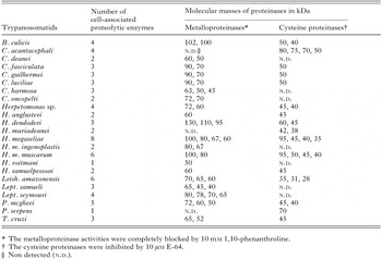
Table 2. Distribution of metallo- and cysteine proteinase activities in trypanosomatids analysed in this study
 You have
Access
- 24
- Cited by