Introduction
Human lymphatic filariasis is one among the neglected parasitic diseases and is also the second most important leading cause for permanent disability, which forced the World Health Organization to install eradication programmes (World Health Organization, 2001). Parasites cause immune suppression to establish themselves in lymphatic tissues and dwell in them for a prolonged period. Parasitic nematodes are often exposed to reactive oxygen species (ROS) and have antioxidant systems, which counteract the host immune system, to cope with the tremendous oxidative stress (Chiumiento & Bruschi, Reference Chiumiento and Bruschi2009). These antioxidant enzymes play a major role in defence and have been reported to be protective anti-parasitic vaccines in many parasitic diseases (LoVerde et al., Reference LoVerde, Carvalho-Queiroz and Cook2004). Hence, they have been suggested to be useful targets for human therapeutic intervention.
Based on our earlier studies on multiple-antigen vaccines (Anand et al., Reference Anand, Murugan, Prabhu, Anandharaman, Reddy and Kaliraj2008; Vanam et al., Reference Vanam, Pandey, Prabhu, Dakshinamurthy, Reddy and Kaliraj2009), we deduced an approach of selecting two targets in tandem that are vital in defending the parasites against free-radical damage. Such a measure would hamper the ability of parasites to endure the hostile immunological environment of the host for a long time, as reported earlier in Schistosoma mansoni infection (LoVerde et al., Reference LoVerde, Carvalho-Queiroz and Cook2004).
Thioredoxin (TRX) and glutathione (GSH) systems are the major thiol-dependent redox pathways present in nematode cells. Brugia malayi TRX was found to be constitutively expressed throughout the parasite's life cycle, and was suggested to play a role in host immune evasion (Kunchithapautham et al., Reference Kunchithapautham, Padmavathi, Narayanan, Kaliraj and Scott2003). Further, it has been shown to confer 63% protection (Madhumathi et al., Reference Madhumathi, Prince, Anugraha, Kiran, Rao, Reddy and Perumal2010) in filarial experimental models. Accordingly, Wuchereria bancrofti thioredoxin (WbTRX), a representative of the 16-kDa family of TRX proteins from filarial nematodes, was selected for this study to be used for single- and multiple-mode vaccinations. Although TRX proteins lack secretory signal sequences, they are secreted like mammalian thioredoxins and can be readily detected in the excretory–secretory products. Another interesting thiol-specific antioxidant protein utilized by nematodes is thioredoxin peroxidase/thiol-specific antioxidant (TPX/TSA). Filarial TPX was found to play a role in protecting nematode cells against free radicals (Ghosh et al., Reference Ghosh, Eisinger, Raghavan and Scott1998).
The protective efficacy of B. malayi transglutaminase (BmTGA) was enhanced significantly (74%) in multiple antigen vaccination mode along with BmTPX (Vanam et al., Reference Vanam, Pandey, Prabhu, Dakshinamurthy, Reddy and Kaliraj2009). Similarly, mice immunized with DNA vaccines of B. malayi abundant larval transcript-2 (BmALT2) and BmTPX in a cocktail mode cleared microfilariae to a significant level of 78% in our previous study (Anand et al., Reference Anand, Murugan, Prabhu, Anandharaman, Reddy and Kaliraj2008). Thus, vaccination in a cocktail fashion has been shown to elevate the protective immune response in filarial animal models.
Since these antioxidant targets (TRX and TPX) play a crucial role in protecting the parasites from hosts, we hypothesized that using them in multiple mode vaccination would be effective. Also, since both the antigens are expressed in all stages of the parasite and provide synergistic antioxidant benefit for parasite survival, there is greater advantage in using them together. Hence, in order to investigate if the combination of two antioxidants – WbTRX and W. bancrofti thioredoxin peroxidase (WbTPX) – could effectively reduce the parasite load by blocking the major antioxidant system, immune responses and prophylactic efficacy of the proteins in multiple mode were studied in animal models of experimental filariasis.
Materials and methods
Immunization of mice and sera collection
Six- to 8-week-old female BALB/c mice were obtained from Tamilnadu Veterinary and Animal Sciences University, Chennai. All experiments were performed in accordance with Indian Animal Ethics Committee regulations. A group of six mice was injected via the intraperitoneal route with 25 μg of recombinant proteins in 50 μl of phosphate-buffered saline (PBS) along with alum at 1:1 ratio. The control mouse group only received alum in PBS. The same dose of booster was given on days 7, 14 and 21 and the blood was collected on days 0, 14, 28 and 42. Sera were separated and stored at − 80°C.
Expression and purification of recombinant proteins
WbTRX (Accession no. AAN34,968) and WbTPX (Accession no. ACL82,593) genes in pRSET B vector, cloned in NdeI and HindIII sites were transformed and proteins were expressed in Escherichia coli (GJ1158) using 100 mm NaCl as inducer at 0.6 OD for 3 h. Soluble proteins were purified in native conditions by ion exchange chromatography using Q-Sepharose (Amersham Biosciences, Uppsala, Sweden) and analysed by 12% sodium dodecyl sulphate–polyacrylamide gel electrophoresis (SDS-PAGE). Recombinant antigens were purified from lipopolysaccharide (LPS) using Pierce High Capacity Endotoxin Removal Resin (Pierce Biotechnology, Rockford, Illinois, USA) as per manufacturer's instructions. Proteins were further confirmed by Western blots using antisera raised in mice (anti-TRX and anti-TPX antibodies).
Detection of antigen in different life-cycle stages
Production levels of W. bancrofti native TRX and TPX antigens in the microfilarial (mf), L3 and adult stages of the filarial parasites were tested with antisera raised against recombinant TRX and TPX proteins in mice by enzyme-linked immunosorbent assay (ELISA). Ninety-six-well microtitre plates (Nunc Maxisorp, Nunc A/S,Denmark) were coated with crude worm extract (500 ng/well) and anti-TRX or anti-TPX sera (1:1000) were used as primary antibody. The reactivity with recombinant proteins was used as the positive control. The mean absorbance (+2SD) of alum control sera was taken as the cut-off value and an unrelated filarial protein SXP was used as the negative control. SXP is an acronym used by the authors as a diagnostically significant microfilarial antigen. Goat anti-mouse-IgG ALP (alkaline phosphatase) conjugate was used as secondary antibody. The colour was developed using p-nitrophenyl phosphate substrate (1 mg/ml) in substrate buffer (100 mm Tris-Cl, pH 9.5, 100 mm NaCl, 5 mm MgCl2) and absorbance was read at 405 nm.
Cross-reactivity of antigens
The cross-reactivity of the antioxidants TRX and TPX was assessed by ELISA. Plates were coated with recombinant proteins (100 ng/well) and anti-TRX or anti-TPX sera (1:1000) raised in mice was used as primary antibody. The alum immunized serum was used as the negative control and the reactivity of respective antigens was taken as reference. The plates were developed as described above.
Measurement of antibody responses
Total IgG levels in the sera were determined by ELISA as follows. Plates were coated with 100 ng of antigen (100 μl/well). After washing and blocking with 5% skimmed milk powder, a serial twofold dilution (1:500–1:128,000) of antisera was used. The antibody titre was assessed as the highest serum dilution giving an absorbance (0.15) higher than that of alum control sera. The plate was developed as described above. For estimating IgG isotypes, mice sera (1:100) from different immunization groups were incubated for 1 h at 37°C, with respective proteins coated on ELISA plates. The IgG isotype binding was detected using secondary rabbit anti-mouse IgG specific for each subclass (Pierce Isotyping kit; Pierce Biotechnology) as per the manufacturer's instructions. The absorbance was read at 405 nm.
Measurement of cellular responses
Immunized mice were sacrificed after a peak titre was attained and the spleens were removed aseptically. Splenocytes were separated and washed twice with fresh culture medium (RPMI 1640; Sigma, St. Louis, Missouri, USA). Lysis buffer (0.1% ammonium chloride) was added to the pellet to remove the red blood cells (RBCs) and the cells were counted. The single-cell suspension was cultured in triplicate in 96-well plates at 2 × 10⁵ cells per well in RPMI 1640 medium (100 μl/well) supplemented with gentamycin (80 μg/ml) (Ranbaxy Laboratories, New Delhi, India), 25 mm HEPES (Wipro GE Healthcare Chennai, India), 2 mm glutamine (Wipro GE Healthcare) and 10% fetal bovine sera. Cells were then stimulated in vitro with antigen (0.1, 1, 5 and 10 μg/well) or positive control concanavalin A (Con A) (1 μg/well) and incubated at 37°C with 5% CO2 for 72 h. Wells with medium alone were used as unstimulated controls. Cultures were pulsed with [3H]thymidine (USB, Amersham Pharmacia) at 0.5 μCi per well, 18 h before harvesting. Cells were harvested on to glass-fibre discs and thymidine incorporation was measured by a liquid scintillation counter. Proliferative responses were expressed as stimulation index (SI) calculated as counts per minute (cpm) of cells stimulated with antigen divided by cpm of unstimulated cells. All cultures were taken in triplicate and the results expressed as mean SI ± standard error of the mean (SEM).
For cytokine assay, the culture supernatants were centrifuged at 5000 rpm for 15 min, filtered through 0.22-μm pores and assayed for cytokine levels using sandwich ELISA for interleukin (IL)-5 and interferon (IFN)-γ (Pierce Biotechnology) according to the manufacturer's instructions. All concentrations were derived from standard curves and data expressed in pg/ml after deducting the values of unstimulated cultures.
Immunization and protection in multimammate rats
Four- to 6-week-old male Mastomys coucha, known as the multimammate rat or African soft-furred rat, were used in this study, with six rats per group, as they are well known permissive models for filarial parasites (Sänger et al., Reference Sänger, Lämmler and Kimmig1981; Lok & Abraham, Reference Lok and Abraham1992). Rats were obtained from the Mahatma Gandhi Institute of Medical Sciences, India, and were handled in accordance with the institutional guidelines approved by an institutional animal care committee. Each rat was immunized with 50 μg of recombinant proteins, either as single antigens TRX or TPX or in combination, TRX + TPX (25 μg each) in alum. Four doses at weekly intervals were administered intraperitoneally. The control group received PBS alone in alum. Sera collected periodically after immunization were used to check the antibody titre by ELISA. For Brugia malayi infection, the mosquito colony and parasites were maintained as described previously (Thirugnanam et al., Reference Thirugnanam, Pandiaraja, Ramaswamy, Murugan, Gnanasekar, Nandakumar, Reddy and Kaliraj2007). Twelve days after infection, B. malayi L3 larvae were obtained by crushing the insects (Suzuki & Seregeg, Reference Suzuki and Seregeg1979) and carefully removing the L3 s. Larvae were counted under a microscope and used for the protection study.
The protection of rats was undertaken by a micropore chamber method as described previously (Abraham et al., Reference Abraham, Lange, Yutanawiboonchai, Trpis, Dickerson, Swenson and Eberhard1993; Chenthamarakshan et al., Reference Chenthamarakshan, Reddy and Harinath1995). Each group of six rats was immunized with 50 μg of recombinant proteins in alum intraperitoneally and sera were collected at 1-week intervals. Antibody titre was measured as described above. Two weeks after the final booster immunization, the animals were challenged by intraperitoneal implantation of a micropore chamber containing 20 live B. malayi L3 larvae in RPMI 1640 media. After 48 h the micropore chamber was harvested and the contents were removed on to a glass slide and examined microscopically for cytotoxicity. The group immunized with alum alone was used as negative control. The number of live and dead L3 was enumerated by observing movement of L3 under a Nikon microscope. The percentage protection was expressed as [(the average number of worms recovered from the control animals minus average number of worms recovered from the vaccinated animals)÷average number of worms recovered from the control animals] × 100 (Gnanasekar et al., Reference Gnanasekar, Rao, He, Mishra, Nutman, Kaliraj and Ramaswamy2004). Experiments were performed three times and data were presented as means ± SEM.
Data analysis
All statistical analyses were done using Graphpad prism software version 5 (GraphPad Software, Inc., California, USA). Comparisons between two individual data points were made using a non-parametric Mann–Whitney test. For multiple comparisons, a non-parametric Kruskal–Wallis test was used along with Bonferroni's post test. For T-cell proliferation studies, two-way analysis of variance (ANOVA) was used. A probability (P) value ≤ 0.05 was considered statistically significant.
Results
Recombinant proteins and antigen reactivity
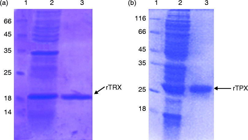
Recombinant proteins TRX and TPX were expressed as 20-kDa and 25-kDa fusion proteins without histidine fusion tag in E. coli (GJ1158) (fig. 1). The proteins were purified in non-denaturing conditions by ion exchange chromatography which yielded ~3–5 mg/l culture.
(colour online) Expression and purification of (a) rTRX (recombinant thioredoxin) and (b) rTPX (recombinant thioredoxin peroxidase) by PAGE. Lanes: 1, molecular marker; 2, induced culture of E. coli (GJ1158) showing 20-kDa rTRX and 25-kDa rTPX; 3, purified rTRX and rTPX.

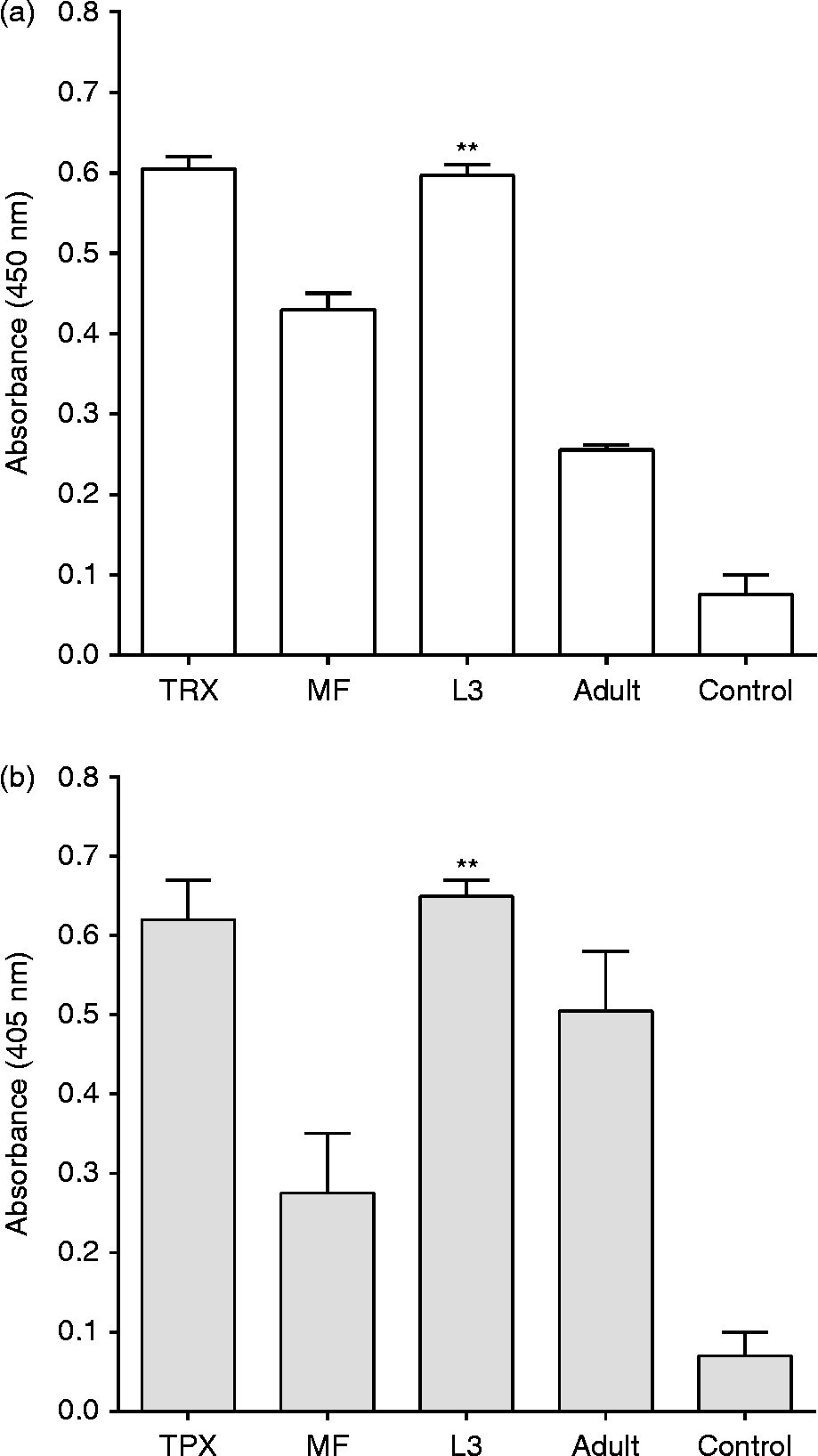
The expression analysis of native antigens in different stages of the parasite showed significantly higher production (P= 0.02 for TRX and P= 0.004 for TPX) of antioxidants TRX and TPX in the L3 stage when compared to controls (0.6 and 0.65 OD, respectively), which correlates with previous observations (fig. 2). However, both proteins were found to be expressed at different levels in all stages, as reported earlier (Ghosh et al., Reference Ghosh, Eisinger, Raghavan and Scott1998; Blaxter et al., Reference Blaxter, Daub, Guiliano, Parkinson and Whitton2002; Kunchithapautham et al., Reference Kunchithapautham, Padmavathi, Narayanan, Kaliraj and Scott2003). TPX showed almost similar expression in adult and mf stages (0.35–0.43 OD; P= 0.16) while TRX showed higher expression in mf (0.45 OD) compared to adult worms (0.25 OD)(P= 0.0138).
Reactivity of mouse sera using (a) anti-TRX and (b) anti-TPX with crude extracts from microfilariae (MF), infective third-stage larvae (L3) and adult worms of B. malayi using ELISA: mean absorbances of antigens with sera containing alum was taken as the cut-off value and filarial diagnostic protein (SXP) was used as a negative control. **Significant differences for TRX (P < 0.01) and TPX (P < 0.01) for L3 versus control.
Cross-reactivity
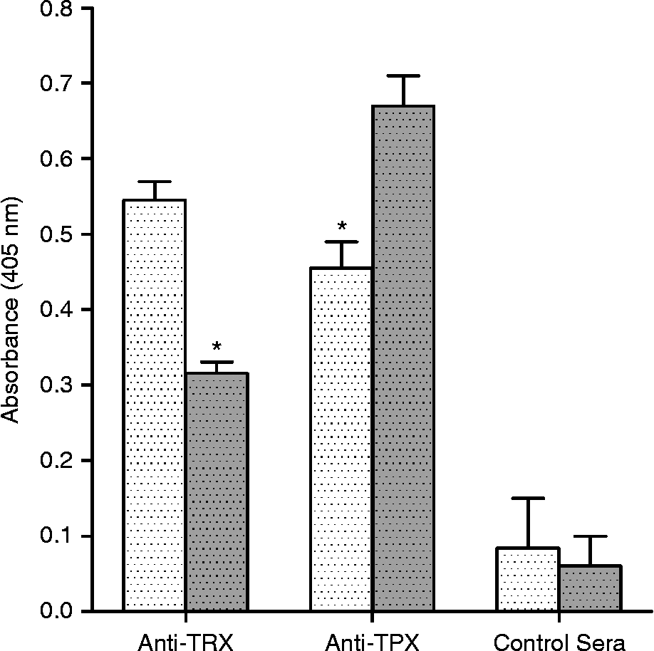
Antigens showed significantly higher reactivity with their respective antisera, which were taken as reference values. The mean absorbance of TRX and TPX with anti-TPX and anti-TRX, respectively, was significantly higher than with the control sera, indicating cross-reactivity (P < 0.01 for TRX vs anti-TPX and P < 0.05 TPX vs anti-TRX) (fig. 3). This shows that the antioxidants share common epitopes recognized mutually by their antisera.
Cross-reactivity of anti-TPX sera with TRX (white dotted bars) and anti-TRX sera with TPX (grey dotted bars), respectively, using ELISA: immunized sera with alum were used as negative controls. Significant differences shown as P< 0.05 (*).
Antibody and cellular responses in mice
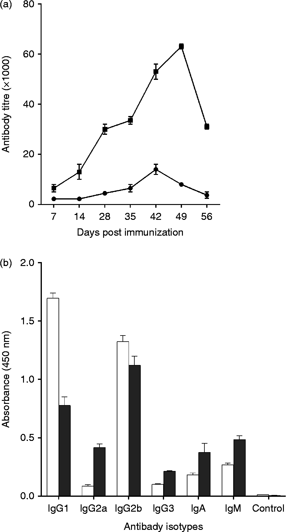
The antibody response was measured in terms of peak titre. Both the recombinant antigens induced significantly high levels of antibodies compared to the alum control. TRX induced a peak titre of 16,000 on day 42. The antibody levels showed a steady increase until day 49 for TPX, with a peak titre of 64,000, and declined by day 56 (fig. 4a). The antibody isotype analysis showed a predominant IgG1 followed by IgG2b level in anti-TRX sera, whereas IgG2b levels were predominant followed by IgG1 and IgG2a in anti-TPX sera (fig. 4b). TPX also showed higher IgG2a, IgA and IgM subtypes compared to TRX.
(a) Antibody titres (mean ± SEM) induced by rTRX (black dots) and rTPX (black squares) immunization in mice. (b) Antibody subtypes elicited by rTRX (white bars) and rTPX (black bars) mice, with normal rabbit serum used as a control.
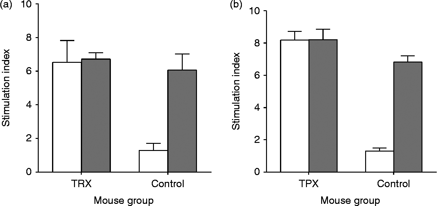
To study the cellular responses of the antigens, mice were primed either with TRX or TPX and spleen cells were stimulated in vitro with respective antigens, mitogen or the positive control, Con A. Both antigens showed significantly (P< 0.0001) higher proliferation (mean SI = 6.2–8.1) at concentration 1–5 μg/ml compared to the alum control (fig. 5). Con A induced proliferation in both immunized and control mice. Thus the antioxidants were found to induce potent cellular responses in mice.
Splenocyte proliferation assay in mice immunized with (a) rTRX and (b) rTPX (grey bars) plus positive control, Con A (white bars): stimulation indices (mean ± SEM) with cell proliferation from alum-immunized groups taken as negative controls.
Cytokine responses were analysed by measuring the IL-5 and IFN-γ levels in the culture supernatants of mice splenocytes. Both TRX and TPX induced high levels of IFN-γ (457.1 ± 32.02 pg/ml and 534.9 ± 38.34 pg/ml, respectively) in their respective groups, whereas IL-5 level was higher in the TPX-immunized group (40.71 ± 4.29 pg/ml) compared to the TRX group (18.77 ± 1.23 pg/ml). The alum control group showed 0.43 ± 0.05 pg/ml of IL-5 and 32.58 ± 0.14 pg/ml IFN-γ. This shows that TPX induces a mixed Th1/Th2 response.
Antibody titre and immunoprotection in Mastomys
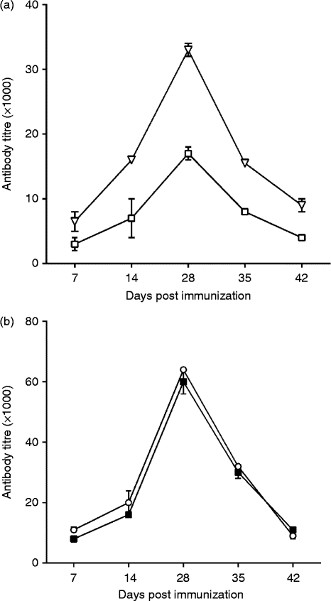
The antibody titre induced by TRX and TPX as single antigens and in combination was analysed in M. coucha. TRX when immunized alone induced a peak titre of 16,000 at day 28, whereas when immunized along with TPX, it showed a twofold increase of 32,000 (fig. 6). On the contrary, TPX induced a high peak antibody titre of 64,000, both when immunized individually and in combination with TRX. This suggests that TPX contributes to enhancing the levels of total IgG against TRX.
Antibody titres (mean ± SEM) induced by rTRX and rTPX as a single or cocktail mode in multimammate rats (Mastomys coucha): (a) total IgG against rTRX in the anti-TRX and anti-(TRX + TPX) sera (open squares,TRX; open triangles, TRX + TPX); (b) total IgG against rTPX in the anti-TPX and anti-(TRX + TPX) sera (closed squares, TPX; open circles, TRX + TPX).
The immunoprophylactic potential of TRX and TPX as a single antigen or in combination was studied by a micropore chamber experiment in M. coucha against L3 larvae. The chamber was recovered after 48 h and the numbers of live and dead L3 larvae were counted in the rats. Mean worm recovery was found to be 8.25 ± 0.67 and 7.25 ± 0.31 in the TRX and TPX immunized groups, respectively, compared to 19.01 ± 0.42 in the alum control group. Rats immunized with a mixture of TRX and TPX antigens showed a mean worm recovery of 5.5 ± 0.62. The percentage reduction in worm burden in the combination group (TRX + TPX) compared to alum control was 71.07%, which was significantly higher than that of the individual proteins, TRX, 56.6% (P < 0.001) and TPX, 61.86% (P < 0.02).
Discussion
Neglected tropical diseases cause havoc in underdeveloped countries by imparting long-term disability during the productive phase of the population. Current drugs, namely diethylcarbamazine (DEC) and albendazole, used for lymphatic filariasis control through mass drug administration (MDA) are not curative. These drugs act only on the microfilarial stages and do not eradicate infective-stage larvae or adult parasites. Moreover, numerous rounds of mass drug treatment will be necessary to reduce the levels of infection below those necessary to sustain transmission(Hoerauf, 2006).
Filarial parasites trigger a range of inflammatory damage in the host that ranges from lymphatic dysregulation to elephantiasis, along with secondary infections, leading to complete deterioration of the quality of life in patients with chronic pathology. Filarial nematodes survive in the immune tissues of the human host for many years under sustained stress from multiple assaults by immune cells. Hence, weakening the stress tolerance of the parasites by targeting their antioxidant enzymes will be a novel strategy in the control of filariasis (Chiumiento & Bruschi, Reference Chiumiento and Bruschi2009). LoVerde et al. (Reference LoVerde, Carvalho-Queiroz and Cook2004) has shown the protective role of antioxidants against challenge infection in S. mansoni.
The well-known antioxidant systems in mammals include enzymes such as superoxide dismutase (SOD), glutathione peroxidase (GPx) and various catalases (CAT). The list is limited in nematode parasites, including enzymes that deal exclusively with hydrogen peroxide which are less characterized (Henkle-Dührsen & Kampkötter, Reference Henkle-Dührsen and Kampkötter2001). Our study tends to focus on the protective immune response of the new family of peroxiredoxin (PRXs) enzymes which are abundantly present in parasitic nematodes (McGonigle et al., Reference McGonigle, Dalton and James1998). Efforts from genome sequencing and expressed sequence tag (EST) projects have shown that peroxiredoxins are distributed widely among both helminth and protozoan parasites (Blaxter et al., Reference Blaxter, Daub, Guiliano, Parkinson and Whitton2002).
Thioredoxins, in general, execute their function of protecting cytosolic proteins from aggregation or inactivation via oxidant-mediated formation of intra- or intermolecular disulphides. Filarial TRX has been shown to be an important source of reducing equivalents required to maintain the activity of antioxidant enzymes (Kunchithapautham et al., Reference Kunchithapautham, Padmavathi, Narayanan, Kaliraj and Scott2003). In addition, TRX proteins show immune regulatory effects by promoting cytokine release and they also control the DNA binding activity of immunologically active transcription factors.
Parasitic TPX is another antioxidant that is required for the survival of filarial parasites. Studies on thioredoxin peroxidase of Dirofilaria immitis suggest that, in addition to its antioxidant property, D. immitis TPX may also be a protective antigen (Klimowski et al., Reference Klimowski, Chandrashekar and Tripp1997). Brugia malayi TPX is a 25-kDa protein found to be present in all stages of the parasite (Ghosh et al., Reference Ghosh, Eisinger, Raghavan and Scott1998). Here, we report the protective responses of two antioxidant enzymes (WbTRX and WbTPX) in tandem approach when immunized as a cocktail in murine experimental filariasis.
Native antigens were found to be expressed at high levels in the infective L3 stage, confirming earlier reports (Ghosh et al., Reference Ghosh, Eisinger, Raghavan and Scott1998; Blaxter et al., Reference Blaxter, Daub, Guiliano, Parkinson and Whitton2002). Targeting the antigens highly expressed in the infective stage would enhance the parasite clearance by preventing the initial parasite establishment. Further, the expression of antioxidants in all stages of the parasite makes them ideal targets, since blocking their expression may completely hinder parasite survival.
Since both W. bancrofti TRX (TryX_like_family: cd02964) and TPX (PRX_Typ2cys family: cd03015) belong to the thioredoxin superfamily and share homology, having a common structural feature (the thioredoxin fold), the possible immunological cross-reactivity was analysed. Antigens showed considerable cross-reactivity which would enhance their mutual immunogenic potential. When cross-reacting antigens are immunized in a multivalent mode there is a possibility of synergistic enhancement of ADCC (antibody dependant cellular cytotoxicity), leading to parasite clearance.
Preliminary immune responses were tested in a mouse model to investigate the possible protective efficacy of the antigens. The isotype analysis showed a high level of IgG1 isotype induced by anti-TRX sera, and IgG2b isotype for anti-TPX sera, correlating with previous observations (Vanam et al., Reference Vanam, Pandey, Prabhu, Dakshinamurthy, Reddy and Kaliraj2009; Madhumathi et al., Reference Madhumathi, Prince, Anugraha, Kiran, Rao, Reddy and Perumal2010). Interestingly, both IgM and IgA levels were higher for TPX than for TRX, both of which have been reported to be involved in parasite clearance (Grezel et al., Reference Grezel, Capron, Grzych, Fontaine and Lecocq1993; Rajan et al., Reference Rajan, Ramalingam and Rajan2005). It has been established that infective (L3) larval and adult stages of the parasite primarily induce Type 2 responses, which are known to be protective against helminth infections. Also, a predominant IgG1 and IgM isotype has been proved to be indicative of a protective response (Lawrence, 2001). The majority of filarial vaccine studies have shown that the Th2 response, mostly mediated through IL-5, is essential for protection both in vaccine-induced and in primary resistance to infection.
TRX and TPX induced both IL-5 and IFN-γ, showing a classical mixed Th1/Th2 profile. Since there is a modified Th2 response in filariasis (Maizels & Yazdanbakhsh, Reference Maizels and Yazdanbakhsh2003), induction of a balanced response of both Th1 and Th2 types is essential for the skewed immune system to return to normal. Various reports have attributed IL-5 elevation to parasite clearance in murine models (Martin et al., Reference Martin, Al-Qaoud, Ungeheuer, Paehle, Vuong, Bain, Fleischer and Hoerauf2000; Lawrence, Reference Lawrence2001; Ramalingam et al., Reference Ramalingam, Ganley, Porte and Rajan2003; Hoerauf et al., Reference Hoerauf, Satoguina, Saeftel and Specht2005). Also, previous studies in IFN-γ knockout mice have shown that the absence of IFN-γ can increase the recovery of infective larvae in mice, suggesting its role in parasite killing (Babu et al., Reference Babu, Ganley, Klei, Shultz and Rajan2000).
The use of multiple antigens in a cocktail mode for a complex multicellular parasite would be necessary to achieve a high level of protection (Rainczuk et al., Reference Rainczuk, Scorza, Spithill and Smooker2004; Mendez et al., Reference Mendez, Zhan, Goud, Ghosh, Dobardzic, Wu, Liu, Deumic, Dobardzic, Liu, Bethony and Hotez2005). A recent study by Anand et al. (Reference Anand, Kodumudi, Reddy and Kaliraj2011) showed that an abundant larval transcript (ALT) and venom allergen homologue (VAH) cocktail vaccine conferred significantly high protection. However, the selection of antigens for a cocktail mix is crucial, since not all antigen mixtures are effective (Vanam et al., Reference Vanam, Pandey, Prabhu, Dakshinamurthy, Reddy and Kaliraj2009).
The promising preliminary results with TRX and TPX led further, to the study of their efficacy in a higher filarial model. In order to validate their prophylactic potential in combination, protection studies were carried out against B. malayi infective L3 larvae in the filarial model, M. coucha. Since W. bancrofti is strictly specific for human hosts and due to the high level of sequence similarity (>95%) between W. bancrofti and B. malayi antigens, the proteins were tested against B. malayi larvae.
The antibody titre in M. coucha indicated that W. bancrofti TPX is highly immunogenic. Interestingly, TPX greatly augmented the humoral responses induced against TRX when given as a cocktail, possibly due to the shared epitopes. The presence of cross-reactive epitopes is beneficial, as both the enzymes of the parasite are effectively targeted by the antisera. Antibodies may also act on the thioredoxin system of other parasites sharing conserved epitopes, making the cocktail a potential broad-spectrum vaccine.
The protection study in Mastomys confirmed the synergistic activity of both the antioxidants in parasite clearance. It should be noted that WbTPX, which induced a high titre, also showed a significantly higher percentage reduction in worm burden, presumably by ADCC, as expected (Lawrence, Reference Lawrence2001), compared to WbTRX. The elevated protection (71%) conferred by the multiple antigen group (TRX + TPX) suggests that targeting the antioxidant system as a whole, instead of individual proteins, is a promising approach in managing parasitic infections. Hence, a rational combination of secretory or surface antigens vital for parasite survival and spanning all stages (especially the infective stage) can be used in the multiple antigen vaccine strategies for effective clearance of filarial parasites.
Acknowledgements
P.R.P. wishes to acknowledge the Department of Biotechnology (DBT), New Delhi, India, for Junior/Senior Research Fellowship. The authors are thankful to Department of Biotechnology (DBT) (grant no: BT/01/COE/06/13), Government of India, for financial assistance to carry out this project.