Introduction
Fascioliasis, an infectious parasitic disease caused by Fasciola hepatica or F. gigantica, affects millions of people worldwide. Up to 17 million people are infected and around 91.1 million are at risk of infection (Keiser & Utzinger, Reference Keiser and Utzinger2005). Today, fascioliasis is recognized as an emerging and re-emerging vector (ungulate)-borne disease with the widest latitudinal, longitudinal and altitudinal distribution known for any zoonotic disease (Mas-Coma et al., Reference Mas-Coma2005). Hence, the World Health Organization has classified fascioliasis as an important human parasitic disease that merits international attention (Mas-Coma et al., Reference Mas-Coma2005).
Most of the people affected by this zoonosis are in the Andean Region of South America (Marcos & Terashima, Reference Marcos and Terashima2007; Marcos et al., Reference Marcos, Terashima and Gotuzzo2008a), where a prevalence of up to 71% has been reported by serology (Marcos et al., Reference Marcos, Maco, Florencio, Terashima, Samalvides, Miranda, Tantaleán, Espinoza and Gotuzzo2005a). In particular, in past decades more than 1700 people in Peru have been reported to be infected by F. hepatica (Marcos et al., Reference Marcos, Terashima, Leguia, Canales, Espinoza and Gotuzzo2007a). Fascioliasis is an important public health problem in these regions and deserves more attention.
The most common clinical complications caused by fascioliasis are biliary obstruction, cholecystitis, recurrent cholangitis, liver abscesses and subcapsular haemorrhages (Arjona et al., Reference Arjona, Riancho, Aguado, Salesa and Gonzalez-Macias1995; MacLean, Reference MacLean, Guerrant, Walker and Weller1999; Marcos et al., Reference Marcos, Tagle, Terashima, Bussalleu, Ramirez, Carrasco, Valdez, Huerta-Mercado, Freedman and Gotuzzo2008b), although half of the chronically infected patients can be asymptomatic in endemic areas (Marcos et al., Reference Marcos, Maco, Terashima, Samalvides and Gotuzzo2004).
Fasciola hepatica can produce proline (Campbell et al., Reference Campbell, Sheers, Moore, Edwards and Montague1981; Modavi & Isseroff, Reference Modavi and Isseroff1984), causing bile duct hyperplasia (Wolf-Spengler & Isseroff, Reference Wolf-Spengler and Isseroff1983) with deposition of collagen types I and III (Mark & Isseroff, Reference Mark and Isseroff1983). Similar changes are produced in cirrhosis of the liver and other pathological conditions, including wound healing. For many years, it has been speculated that liver fibrosis is present in fascioliasis but the studies have been limited to animals. For example, in a recent study carried out in Peru, half of a sample of 30 cattle livers examined by histology had liver cirrhosis. Interestingly, the number of parasites was associated with the degree of fibrosis (Marcos et al., Reference Marcos, Yi, Machicado, Andrade, Samalvides, Sanchez and Terashima2007b). In Zambia, fibrosis and calcifications in the livers of cattle were associated with Fasciola infection, with the more severe findings in the vicinity of the bile ducts (Phiri et al., Reference Phiri, Phiri, Sikasunge, Chembensofu and Monrad2006). In two other animal studies, thickened walls of bile ducts and severe cirrhosis were found by microscopic examination (Haridy et al., Reference Haridy, Ibrahim, Morsy and El-Sharkawy1999; Shirai et al., Reference Shirai, Sato, Shibuya, Naito and Tsukise2006). In goats, the number of parasites was associated with the severity of the liver lesions as well (e.g. hepatic calcareous granuloma), including marked cirrhosis and even death (Perez et al., Reference Perez, Martin de las Mulas, Carrasco, Gutierrez, Martinez-Cruz and Martinez-Moreno1999). In humans, liver cirrhosis has been reported in children (Almendras-Jaramillo et al., Reference Almendras-Jaramillo, Rivera-Medina, Seijas-Mogrovejo and Almendras-Jaramillo1997; Marcos et al., Reference Marcos, Maco, Castillo, Terashima, Zerpa and Gotuzzo2005b) and adults (Heredia et al., Reference Heredia, Bordas, Mondelo and Rodes1984; Sanchez-Sosa et al., Reference Sanchez-Sosa, Rojas-Ortega, Reed-San Roman and Torres-Santana2000) especially when those livers had massive infections.
In the diagnosis, a novel serological test has been developed recently for human fascioliasis in Peru. The enzyme-linked immunosorbent assay (ELISA) against the Fas2 antigen of the adult parasite has a sensitivity of 92.4%, specificity 83.6%, and a negative predictive value of 97.2% (Espinoza et al., Reference Espinoza, Maco, Marcos, Saez, Neyra, Terashima, Samalvides, Gotuzzo, Chavarry, Huaman, Bargues, Valero and Mas-Coma2007). This ELISA is currently used in the clinical setting as a confirmatory test when a patient has a clinical picture consistent with fascioliasis (Marcos & Terashima, Reference Marcos and Terashima2007).
Methods
In the light of this evidence, we hypothesized that F. hepatica infection might be present in subjects with established cirrhosis, and we assessed this hypothesis using the highly sensitive and specific serological test (Fas2 ELISA) to detect antibodies of F. hepatica in stored serum of cirrhotic subjects in the Gastroenterology Service of the Hospital Nacional Cayetano Heredia in Lima, Peru. Statistical analysis was performed using SPSS 9.0 for Windows (SPSS Inc., Chicago, Illinois USA). Proportions were compared using the χ2 test. A value of P < 0.05 was considered as the critical level of significance.
Results and discussion
The study was carried out in 44 serum samples from cirrhotic patients collected over a period of 6 years (1998–2003). Out of the 44 sera, 34 had serology to hepatitis B virus: hepatitis B surface antigen (HBsAg), 8.8% (+); hepatitis B core antibody (anti-HBc), 32.5% (+). Thirty-three sera were tested for hepatitis C antibodies (anti-HCV): 9.1% (+). All serum samples were also evaluated by Fas2 ELISA (Espinoza et al., Reference Espinoza, Timoteo and Herrera-Velit2005, Reference Espinoza, Maco, Marcos, Saez, Neyra, Terashima, Samalvides, Gotuzzo, Chavarry, Huaman, Bargues, Valero and Mas-Coma2007); 9.1% (n = 44) had antibodies to F. hepatica Fas2 antigen. The optical densities measured at 450 nm (OD450) by Fas2 ELISA of each patient were: case 1 = 0.415, case 2 = 0.313, case 3 = 0.432, and case 4 = 0.223. The test cut-off is 0.200 OD450. Fas2 ELISA is based on the capture of circulating IgG by a 25 kDa cysteine proteinase Fas2 (Córdova et al., Reference Córdova, Herrera, Ñopo, Bellatin, Náquira, Guerra and Espinoza1997, Reference Córdova, Reategui and Espinoza1999; Espinoza et al., Reference Espinoza, Timoteo and Herrera-Velit2005).
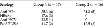
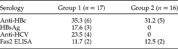
All Fas2 ELISA positive cases were males and their age mean was 63 years. In order to further examine these data, we compared the characteristics of cirrhotic patients with a possible aetiology and cirrhotic patients with unknown aetiology: Group 1, with chronic viral hepatitis and a history of heavy alcohol consumption (n = 17); and Group 2, without chronic viral hepatitis or a past history of heavy alcohol consumption (n = 16). The Fas2 ELISA was positive in 11.7% of Group 1 and 12.5% of Group 2 (P = 0.9) (table 1). The proportion of patients who came from endemic areas was similar; 55.5% of Group 1, and 62.5% of Group 2 (P = 0.68).
Serology (%) of patients with liver cirrhosis with a chronic (Group 1) and without a chronic (Group 2) viral infection; n=number of patients examined.
Out of the four cases positive by Fas2 ELISA, one had co-infection with hepatitis C; the second and third patients had neither hepatitis B nor C infection nor a past history of heavy alcohol consumption; and the fourth was negative to hepatitis C and B virus and had a history of mild alcohol consumption.
Interestingly, similar percentages for HCV, HBsAg and F. hepatica infection were seen in this series of cirrhotics. However, the high number of patients with cirrhosis of unknown aetiology (Group 2) suggests the involvement of other unknown factors besides viral infection and alcohol consumption (Barham et al., Reference Barham, Figueroa, Phillips and Hyams1994). The serum samples were collected without a matched control. Most patients had been living in the city of Lima (where fascioliasis is not endemic) for many years when blood was drawn, but most of them came originally from the highlands where they may have been infected many years ago. These preliminary results, even with the limitation of a small sample size, are a basis for a future clinical age-matched prospective study to analyse this finding on a greater scale.
Fasciola hepatica infection in cirrhotic patients is suggested by the presence of circulating anti-Fas2 IgG antibodies. Infected humans and animals elicit an early specific humoral immune response against the parasite by raising circulating specific anti-Fas2 IgG antibodies (Córdova et al., Reference Córdova, Herrera, Ñopo, Bellatin, Náquira, Guerra and Espinoza1997), to the extent that Fas2 is used as a sensitive and specific marker for human infection by F. hepatica (Espinoza et al., Reference Espinoza, Timoteo and Herrera-Velit2005, Reference Espinoza, Maco, Marcos, Saez, Neyra, Terashima, Samalvides, Gotuzzo, Chavarry, Huaman, Bargues, Valero and Mas-Coma2007). However, we do not know if cirrhotic patients with a positive Fas2 ELISA bear an active or resolved infection, since no coprological examination was performed at the time of clinical examination. In this study, however, the presence of IgG antibodies against Fas2 is strongly suggestive of F. hepatica infection.
In conclusion, whether F. hepatica infection, by itself or in addition to another disease or condition (e.g. HCV, HBV or alcohol consumption), is associated with the development of cirrhosis, is still an open question. Even though these preliminary results need consolidation, they do add a new dimension to involvement of this parasitic infection in chronic liver diseases in endemic countries. We propose detecting F. hepatica infection in patients with a liver disease who attend the gastroenterology services of hospitals in countries where this parasitic disease is endemic.
Acknowledgements
We are grateful to Olga Timoteo for performing Fas2 serology. There is no grant source to acknowledge and the authors have no conflicts of interest to declare.