Introduction
In the clinical field of otorhinolaryngology, tympanic membrane perforation is a relatively common disease. The main clinical features are hearing loss, ear purulence, earache, tinnitus and ear fullness. Myringoplasty, also known as type I tympanoplasty, is an important part of tympanoplasty. By reconstructing the tympanic membrane, myringoplasty can restore the anatomy and function of the tympanic membrane to prevent repeated middle-ear infection and improve the hearing of the affected ears.Reference Wullstein1
Microscopy surgery is the main method of otology procedures. In the past, endoscopy was usually used as a tool to assist in exploring the ear cavity.Reference Marchioni, Villari, Mattioli, Alicandri-Ciufelli, Piccinini and Presutti2–Reference Bennett, Zhang, Labadie and Noble5 With the popularisation of minimally invasive technology and the development of endoscopic surgery, endoscopy has gradually become widely used in otology.Reference Pap, Id-Orcid, Toth, Gede, Hegyi and Szakacs6 Endoscopic surgery has the benefits of a wide field of vision, minimal trauma and a short operation time without the bone removal and with less blood loss than in myringoplasty, attic cholesteatoma surgery, stapes surgery, second-look tympanotomy and reconstruction of ossicles surgery.Reference Glikson, Yousovich, Mansour, Wolf, Migirov and Shapira7–Reference Vincenti, Pasanisi, Bacciu and Bacciu9 These benefits make endoscopic surgery especially suitable for middle- and inner-ear surgery. The myringoplasty in this study was performed under endoscopy.
To expose the operation field, it is usually necessary to make a circular incision through the annulus tympanicus to form a tympanomeatal flap.Reference Tseng, Lai, Wu, Yuan and Ding10 However, some patients will occasionally develop external auditory canal scars, external auditory canal atresia and lateralised tympanic membranes after surgery, which have a certain impact on the patient's quality of life. Often a second operation is needed to expand the external auditory canal and to reconstruct the external auditory canal.Reference Bajin, Yilmaz, Gunaydin, Kuscu, Sozen and Jafarov11–Reference Sperling and Kay13
The feasibility of ear surgery under endoscopy has been recognised by some surgeons, but most studies have focused on the comparison of microscopic and endoscopic surgery. There are few studies comparing the difference between the transcanal approach and tympanomeatal flap approach in endoscopic myringoplasty. We analysed patients with small- and medium-sized perforations in recent years and compared the difference in clinical data between the transcanal approach group and the tympanomeatal flap approach group.
Materials and methods
From 2015 to 2017, 132 cases of chronic suppurative otitis media were analysed retrospectively. The patients were divided into two groups according to the operation method: the no tympanomeatal flap approach group (group A, 56 ears) and the tympanomeatal flap approach group (group B, 76 ears).
The inclusion criteria were: (1) the patient was diagnosed with chronic suppurative otitis media by clinical examination; (2) the patient had ear surgery for the first time on the affected side; (3) no cholesteatoma, blood diseases, cancer or other diseases were present that may have affected the research data; (4) the ossicular chain was complete with no pathological changes in the tympanum or tympanic sinus; and (5) the patient provided informed consent.
The exclusion criteria were: (1) patients with stenosis of the external auditory canal; (2) patients with marginal perforation; and (3) patients with a large perforation (perforation diameter more than 5 mm).
Equipment and instruments
Endoscopy was performed using a Karl Storz (Neuhausen, Germany) hard tube ear endoscope, with outer diameters of 2.5 mm and 3.0 mm, angles of 0° and 30°, and length of 14 cm. The equipment had a cold light source and an endoscopic imaging system, and conventional ear microsurgery instruments were used.
Operative method
The patient was in a supine position under general anaesthesia, with the affected ear facing up and routinely sterilised. The tragal perichondrium of the affected ear was taken, and a glue-bonded tragus incision was made to treat the edge of the perforation and make a fresh wound. There was no tympanomeatal flap in group A. In group B, the tympanomeatal flap was performed at a distance of approximately 5 mm from the outer side of the annulus tympanicus. The tympanum was explored to confirm that there was no diseased tissue in it, that the tympanic opening of Eustachian tube was unobstructed and that the ossicular chain was complete and active. The middle-ear cavity was filled with gelatine sponge. The spare tragal perichondrium was used to repair the tympanic membrane. The tympanic membrane was separated from the manubrium of the malleus so that the perichondrium was located under the malleolus. Patients in Group A were not treated with a tympanomeatal flap. Patients in group B were treated with the tympanomeatal flap. Finally, the external ear canal was filled with gelatine sponge (Figure 1).
Images of operation method of group A showing: (a) original perforation, (b) the perforated edges that are treated for fresh wounds, (c) the underlay method for myringoplasty (cartilage-perichondrium autografting placement with a suction), and (d) successful placement of cartilage-perichondrium autografting.

Post-operative follow up
Patients took oral antibiotics for the duration of the first week after surgery. Follow-up was performed at one week and at one, two, three and six months after the operation, and the total length of follow up was six months. The external ear canal packing was removed one week after the operation.
Observation indicators
The time of operation, the amount of intra-operative blood loss, the degree of hearing improvement, the rate of hearing improvement, the dry ear time (when the external auditory canal and the operating cavity were dry) after operation and the success rate of tympanic membrane repair were recorded.
Hearing test
At each follow up, the patient's hearing level was evaluated by pure tone audiometry with a pure tone audiometer. According to standard test protocols, the test was performed in an acoustic room without environmental noise. The air and bone conduction thresholds were calculated at 500, 1000 and 2000 Hz, respectively. The above frequencies represented the range of speech frequency, and the increase of these frequency thresholds would have clinical significance. The air–bone gap (ABG) was calculated by subtracting the bone conduction threshold from the air conduction threshold. A post-operative ABG of equal to or less than 20 dB HL indicated hearing improvement.
Statistical processing
Data were processed using SPSS® (version 18) statistical software. Count data were described as the number of cases and percentage. Measurement data were described as the mean ± standard deviation. Count data were analysed using the chi-square test or Fisher's exact test. Measurement data were analysed using a t-test. Statistical significance was set at p < 0.05.
Results
Demographics and baseline characteristics of the two groups
There were no significant differences between the two groups in age, gender, side, perforation size distribution, duration of disease or initial air conduction (Table 1).
Demographics and baseline characteristics of patients in the two groups

*Chi-square test; †independent-sample t-test. SD = standard deviation; TM = tympanic membrane
Comparison of intra-operative data between the two groups
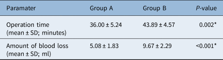
The average operation time of group A was 36.00 ± 5.24 minutes, which was significantly shorter than the average operation time of group B, which was 43.89 ± 4.57 minutes (p = 0.002). The blood loss of group A was 5.08 ± 1.83 ml, which was significantly less than that of group B (9.67 ± 2.29 ml; p < 0.001) (Table 2).
Intra-operative outcomes between the two groups

* Independent-sample t-test. SD = standard deviation
Hearing condition of the two groups
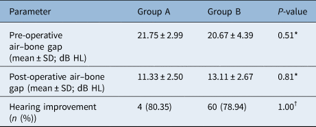
Before the operation, the ABG was 21.75 ± 2.99 dB HL in group A and 20.67 ± 4.39 dB HL in group B (p = 0.51), and there was no significant difference between the two groups. Six months after the operation, the ABG was 11.33 ± 2.50 dB HL in group A and 13.11 ± 2.67 dB HL in group B (p = 0.81), and there was no significant difference. At the end of the follow up, 45 patients (80.35 per cent) in group A and 60 patients (78.94 per cent) in group B had improved hearing. There was no significant difference between the two groups (p = 1.00; Table 3).
Comparison of hearing outcomes between the two groups

*Independent-sample t test; †chi-square test
Post-operative basic conditions of the two groups
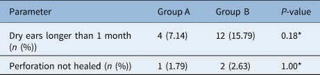
There were 4 cases (7.14 per cent) in group A and 12 cases (15.79 per cent) in group B with a dry ear time exceeding 1 month, and the difference was not statistically significant (p = 0.18). One case (1.79 per cent) in group A and two cases (2.63 per cent) in group B had an unhealed perforation, and the difference was not statistically significant (p = 1.00; Table 4).
Post-operative common data of patients in the two groups

* Chi-square test
Post-operative complications
One patient in group A and three patients in group B had persistent otorrhoea. Post-operative CT examination also confirmed middle-ear infection, but it was resolved after treatment with otic and oral antibiotics. None of the patients experienced sensorineural hearing loss, taste changes or had an implantation cholesteatoma in the two groups.
Discussion
The rise of otology surgery began in the 19th century, but breakthroughs were made in the middle of the 20th century. In 1952, Wullstein used operative microscopy in otology, which laid the foundation for otology microsurgery. Subsequently, surgical microscopy was widely used, which continuously improved middle-ear microsurgery technology.
The surgeon can only observe the field of view in front of the objective axis when using microscopy. To better expose the surgical field, they must elevate a tympanomeatal flap to better show the tympanic cavity. During ear microsurgery, classical operations, as devised by William House and Ugo Fisch, invariably elevated a tympanomeatal flap during tympanoplasty. Endoscopy gives a 0°, 30° or 70° lens to choose from and a wide field of vision. It is not affected by ear canal stenosis or bending. The endoscope can reach the tympanic membrane through the ear canal and even cross the tympanic membrane to further observe the pathological changes in the tympanum. Therefore, a question worthy of consideration is whether the elevation of a tympanomeatal flap is necessary for endoscopy.
Transtympanic myringoplasty, the technique of not elevating a tympanomeatal flap, was first reported by Shea in 1960.Reference Shea14 Singh et al. first reported 25 cases of chronic suppurative otitis media where patients underwent microscopic tympanoplasty with the transtympanic myringoplasty technique in 2006, and all of them achieved good results.Reference Singh, Sharma and Singh15 In 2010, Alzoubi et al. compared the difference between transtympanic myringoplasty and the normal tympanomeatal flap technique in microscopic tympanoplasty. They found that the former was better than the latter in terms of hearing improvement for small- to medium-sized perforations.Reference Alzoubi, Tarifi, Khader and de Carpentier16 Comparatively speaking, there are more reports of the transtympanic myringoplasty technique under endoscopy, which is generally called endoscopic transcanal myringoplasty. Ahmed et al. reported 36 cases of endoscopic transcanal myringoplasty in 1992, and all achieved good results. In 2001, Raj et al. compared the differences between a microscopic conventional myringoplasty group and an endoscopic transcanal myringoplasty group, with 20 patients in each group. The patients in the two groups achieved the same surgical effect.Reference Raj and Meher17 Since then, reports of the comparison between microscopic conventional myringoplasty and endoscopic transcanal myringoplasty have gradually increased. In 2016, Tseng et al. reported the surgical effect of endoscopic transcanal myringoplasty for anterior tympanic membrane perforation.Reference Tseng, Lai, Wu, Yuan and Ding18 However, there are few articles comparing transcanal myringoplasty and conventional myringoplasty under endoscopy. Only one paper was found, which was reported by Tseng et al.Reference Tseng, Lai, Wu, Yuan and Ding19
Before the operation, the ABG was 21.75 ± 2.99 dB HL in group A and 20.67 ± 4.39 dB HL in group B (p = 0.51), and there was no significant difference between the two groups. Six months after the operation, the ABG was 11.33 ± 2.50 dB HL in group A and 13.11 ± 2.67 dB HL in group B (p = 0.81), and there was no significant difference. The hearing results in this study are consistent with those reported by Tseng et al.Reference Tseng, Lai, Wu, Yuan and Ding19 In this study, the average operation time of group A was 36.00 ± 5.24 minutes, which was significantly lower than that of group B, which was 43.89 ± 4.57 minutes; p = 0.002). According to previous reports, the operation time of the group without a tympanomeatal flap is shorter than that of the group with a tympanomeatal flap, but the specific difference is not particularly consistent.Reference Alzoubi, Tarifi, Khader and de Carpentier16,Reference Tseng, Lai, Wu, Yuan and Ding20 We think that the specific time gap is related to the proficiency of the operator.
• The no tympanomeatal flap technique is suitable for small and medium perforations
• Tragal perichondrium was used to repair the tympanic membrane
• Blood loss was less in the no tympanomeatal flap group
• Average operation time was shorter in the no tympanomeatal flap group
• The tympanomeatal flap group and no tympanomeatal flap group achieved the same surgical results
Endoscopic transcanal myringoplasty requires the operator to be skilled in endoscopy. During the operation, the following aspects need to be completed: (1) fresh incisions with perforated edges should be made; (2) a check that the perichondrium is large enough; (3) the grafts and perforated edges should be well sealed; and (4) regular observation should occur after surgery. Although the transcanal myringoplasty technique has many advantages, not all tympanic membrane repair methods can be used. This method is not recommended if the tympanic mucosa has lesions, the external auditory canal is too narrow, or the anterior subtympanic perforation or tympanic membrane edge is too small.
Conclusion
The endoscopic transcanal myringoplasty technique is easy to perform. It reduces the operation time and leads to the same surgical results achieved in the tympanomeatal flap approach group. However, we need to increase the follow-up time of the two groups and conduct further prospective studies on their subjective and objective factors, results and theoretical basis to better understand the effect of endoscopic transcanal myringoplasty technology.
Acknowledgements
This study was supported by the National Natural Science Foundation of China (grant number: 81670933).
Competing interests
None declared