Kawasaki disease is the most common vasculitis in children resulting in coronary artery aneurysms in 25% of patients as its main complication. Reference Dietz, Tacke and Kuipers1
Myocarditis in the acute phase can result in ventricular systolic and diastolic dysfunctions and reduced strain values; however, it shows significant recovery after intravenous immunoglobulin administration. Reference Dionne2 Cardiac magnitude resonance has significant diagnostic value in Kawasaki disease and can be used to assess ventricular size and function and tissue oedema/inflammation, ischaemia, and fibrosis. Reference Pilania, Jindal, Bhattarai, Naganur and Singh3
Case presentation
A 9-year-old previously healthy girl was first admitted to a local hospital with prolonged high-grade fever and headache from a week before admission. Physical exam showed, tachycardia disproportionate to fever severity, with a pulse rate of 150 bpm, blood pressure of 110/70 mmHg, and temperature of 39. Lab data showed increased white blood cells: 16,410 cells/mm3, with neutrophil predominance: 65%. Erythrocyte sedimentation rate and C-reactive protein levels were normal. ECG showed no pathologic changes except sinus tachycardia. She underwent lumbar puncture, which came back normal.
For further workup of prolonged unexplained fever, echocardiography was performed, which showed preserved left ventricular systolic function, normal right ventricular size and function, and significant dilation of the proximal part of coronary arteries. With the diagnosis of Kawasaki disease, high dose aspirin (80–100 mg/kg/d) and intravenous immunoglobulin were administered, and the patient was referred to our tertiary centre for further workup. She experienced a transient episode of strabismus by this time, which resolved in 2–3 hours. Brain CT angiogram for embolic attack was negative, and infliximab was administered 5 mg/kg intravenously.
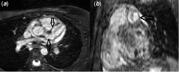
Cardiac magnitude resonance was performed on day 35 of initial admission and day 5 of referral. It showed a normal left ventricular size with mildly reduced systolic function, Left Ventricular Ejection Fraction (LVEF): 52%, oedema/inflammation in basal to the mid septum, with an increased regional T2 value of 66 ms, and high regional T1 value of 1115 ms with total extracellular volume of 27%. There was mid-wall fibrosis in the basal to the mid septum (Fig 1). Significant aneurysmal dilation of coronary arteries was evident with partial luminal thrombosis in whole heart and cine images (Fig 2, supplementary videos 1, 2). There were right coronary artery dilation from proximal to distal part (maximum diameter: 10 mm) and left anterior descending dilation from proximal to mid-segment (maximum diameter: 18 mm) with partial thrombosis.
Short axis (a) and 4 chamber long axis (b) T2 based Images showing Myocardial edema in the mid septal wall. Short axis (c) and 4 chamber long axis (d) Late Gadolinium Enhancement (LGE) images showing LGE in the mid septal wall in favour of active myocarditis.

(a) Axial and (b) Short axis view of the whole heart sequence showing aneurysmal dilation of the Left Main (LM) and Left Anterior Descending (LAD) arteries with partial thrombosis.

The patient was discharged with preserved LVEF of 50% and with the following medications: aspirin 80 mg (5 mg/kg/d), warfarin 2.5 mg daily, prednisolone 5 mg daily, and carvedilol 3.125 mg every 12 hours.
According to the patient’s aneurysmal coronary arteries and myocardial injury, long-term follow-up with subsequent echocardiography every 6 months was planned.
Discussion
We present a case of Kawasaki with giant coronary artery aneurysm and concomitant active myocarditis, which is in keeping with non-ischaemic active myocardial injury. Employing cardiac magnitude resonance showed the comprehensive effects of Kawasaki disease in the heart, from coronary aneurysms to myocardial injury. Our patient was referred for cardiac magnitude resonance in the sub-acute phase of the disease in the presence of normal erythrocyte sedimentation rate and C-reactive protein levels and after intravenous immunoglobulin and infliximab administration. The cardiac magnitude resonance showed ongoing active myocarditis and myocardial injury. However, ischaemic myocardial injury is the most prevalent type of fibrosis in the late phase of Kawasaki disease patients.
In Kawasaki disease, acute systemic inflammatory process results in disruption of the medial and internal elastic layer of the major coronary vessel, causing vasculopathy and aneurysm formation. As a result of simultaneous stagnant coronary blood flow and active inflammation, myo-intimal proliferation and/or layering thrombosis can happen in the vessel lumen, leading to ischaemic cardiomyopathy. Reference Dietz, Tacke and Kuipers1
According to echocardiographic studies, induced myocarditis is universal in all patients in the acute phase causing transient left ventricular dysfunction in more than 50% of the patients. It usually occurs around the coronary arteries due to inflammatory cell shedding from perivasculitis and shows almost complete recovery after timely intravenous immunoglobulin administration. Reference Dionne2 Rarely, severe myocarditis can show degenerative changes and lead to cardiomyopathy. Pathological findings in patients with Kawasaki disease are mostly related to oedema and inflammation. Myocardial cell necrosis is unusual as opposed to viral myocarditis. Reference Pilania, Jindal, Bhattarai, Naganur and Singh3
It has been shown that myocardial fibrosis following Kawasaki disease is primarily consistent with myocardial infarction from coronary artery aneurysm, leading to ischaemic cardiomyopathy with subendocardial late gadolinium enhancement patterns in the coronary territories. However, myocardial fibrosis may develop secondary to Kawasaki disease-induced myocarditis affecting the patient’s long-term outcome, the likelihood of mortality, sudden cardiac death, and arrhythmias. These patients are prone to developing cardiomyopathy over time. Reference Dusenbery, Newburger, Colan, Gauvreau, Baker and Powel4
According to the American Heart Association (AHA) scientific statement for treatment and management of Kawasaki disease, patients with definite Kawasaki disease should receive high dose aspirin (80 mg/kh/d) accompanied with intravenous immunoglobulin 2 gr/kg in the acute phase. In patients with limited response to intravenous immunoglobulin, support with other anti-inflammatory agents, for instance, infliximab, is recommended to reduce resistance to treatment and improve outcomes. For management of patients with giant coronary aneurysms (absolute dimension >= 8mm), therapy with low dose aspirin and warfarin is recommended, and prednisolone for its anti-inflammatory effects. Reference McCrindle, Rowley and Newburger5
Cardiac magnitude resonance is a valuable modality for evaluating Kawasaki disease activity and cardiomyopathies in active and chronic phases of Kawasaki disease in both diagnostic and prognostic pathways.
Supplementary material
To view supplementary material for this article, please visit https://doi.org/10.1017/S1047951122002797
Financial support
This research received no specific grant from any funding agency, commercial, or not-for-profit sectors.
Conflicts of interest
None.