INTRODUCTION
The primary cause of death in schistosomiasis is the formation of liver egg granulomas and secondary hepatic fibrosis. Studies have shown that granuloma inflammatory reaction and fibrosis in liver tissue continued to be aggravated, even though efficacious schistosomicides were given (Cioli and Pica-Mattoccia, Reference Cioli and Pica-Mattoccia2003; Southgate et al. Reference Southgate, Rollinson, Tchuem Tchuente and Hagan2005; Gryseels et al. Reference Gryseels, Polman, Clerinx and Kestens2006). The intervention and control of such aggravation during or before the formation of granuloma, or at the early stage of fibrosis become another key therapeutic strategy after efficacious treatment of praziquantel. However, so far, no anti-fibrosis drug with low toxicity and high efficiency has been applied to prevent or reverse the hepatic fibrosis in schistosomiasis.
Liver fibrosis is characterized by 2 major events (Zhang, Reference Zhang, Zhang, Jin, Zhou, Xie, Guo, Zhang and Qian2001): proliferation of hepatic stellate cells (HSCs) and an increase of synthesis of extracellular matrix, particularly collagen type I (Col I) and collagen type III (Col III). Fibrotic liver injuries result in activation of macrophages and release of fibrogenic cytokines, such as transforming growth factor-beta1 (TGF-β1) and so on, which activate HSCs to fibrogenic myofibroblast-like cells that produce much of the excess Col I and III (Matsuoka et al. Reference Matsuoka, Zhang and Tsukamoto1990).
The hepatosplenic schistosomiasis is characterized by a granulomatous inflammatory response and fibrosis evoked by SEA (soluble egg antigen) secreted from eggs of S. japonicum (Boros, Reference Boros1989). Macrophage cells are important in granuloma formation as they constitute one of the main cell types in granulomas (Mansy, Reference Mansy1998). HSCs have a major role in hepatic fibrosis of human schistosomiasis (Chang et al. Reference Chang, Ramalho, Ramalho, Martinelli and Zucoloto2006). It remains unclear whether macrophages, in hepatic fibrosis of schistosomiasis, can be induced to produce fibrogenic cytokines by SEA, and whether macrophage-conditioned medium elicited by SEA can promote HSCs proliferation and collagen synthesis.
Paeony (Paeoniae radix) root is one of the most well-known herbs in China. Paeoniflorin (PAE, C23H28O11) is known to be one of the principal bioactive components of paeony root. PAE has been reported to have immunoregulatory (Tsuboi et al. Reference Tsuboi, Hossain, Akhand, Takeda, Du, Rifa'i, Dai, Hayakawa, Suzuki and Nakashima2004) and anti-inflammatory (Yamahara et al. Reference Yamahara, Yamada, Kimura, Sawada and Fujimura1982) effects. Preparations of many traditional Chinese herbs used in anti-hepatic fibrosis contain paeony root. However, the mechanism by which it elicits anti-hepatic fibrosis in schistosomiasis has not been elucidated.
In this report we present evidence, for the first time, that SEA can induce the production of TGF-β1 from mouse peritoneal macrophages (PMφs), and that the TGF-β1 in mouse peritoneal macrophage-conditioned medium (PMCM) induced by SEA can promote mouse HSCs proliferation and collagen synthesis from HSCs. Furthermore, PAE can inhibit HSC proliferation and collagen synthesis by down-regulating Smad3 gene expression and phosphorylation through TGF-β signalling.
MATERIALS AND METHODS
SEA preparation
SEA was prepared using the method described previously (Harn, Reference Harn, Mitsuyama and David1984; Li et al. Reference Li, Di, Fu, Tang, Zhang, Cai and Wu1984). Freeze-dried eggs (60 mg) were mixed with an appropriate volume of silicon dioxide and 15 ml of sterile PBS (0·01 m, pH 7·2), and ground until no intact egg could be seen under the microscope. Following centrifugation of the mixture for 20 min at 2000 g, at 4°C, the crude supernatant was harvested and ultracentrifuged for 90 min at 100 000 g, at 4°C. The final supernatant (containing SEA) was sterilized by being passed through a 0·2 μm syringe filter, and then the SEA protein concentration was determined by the Lowry method (Lowry et al. Reference Lowry, Rosebrough, Farr and Randall1951).
Isolation and culture of PMφs
Male BALB/c mice, 6–8 weeks old (18–20 g), were obtained from the Laboratory Animal Center of Anhui Medical University (Anhui, China). PMφs were obtained according the method previously described (Takakura et al. Reference Takakura, Kiyohara, Murayama, Miyazaki, Miyoshi, Shinomura and Matsuzawa2002). Cellular viability was determined in each experiment by the trypan-blue exclusion test. The purity of the cell population was determined by morphology and non-specific esterase staining.
The purified PMφs were cultured in Dulbecco's modified Eagle's medium (DMEM, Gibco, Grand Island, NY, USA) containing high glucose and 100 IU/ml penicillin and 100 μg/ml streptomycin, supplemented with 10% (v/v) fetal bovine serum (FBS, Hangzhou Sijiqing Biological Engineering Materials Co. Ltd, China) for 48 h, and then they were washed twice with PBS and serum-starved for 24 h with serum-free DMEM. The PMφs were applied for the following experiments.
Isolation and culture of HSCs
Mouse HSCs were isolated according to the method described by Geerts et al. (Reference Geerts, Eliasson, Niki, Wielant, Vaeyens and Pekny2001). Cellular viability was determined in each experiment by the trypan-blue exclusion test. The purity of the HSCs in primary culture was assessed by typical morphological features, mainly the presence of vitamin A droplets, and the positive immunocytochemical stain for desmin.
The HSCs were plated on uncoated plastic at a density of 1×10⁸ cells/l in DMEM supplemented by 10% FBS. After primary culture for 3–4 days, the HSCs were washed twice with PBS and serum-starved for 24 h with serum-free DMEM. The HSCs were applied for the following experiments.
PAE and SB-431542 cytotoxicity assays
To determine the potential cytotoxicity of PAE (Xuancheng BaiCao Plants Industry and Trade Co. Ltd, China) or SB-431542 (s4317, Sigma-Aldrich, Inc., St Louis, MO, USA) to HSCs, concentration-dependent cytotoxicity assays (Lappalainen et al. Reference Lappalainen, Jaaskelainen, Syrjanen, Urtti and Syrjanen1994) were initially performed at various concentrations of PAE or SB-431542 as used following other experiments. Cellular viability was assessed by morphology and trypan-blue exclusion test, and cell injury was quantitatively assessed by measurement of lactate dehydrogenase released from damaged or destroyed cells into the supernatants. Lactate dehydrogenase activity was measured using the Lactate Dehydrogenase Detection Kit (A020, Nanjing Jiancheng Bioengineering Institute, Nanjing, China) according to the directions of the manufacturer. The assays were performed in sextuplicate from at least 3 independent experiments.
Measurement of PMCM-stimulated HSC proliferation
PMφs were cultured with SEA at various concentrations (0, 2·5, 5, 10, 20, 40 mg/l) in serum-free DMEM for 48 h. The PMφs culture supernatant was collected and centrifuged for 7 min at 450 g, at 4°C and filtered with a 0·45 μm membrane filter. The TGF-β1 content in the supernatants was detected by enzyme-linked immunosorbent assay (ELISA) using TGF-β1 Emax ImmunoAssay System (G7590, Promega Corporation, USA). The optimal SEA concentration was selected according to the TGFβ1 content. Then stimulated PMCM was induced from PMφs with SEA at the optimal concentration and unstimulated PMCM was induced without SEA.
HSCs were cultured with stimulated PMCM or SEA at various dilutions (1:0, 1:2, 1:4, 1:8, 1:16, 1:32 v/v) in DMEM containing 0·5% FBS for 48 h. Then HSC proliferation was detected by 3-(4,5-dimethylthiazol-2-yl)-2,5-diphenyltetrazolium bromide (MTT, Sigma-Aldrich, Inc., St Louis, MO, USA) colorimetric assay (Zhang et al. Reference Zhang, Zhang, Jin, Zhou, Xie, Guo, Zhang and Qian2001).
Measurement of PMCM-stimulated collagen secretion by HSCs
HSCs were cultured with stimulated PMCM or SEA at various dilutions (1:0, 1:2, 1:4, 1:8, 1:16, 1:32 v/v) in DMEM containing 0·5% FBS for 72 h. Then the HSC culture media were collected, and centrifuged for 20 min at 450 g, at 4°C. The Col I or III concentration in supernatants was then determined by ELISA using Col I or III ELISA kits (Senxiong Technology Enterprise Co. Ltd, Shanghai, China) according to the directions of the manufacturer.
To extract cell layer proteins, the HSCs adhered to the bottom of the plates were lysed and the cell proteins were extracted using the method described previously (Vetter et al. Reference Vetter, Chen, Chang, Che, Liu and Chang2003). HSCs layer protein concentration was then measured by the Lowry method (Lowry et al. Reference Lowry, Rosebrough, Farr and Randall1951). Collagen secretion was determined by taking the ratio of the concentration of Col I or Col III to the concentration of HSC layer proteins. Data were expressed as collagen relative contents.
We selected stimulated PMCM at 1:2 as the optimum peritoneal macrophage-conditioned medium (OPMCM) for the following experiments.
Modulation of HSC proliferation and Col I and III secretion by TGF-β1 in PMCM
HSCs were cultured with SEA at 1:2 or unstimulated PMCM at 1:2 or OPMCM in DMEM containing 0·5% FBS for 48 h. The HSCs, with addition of unstimulated PMCM or OPMCM, were cultured in the presence or absence of 10 μg/ml monoclonal anti-TGF-β1 antibody (MAB240, R & D Systems, Inc., Minneapolis, USA). Conditioned media were collected and stored at −70°C until use. HSC proliferation was detected by use of the MTT colorimetric assay (Zhang, Reference Zhang, Yu, Gao, Wei, Ju and Xu2004).
Col I or III concentrations in the HSC-conditioned medium were measured by ELISA. Collagen secretion was determined by taking the ratio of the concentration of Col I or Col III to the concentration of HSC layer proteins. Data were expressed as collagen relative contents.
Modulation of OPMCM-stimulated HSC proliferation by PAE
Mouse HSCs were treated with OPMCM for 12 h before being treated with PAE (0, 7·5, 15, 30, 60, 120 mg/l) or colchicine (Sigma-Aldrich, Inc., St Louis, MO, USA, 1 μmol/l, as a positive control) for 48 h. Then HSC proliferation was detected by MTT colorimetric assay (Zhang, Reference Zhang, Zhang, Jin, Zhou, Xie, Guo, Zhang and Qian2001). The absorbance (A) was read on an ELISA reader at the test wavelength of 570 nm and reference wavelength of 630 nm. The inhibition rate of PAE on HSC proliferation stimulated by OPMCM was calculated by the following formula:

Modulation of OPMCM-stimulated HSC Col I and III secretion by PAE
Mouse HSCs were treated with OPMCM for 12 h before being treated with PAE (0, 7·5, 15, 30, 60, 120 mg/l) or SB-431542 (10 μmol/l, as a positive control) for 72 h. Col I or Col III concentrations in the supernatants were then determined by ELISA. Collagen secretion was determined as relative contents by taking the ratio of the concentration of Col I or III to the concentration of HSC layer proteins. The inhibition rate of PAE on collagen secretion by OPMCM-stimulated HSCs was calculated by the following formula:

HSC RNA purification and reverse transcription
Mouse HSCs were treated with or without OPMCM for 12 h before being treated with or without a variety of concentrations of PAE or SB-431542 for another 12 h to determine mRNA levels. Total RNA was isolated using Trizol reagent (15596-026, Invitrogen Corporation) following the manufacturer's instruction. Then DNase I digestion and first-strand cDNA synthesis were performed using Reverse Transcription System (A3500, Promega Corporation) according to the manufacturer's protocol.
Polymerase chain reaction (PCR) assay
For each reaction, 100 μl of PCR amplification reaction mix was prepared using Reverse Transcription System (A3500, Promega Corporation) following the manufacturer's instructions. Then, the cDNAs were amplified with various specific primers. Mouse glyceraldehyde-3-phosphate-dehydrogenase (GAPDH) primers were used as an internal control. The amplification was performed by 30 cycles of thermal cycling, each consisted of denaturation at 94°C for 45 s, annealing at 50°C for 45 s, and extension at 72°C for 45 s followed by a final extension step for additional 10 min. PCR products were quantified by use of mouse GAPDH internal standards.
The primers used in this study included: mouse Col I (α1) (GenBank, Accession number: NM007742), forward, 5′-GCCCGGAAGAATACG-3′, reverse, 5′-ACATCTGGGAAGCAAA-3′ (product size 204 bp); mouse Col III (α1) (GenBank, Accession number: BC052398), forward, 5′-GCTGCCATTGTTGGAGTTG-3′, reverse, 5′-TGCTTACGTGGGACAGTCAT-3′ (product size 335 bp); mouse Smad2 (GenBank, Accession number: NM010754), forward, 5′-GACTACACCCACTCCATTCC-3′, reverse, 5′-CACTTAGGCACTCAGCAAAC-3′ (product size 472 bp); mouse Smad3 (GenBank, Accession number: AF016189), forward, 5′-CTGGCTACCTGAGTGAAGATG-3′, reverse, 5′-TGGGAGACTGGACGAAAA-3′ (product size 411 bp); mouse Smad4 (GenBank, Accession number: NM008540), forward, 5′-GTGGCTGGTCGGAAAGG-3′, reverse, 5′-GTGCTGGTGGCGTTAGA-3′ (product size 380 bp); mouse Smad7 (GenBank, Accession number: AF015260), forward, 5′-ACTCGGTGCTCAAGAAACTC-3′, reverse, 5′-CCCAGGCTCCAGAAGAAG-3′ (product size 479 bp); mouse GAPDH (GenBank, Accession number: BC095932), forward, 5′-TCAACGGCACAGTCAAGG-3′, reverse, 5′-AAGTCGCAGGAGACAACC-3′ (product size 691 bp).
Western blotting
Mouse HSCs were treated with or without OPMCM for 12 h before being treated with or without a variety of concentrations of PAE or SB-431542 for another 24 h to analyse OPMCM-regulated protein levels. The treated HSCs were lysed and the cell proteins were extracted using the method described previously (Vetter, Reference Vetter, Chen, Chang, Che, Liu and Chang2003). Total HSC protein concentrations in the supernatants were then measured by the Lowry method (Lowry et al. Reference Lowry, Rosebrough, Farr and Randall1951).
The HSC proteins from each sample were analysed by SDS-polyacrylamide gel electrophoresis and transferred onto a nitrocellulose membrane by semi-dry transfer. Blots were probed overnight at 4°C with the following primary antibodies: anti-Col Iα1, anti-Col IIIα1, anti-Smad2/3, anti-phospho-Smad2/3, anti-Smad4, anti-Smad7 (all these primary antibodies from Santa Cruz Biotechnology Inc., Santa Cruz, CA, USA), and anti-β-actin antibodies (BM0627, Wuhan Boster Biological Technology Ltd). This was followed by incubation with the appropriate horse radish peroxidase-conjugated secondary antibody at various dilutions for 2 h. Detection was achieved by enhanced chemiluminescence using SuperSignal West Femto Maximum Sensitivity Substrate (34094, Pierce Biotechnology, Inc.) and exposed to film. Filters were quantified by use of the JD-801 gel image analysis system (Jiangsu JEDA Science-Technology Development Co. Ltd, China).
Statistical analysis
Data were expressed as means±s.d., and analysed by ANOVA, t-test and Pearson correlation. P<0·05 was considered statistically significant.
RESULTS
Isolation and culture of mouse PMφs and HSCs
The number of isolated PMφs ranged from 2×10⁶ to 3×10⁶ cells per mouse. PMφ viability was higher than 92% and the purity was approximately 96%. The initially isolated PMφs were spherules that adhered to plates, and some spread their pseudopods 4 h after seeding. The HSCs ranged from 3×10⁶ to 6×10⁶ cells per liver. HSC viability was higher than 90% and the purity was approximately 95%. During the first 24 h of HSC culture, the cells rounded up and flattened out. After 2–3 days in culture, HSCs developed a characteristic stellate shape.
Effect of PAE or SB-431542 on HSC viability and growth
HSCs were exposed to PAE or SB-431542 for 72 h, and the cells grew very well without obvious abnormal changes in morphology. Cellular viability was above 92% as determined by the trypan-blue exclusion test. Lactate dehydrogenase activity was measured by spectrophotometry and there was no statistical significance among these groups (P>0·05, respectively, data not shown). HSCs could be treated with PAE at concentrations up to 120 mg/l or with SB-431542 at 10 μmol/l for up to 72 h without impact on cell viability. Thus PAE or SB-431542 could be used in the following experiments.
Effect of SEA on production of TGF-β1 from PMφs
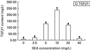
SEA at 10 mg/l could induce the highest production level of TGF-β1 from PMφs (Fig. 1). Therefore, SEA at 10 mg/l was regarded as the optimal SEA concentration. The proliferation of PMφs induced by SEA had no statistical significance among various concentrations (0, 2·5, 5, 10, 20, 40 mg/l) (data not shown).
Effect of various concentrations SEA on secretion of TGF-β1 from PMϕs. PMϕs were exposed to SEA (0, 2·5, 5, 10, 20, 40 mg/l) for 48 h. TGF-β1 content in the supernatants was assayed by ELISA. ■ There was statistical significance among all groups (P<0·05). Each bar represents the mean and standard deviation of triplicate determinations and data presented are representative of 3 independent experiments.

PMCM promotes HSC proliferation and collagen secretion
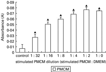
When cells were cultured with various dilutions of stimulated PMCM, HSCs proliferated remarkably compared with the control (P<0·01) (Fig. 2). The proliferation of HSCs induced by SEA had no statistical significance among various dilutions (1:0, 1:2, 1:4, 1:8, 1:16, 1:32 v/v) (data not shown).
Effect of PMCM on HSC proliferation. HSCs were cultured with or without stimulated PMCM for 48 h, and then HSC proliferation was detected by MTT colorimetric assay. Control, DMEM containing 0·5% FBS; stimulated PMCM dilution, stimulated PMCM:DMEM (v/v) containing 0·5% FBS. ▲P<0·01 compared with control group. Each bar represents the mean and standard deviation of triplicate determinations and data presented are representative of 3 independent experiments.

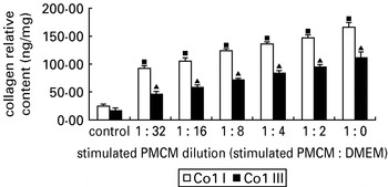
When cells were cultured with various dilutions of stimulated PMCM, Col I or Col III secretion from PMCM-stimulated HSCs was remarkable compared with the control (P<0·01, respectively). Col I or Col III protein was increased by almost 7-fold, respectively (Fig. 3). The secretion of Col I or Col III from HSCs induced by SEA had no statistical significance among various dilutions (1:0, 1:2, 1:4, 1:8, 1:16, 1:32 v/v) (data not shown).
Effect of stimulated PMCM on Col I or III secretion from HSCs. HSCs were cultured with or without stimulated PMCM for 72 h, and then Col I or III concentration in supernatants was determined by ELISA. Control, DMEM containing 0·5% FBS; stimulated PMCM dilution, stimulated PMCM:DMEM (v/v) containing 0·5% FBS. ■P<0·01 compared with the control group and P<0·05 compared among these groups; ▲P<0·01 compared with the control group and P<0·05 compared among these groups. Data from 3 experiments (each in triplicate) were normalized for HSC layer protein concentration per plate as Col I or Col III content (ng) per μg protein.

Effect of TGF-β1 in PMCM on HSC proliferation and Col I and III secretion by HSCs
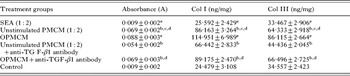
When HSCs were cultured in the presence of SEA alone, HSC proliferation was not observed and there was no difference between the relative contents of Col I or Col III secreted by HSCs compared with the control group (P>0·05, respectively). When HSCs were cultured in the presence of unstimulated PMEM or OPMCM, HSC proliferation was remarkable (P<0·01 vs control group, respectively) and there was a difference between the two groups (P<0·01). In addition, HSC proliferation promoted by unstimulated PMCM or OPMCM was suppressed partly in the presence of anti-TGF-β1 antibody (P<0·01, respectively). Meanwhile, when HSCs were cultured in the presence of unstimulated PMEM or OPMCM, the relative contents of Col I or Col III were markedly enhanced (P<0·01 vs control group, respectively), but there was a difference between the two groups (P<0·01). Furthermore, Col I or III secretion driven by unstimulated PMCM or OPMCM was partly blocked in the presence of anti-TGF-β1 antibody (P<0·01, respectively) (Table 1).
Effect of TGF-β1 in PMCM on HSCs proliferation and Col I and Col III secretion by HSCs (means±s.d., n=6)
(Control group, DMEM containing 0·5% FBS.)

a P>0·05 vs control group
b P<0·01 vs control group
c P<0·01 vs unstimulated PMCM (1:2)+anti-TGF-β1 antibody group
d P<0·01 vs OPMCM group
e P<0·01 vs control group.
Effect of PAE on the inhibition of OPMCM-stimulated HSC proliferation
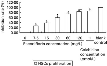
When HSCs were cultured with various concentrations of PAE, HSC proliferation driven by OPMCM was suppressed significantly in a concentration-dependent manner. The inhibition rate of HSCs proliferation was significantly correlated with the concentration change of PAE (Pearson's correlation coefficient r=0·857, P=0·029). PAE at 120 mg/l resulted in the highest inhibition rate of 83·58±3·28% on HSC proliferation, and the inhibitory effect was as strong as that of colchicine at 1 μmol/l (P>0·05) (Fig. 4).
Effect of PAE on proliferation of HSCs stimulated by OPMCM. HSCs were treated with OPMCM for 12 h before being treated with PAE (0, 7·5, 15, 30, 60, 120 mg/l) or colchicine (1 μmol/l) for 48 h, and then HSC proliferation was detected by MTT colorimetric assay. The negative control contained PAE at 0 mg/l; the blank control was PMCM containing neither HSCs nor PAE. •P>0·05 compared with the colchicine group; ▲P<0·01 compared with the control. Each bar represents the mean and standard deviation of triplicate determinations and data presented are representative of 3 independent experiments.

Effect of PAE on the inhibition of Col I or Col III secreted by OPMCM-stimulated HSCs
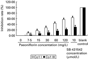
When HSCs were cultured with various concentrations of PAE, Col I or III secretion by OPMCM-stimulated HSCs were depressed significantly in a concentration-dependent manner. The inhibition rate of Col I or III secretion was highly correlated with the concentration change of PAE (Pearson's correlation coefficient r=0·952, P=0·003; r=0·940, P=0·005). PAE at 120 mg/l induced the highest inhibition rate of 57·14±1·78%, and 22·12±0·67% on Col I and III secretion respectively (Fig. 5).
Effect of PAE on Col I or III secreted by OPMCM-stimulated HSCs. HSCs were treated with OPMCM for 12 h before being treated with PAE (0, 7·5, 15, 30, 60, 120 mg/l) or SB-431542 (10 μmol/l) for 72 h, and then Col I or III concentration in supernatants was determined by ELISA. The negative control contained PAE at 0 mg/l; the blank control was PMCM containing neither HSCs nor PAE. ■ There was statistical significance among these groups (P<0·01); ▲ there was statistical significance among these groups (P<0·05). Each bar represents the mean and standard deviation of triplicate determinations and data presented are representative of 3 independent experiments.

Effect of PAE on OPMCM-induced expression of pro-collagen, type I, alpha1 (Col Iα1) and procollagen, typeIII, alpha1 (Col IIIα1)
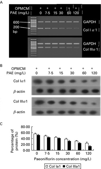
OPMCM induced the expression of Col Iα1, and Col IIIα1 mRNA (Fig. 6A) and protein (Fig. 6B) were significantly reduced in HSCs treated with PAE. Col Iα1 and Col IIIα1 proteins decreased by almost 2-fold and 3·5-fold, respectively (Fig. 6C).
PAE inhibits Col Iα1 and Col IIIα1 gene expression in OPMCM-stimulated HSCs. Cells were treated with OPMCM for 12 h before adding different PAE concentrations ranging from 0 to 120 mg/l for 12 h incubation for RT-PCR analysis or 24 h incubation for Western blotting. (A) Levels of Col Iα1 and Col IIIα1 mRNA were determined using RT-PCR. (B) Levels of Col Iα1 and Col IIIα1 proteins were measured by Western blotting. (C) Values of densitometric scan are means±s.d. of 3 experiments. Experiments were performed in triplicate and data presented are representative of 3 independent experiments.

PAE inhibits OPMCM-induced expression of Smad2/3 and Smad2/3 phosphorylation
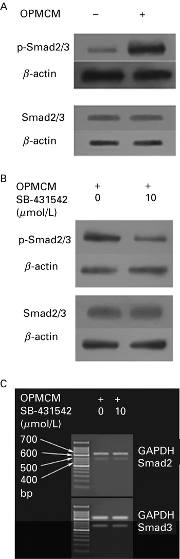
OPMCM induced a significant increase in the level of phosphor-Smad2/3, but exerted no change in Smad2/3 expression in HSCs (Fig. 7A). On the other hand, SB-431542, a selective inhibitor of type I receptor of TGF-β1, acts as a competitive ATP binding site kinase inhibitor and was shown to inhibit the in vitro phosphorylation of Smad2/3 (Callahan et al. Reference Callahan, Burgess, Fornwald, Gaster, Harling, Harrington, Heer, Kwon, Lehr, Mathur, Olson, Weinstock and Laping2002). SB-431542 effectively reduced the phosphorylation of Smad2/3, but had no effect on Smad2/3 protein, Smad2 mRNA and Smad3 mRNA in HSCs (Fig. 7B and C). Thus we presumed it was TGF-β1 in OPMCM that had induced phosphorylation of Smad2/3, and SB-431542 decreased the phosphorylation by inhibiting the receptor of TGF-β1.
SB-431542 specifically down-regulates the expression of Smad2/3 phosphorylation in OPMCM-induced HSCs. (A) Effect of OPMCM on expression of Smad2/3 and the phosphorylation of Smad2/3 (HSCs were treated with or without OPMCM for 36 h before Western blotting). (B) Effect of SB-431542 on expression of the phosphorylation Smad2/3 (upper panel) and Smad2/3 (lower panel) in HSCs induced with OPMCM. (C) Effect of SB-431542 on transcription of Smad2 and Smad3 in HSCs induced with OPMCM. HSCs were treated with OPMCM for 12 h before adding SB-431542 for 12 h incubation for RT-PCR analysis or 24 h incubation for Western blotting.

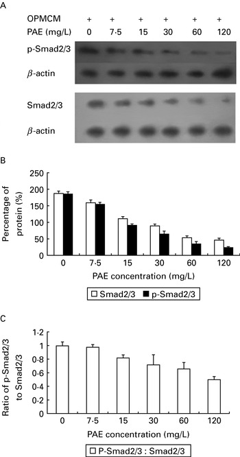
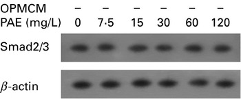
As shown in Fig. 8, levels of Smad2/3 protein and Smad2/3 phosphorylation were markedly down-regulated by PAE in a concentration-dependent manner. To test whether PAE can also inhibit Smad2/3 protein levels at basal state in HSCs, the cells were treated with various concentrations of PAE in the absence of OPMCM (Fig. 9).
Effect of PAE on the expression of Smad2/3 or Smad2/3 phosphorylation. HSCs were treated with OPMCM for 12 h before adding different PAE concentrations ranging from 0 to 120 mg/l for 24 h incubation for Western blotting. (A) The levels of phosphor-Smad2/3 protein (upper panel) and Smad2/3 protein (lower panel) were measured by Western blotting. (B) Values of densitometric scan are means±s.d. of 3 experiments. (C) Ratio of phosphor-Smad2/3 to Smad2/3. Experiments were performed in triplicate and data presented are representative of 3 independent experiments.

Effect of PAE on the expression of Smad2/3 without OPMCM stimulation in HSCs. HSCs were treated with different PAE concentrations ranging from 0 to 120 mg/l for 24 h incubation for Western blotting. PAE had no influence on Smad2/3 at protein levels even when the concentration was increased up to 120 mg/l. Experiments were performed in triplicate (data not shown).

PAE is a specific Smad3 inhibitor
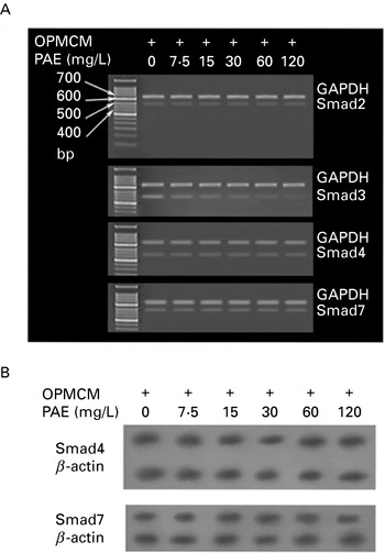
PAE not only inhibited the phosphorylation of Smad2/3, but also reduced the expression of Smad2/3 protein. Is PAE then a Smad3-specific inhibitor? To address this question, we examined by RT-PCR and Western blotting the gene and protein expression involved in the regulation of TGF-β signalling, including Smad2, Smad3, Smad4, and Smad7 mRNAs, and Smad4 and Smad7 proteins. As shown in Fig. 10, except for Smad3 mRNA, PAE had no influence on the mRNA levels of Smad2, Smad4, and Smad7 (Fig. 10A), and the protein levels of Smad4 and Smad7 (Fig. 10B), indicating that PAE was probably a selective inhibitor for Samd3.
PAE specifically down-regulates transcription of Smad3. HSCs were pre-incubated with OPMCM for 12 h before different PAE concentrations were added ranging from 0 to 120 mg/l for 12 h incubation for RT-PCR, or for 24 h for Western blot. (A) The levels of Smad2, Smad3, Smad4, and Smad7 were measured by PT-PCR. (B) The levels of Smad4, and Smad7 were measured by Western blotting.

DISCUSSION
Depending on the cytokine environment, macrophages can differentiate into distinct subsets that perform specific immunological roles. In this regard, the functions of macrophages activated by Th1 cytokines, such as interferon γ, are referred to as classically activated macrophages, whereas macrophages activated by Th2 cytokines, such as interleukin-13 and interleukin-4, are known as alternative activated macrophages (Gordon, Reference Gordon2003). In classically activated macrophages, interferon γ enhances the activity of nitric oxide synthase 2 to generate nitric oxide. In alternative activated macrophages, interleukin-13 and interleukin-4 enhance the activity of arginase and promote the synthesis of polyamine and proline, which can promote fibroblast proliferation and collagen production (Gordon, Reference Gordon2003). In addition, alternatively activated macrophages, by interleukin-13 and interleukin-4, can secrete TGF-β and platelet derived growth factor (PDGF) (Song et al. Reference Song, Ouyang, Horbelt, Antus, Wang and Exton2000; Gordon, Reference Gordon2003; Wynn, Reference Wynn2003).
During Schistosoma infection, with the onset of Schistosoma egg deposition in tissue, the Th1 immune response triggered by adult worms is down-regulted by macrophages, whereas the Th2 immune response develops and dominates (Pearce and MacDonald, Reference Pearce and Macdonald2002). Some components derived from Schistosoma eggs are responsible for the Th1 immune response to Th2 immune response switch during infection, because they can trigger the secretion of inerleukin-4 and interleukin-13 (Noel et al. Reference Noel, Raes, Hassanzadeh Ghassabeh, De Baetselier and Beschin2004). The 2 cytokines then activate macrophages by an alternative pathway, which produces polyamine, proline, TGF-β, and PDGF. These chemokines and growth factors can promote granuloma formation and fibrosis (Song et al. Reference Song, Ouyang, Horbelt, Antus, Wang and Exton2000; Hesse et al. Reference Hesse, Modolell, Laflamme, Schito, Fuentes, Cheever, Pearce and Wynn2001; Wynn, Reference Wynn2003; Noel et al. Reference Noel, Raes, Hassanzadeh Ghassabeh, De Baetselier and Beschin2004). Taken together, Schistosoma eggs can indirectly promote TGF-β secretion by macrophages alternatively activated by egg-induced inerleukin-4 and interleukin-13. The alternative activated macrophages play a key role in SEA-induced granuloma and fibrosis (Hesse et al. Reference Hesse, Modolell, Laflamme, Schito, Fuentes, Cheever, Pearce and Wynn2001).
In our study, PMφs or HSCs were cultured with SEA, and HSCs were also cultured with stimulated PMCM, which mimicked the process whereby SEA affected liver cells in vivo when schistosomiasis occurred. Our data showed that SEA could directly induce TGF-β1 secretion from PMφs without the presence of any other cells. The precise mechanism is unknown. Does SEA have a function similar to Th2 cytokines and alternatively activated macrophages or may SEA induce TGF-β1 by a certain signal transduction pathway in macrophages? Previous observations reported that liposomes composed of phosphatidylserine could directly induce TGF-β1 secretion from macrophages via the activation of extracellular signal-regulated kinase (Otsuka et al. Reference Otsuka, Tsuchiya and Aramaki2004). The mechanism of TGF-β1 secretion from macrophages stimulated by SEA needs to be investigated in future work.
Our study showed that the OPMCM promoted HSC proliferation more significantly, and had a more significant stimulative effect on Col I and III secretion than did the unstimulated PMCM. To determine whether the increased proliferation of HSC and increased collagen production were due to the elevated TGF-β1 secretion in OPMCM, we tested the effects of TGF-β1 with anti-TGF-β1 antibody. The results indicated that when HSCs were cultured in the presence of SEA alone, HSC proliferation could not be observed; however, unstimulated PMCM could promote HSC proliferation. When HSCs were cultured in the presence of OPMCM, HSCs proliferated more remarkably than did HSCs in the presence of unstimulated PMCM. It suggested that PMφs stimulated by SEA at optimal concentration could secrete more factors. Then we measured the relative contents of Col I and Col III in HSC-conditioned media. Our results showed that cytokines secreted from PMφs could promote the capability of single HSC to secrete collagens. This result was similar to that of our previous survey. In the present work, HSC proliferation and collagen production by unstimulated PMCM and OPMCM were suppressed partly in the presence of anti-TGF-β1 antibody, suggesting that HSC proliferation and collagen production might be related with TGF-β1 and some other factors from macrophages, such as platelet derived growth factor (Chen et al. Reference Chen, Lu, Xie, Zhang, Zhang, Wei, Jin and Guo2005), interleukin-4 (Maher, Reference Maher2001), because anti-TGF-β1 antibody could not completely suppress HSC proliferation and collagen production.
TGF-β1 can initiate TGF-β1-Smad3 signalling of HSCs in liver fibrogenesis, and then activated Smad3/Smad4 complexes translocate into the nucleus and immediately bind and activate targeting genes, including Col Iα1, Col IIIα1 (Verrecchia et al. Reference Verrecchia, Chu and Mauviel2001; Goumans et al. Reference Goumans, Lebrin and Valdimarsdottir2003). So, TGF-β1 is a potent stimulus for collagen synthesis (Roberts et al. Reference Roberts, Sporn, Assoian, Smith, Roche, Wakefield, Heine, Liotta, Falanga, Kehrl and Fauci1986; Varga et al. Reference Varga, Rosenbloom and Jimenez1987). However, recent data indicate that TGF-β1 can play a dual role, both inhibiting and promoting DNA synthesis and proliferation of HSCs (Zhang et al. Reference Zhang, Yu, Gao, Wei, Ju and Xu2004; Bachman and Park, Reference Bachman and Park2005; Purps et al. Reference Purps, Lahme, Gressner, Meindl-Beinker and Dooley2007).
Our investigation showed that TGF-β1 in OPMCM could not only promote collagen production, but also HSC proliferation. Given this, we presumed that SEA could induce secretion of, besides TGF-β1, other factors which also exerted an important influence on HSC proliferation and collagen production (Maher, Reference Maher2001; Chen et al. Reference Chen, Lu, Xie, Zhang, Zhang, Wei, Jin and Guo2005). Taken together, our study suggested that SEA could play a key role in hepatic fibrosis of schistosomiasis by inducing fibrogenic cytokines from PMφs.
PAE could inhibit the proliferation of HSCs, which were pre-incubated with OPMCM, in a concentration dependent manner. Furthermore, PAE could suppress Col I and II secretion from HSCs into culture supernatant, and repress Col Iα1 and IIIα1 expression at gene and protein levels.
Further, the mechanism of the PAE inhibiting expression of Col Iα1 and IIIα1 was investigated. First, we proved that OPMCM could induce phosphorylation of Smad2/3, but have no effect on expression of Smad2/3. Second, we proved that PAE could inhibit not only OPMCM-mediated expression of phosphorylation Smad2/3, but also Smad2/3. SB-431542, however, could inhibit only the phosphorylation of Smad2/3 by binding type I TGF-β receptor (Shi and Massague, Reference Shi and Massague2003). Obviously, PAE has a different mechanism for reduction of extracellular matrix components through TGF-β signalling from SB-431542 which failed to affect either the expression of Smad2 and Smad3 mRNA or the level of Smad2/3 protein. Third, to investigate the specificity of PAE to Smad3, we used RT-PCR and Western blotting to examine the effects of PAE on other Smads, including Smad2, Smad4 and Smad7 (Shi and Massague, Reference Shi and Massague2003), and on Smad4 and Smad7 proteins. Except for Smad3 mRNA decreased, the mRNA levels of Smad2, Smad4 and Smad7, and the protein levels of Smad4 and Smad7 were not changed in HSCs treated with PAE. This indicated that PAE specifically decreased the Smad3 mRNA expression. It suggested that paeoniflorin's attenuating impact on the level of phosphorylated Smad2/3 protein might be through the reduction of Smad3 gene expression. However, the reasons why PAE exhibits an inhibitory effect on the expression of Smad3 gene in the presence of OPMCM in HSCs are unknown, but seem to be involved in cross-talk between signalling pathways.
As we know, TGF-β1 plays an essential role in modulating extracellular matrix gene expression, and a growing body of evidence suggests that this is a Smad3-dependent signalling process. Because of the pleiotropic biological actions of TGF-β1 mediated by multiple signalling pathways, therapies targeting TGF-β1 expression/activation or binding of TGF-β1 to its receptors may potentially induce a number of unwanted side-effects. Agents that target specific signalling pathways downstream of the TGF-β1 receptors are more likely to have the desired effects while avoiding complications. Because Smad3 plays such a critical role in mediating the pathobiology of fibrotic disease, inhibition of Smad3 signalling could be a prime target for intervention in fibrotic conditions.
In our present study, we intended to elucidate the liver fibrogenic mechanisms of SEA and the anti-fibrosis mechanisms of PAE and, furthermore, to find a new target based on Smad3 signalling for challenging hepatic fibrosis of schistosomiasis. We demonstrated, for the first time, that SEA could stimulate HSC proliferation and collagen production by promoting secretion of TGF-β1 from PMφs, and PAE not only repressed HSC proliferation, but also selectively inhibited the expression of Smad3 mRNA, but not Smad2, Smad4, or Smad7, which in turn significantly attenuated the TGFβ1-induced expression of Col Iα1 and IIIα1 in HSCs. These results suggest that PAE exerts its anti-fibrogenic effects by inhibiting HSC proliferation and down-regulated Smad3 gene expression and protein phosphorylation through TGF-β1 signalling. This further confirmed Smad3's capacity as a pivotal mediator for TGF-β1 in modulating fibrogenic function and suggested that inhibition of Smad3 protein function may be a potential and effective therapeutic strategy against fibrogenesis.
The project is funded by the National Natural Science Foundation of China (Grant No. 30571631).