An aortopulmonary window is a rare congenital cardiac anomaly, accounting for from 0.2%–0.6% of all cardiac malformations.Reference Kutsche and Van Mierop1 It causes a significant left-to-right shunt, with pulmonary arterial hypertension and early onset congestive cardiac failure becoming evident within a few days to months of life.Reference Van Son, Puga and Danielson2
At our institution, we observed that few patients having an aortopulmonary window were not diagnosed correctly during the initial echocardiogram, the correct diagnosis being made subsequently at repeat echocardiography, cardiac catheterization, or in the operating room. Failure to diagnose this lesion can result in development of pulmonary vascular obstructive disease, the patient becoming inoperable. The objective of our study, therefore, was to analyze the factors that contributed to our initial failure to diagnosis the presence of an aortopulmonary window. We also analyzed and assessed the additional lesions associated with the windows.
Patients and methods
From May, 2002, to September, 2007, we diagnosed 50 patients with an aortopulmonary window at our institution. Of these, 48 underwent surgical repair. They included 31 boys and 19 girls, giving a ratio of male to female of 1.6 to 1. The mean age at the time of intervention was 1.7 years, with a range from 3 months to 17 years.
We reviewed the clinical features, echocardiographic findings and haemodynamic correlations of all the patients. We also correlated the echocardiographic findings with the per-operative observations.
Results
The diagnosis was made correctly by echocardiography during the initial evaluation in 46 of the 50 patients (92%). In the remaining 4 patients, the diagnosis was made either during repeated pre-operative echocardiographic evaluation, cardiac catheterization, or per-operatively. These 4 patients had the following presentations:
Case 1: An 8-month-old boy was diagnosed as having a large atrial septal defect, albeit with congestive cardiac failure, which could not be explained by the atrial septal defect alone. He was scheduled for follow-up 1 month after the start of medical management. On the repeat echocardiographic evaluation, during the second outpatient visit, he was found to have a large aortopulmonary window. This patient underwent surgery, with successful closure of both the window and the atrial septal defect. He had an uneventful postoperative recovery.
Case 2: A 2-year-old girl had extremely poor echocardiographic windows and was diagnosed to have a large patent arterial duct, mitral regurgitation, and severe pulmonary arterial hypertension. In the operating room, the surgical team found a large aortopulmonary window, with no evidence of a patent arterial duct, necessitating a change in the per-operative management. The post operative course, nonetheless, was uneventful.
Case 3: A 7-month-old boy was diagnosed echocardiographically as having double outlet right ventricle with a large subarterial interventricular communication and pulmonary arterial hypertension. These findings seemed to adequately explain the condition of the patient. Per-operatively, the patient was found to have an additional aortopulmonary window, which was closed surgically with normal postoperative recovery.
Case 4: The last of our cases with an incorrect initial diagnosis was a 17-year-old boy deemed to have a large ventricular septal defect with aortic overriding and muscular infundibular stenosis, albeit with minimal gradient across the right ventricular outflow tract as shown by Doppler interrogation. The low gradient was thought to be due to reduced flow. He had been asymptomatic until 16 years of age, but then had presented with dyspnoea on exertion, easy fatigability, cyanosis, clubbing, palpitations and giddiness. The pulmonary arteries were not noted to be remarkable on chest radiography. He underwent cardiac catheterization preoperatively with a diagnosis of tetralogy of Fallot, and unexpectedly was found to have an additional aortopulmonary window with severe pulmonary arterial hypertension, despite the severe infundibular narrowing. Catheterization data showed irreversible pulmonary hypertension, and he was advised to continue with medical management. In retrospect, his low infundibular gradient, which was earlier thought to be due to reduced flow, was in reality the consequence of the pulmonary arterial hypertension.
Associated lesions
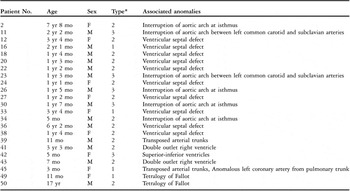
In our series of patients, the aortopulmonary windows were circular and located more or less equidistant between the sinutubular junctions and the bifurcation of the pulmonary trunk in 26 patients, spiral and involving the origin of the right pulmonary artery in 20, and large, extending from the sinutubular junctions to the bifurcation of the pulmonary trunk in the other 4.Reference Mori, Ando, Takao, Ishikawa and Imai3 Of the overall group, 16 had no associated malformations. Another 14 had relatively simple associated lesions, including persistent patency of the arterial duct in 9, defects of the oval fossa in 2, one of whom also had a co-existing persistently patent arterial duct, and patency of the oval foramen in 3, 2 of whom also had a persistently patent arterial duct. In the remaining 23 patients (46%), we found more complex associated malformations.Reference Jansen, Hruda, Rammeloo, Ottenkamp and Hazekamp4 Within this group with more complex associated lesions, the window itself was circular in 5, spiral in 14, and large, extending from the sinutubular junctions to the bifurcation of the pulmonary trunk in the remaining 4.Reference Mori, Ando, Takao, Ishikawa and Imai3 The associated lesions (Table 1) included: a ventricular septal defect in 10 patients, interruption of the aortic arch at the isthmus in 4 patients, interruption of the aortic arch between the common carotid and left subclavian arteries in 2, tetralogy of Fallot in 2, double outlet right ventricle in 2, discordant ventriculo-arterial connections in 2, 1 of whom also had anomalous origin of the left coronary artery from the pulmonary trunk, and superior-inferior ventricles in 1 patient.
Details of patients having associated anomalies with the so-called “complex” variants of aortopulmonary window

*Refers to the type of aortopulmonary window within the categorization of Mori and colleagues.
Discussion
An aortopulmonary window is a communication between the intrapericardial components of the ascending aorta and the pulmonary trunk, sometimes associated with overriding of the origin of the right pulmonary artery, with the presence of two separate arterial valves differentiating the lesion from a common arterial trunk.
We classified the lesions as suggested by Mori and associates,Reference Mori, Ando, Takao, Ishikawa and Imai3 with their Type I lesion being circular, and located between the sinutubular junctions and the bifurcation of the pulmonary trunk, their type II being more distal, taking a spiral form, and involving the origin of the right pulmonary artery, and their rare type III lesion being extensive and involving the entire area between the sinutubular junctions and the bifurcation of the pulmonary trunk. It should be noted that, although the lesion is often described as an “aortopulmonary septal defect”, there is no aortopulmonary septum in the postnatal heart, the intrapericardial components of the aorta and pulmonary trunk possessing their own discrete and separate walls. The lesion, therefore, is better described as an aortopulmonary window.
The lesions can be considered simple when having no associated cardiac defects, or being associated with defects requiring no or only minor intervention, such as a persistent patency of the arterial duct, a right aortic arch, or an atrial septal defect in the oval fossa or patency of the oval foramen. The so-called “complex” lesionsReference Jansen, Hruda, Rammeloo, Ottenkamp and Hazekamp4 have one or more associated complex cardiac anomalies requiring more complex repair, such as interruption of the aortic arch, a ventricular septal defect, cyanotic cardiac diseases, or coronary arterial anomalies. Around half of the patients have such complex defects, posing additional diagnostic and therapeutic difficulties.Reference Kutsche and Van Mierop1
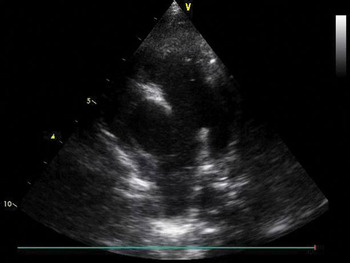
The presence of an aortopulmonary window must be strongly suspected in patients having unexplained early cardiac failure, or signs of significant left-to-right shunting associated with early pulmonary arterial hypertension, with or without functional mitral regurgitation.Reference Comin, Manito, Roca, Castells and Esplugas5 The electrocardiographic and radiological findings are non-specific, emphasizing echocardiography as the most important diagnostic tool (Fig. 1). Use of equipment with scant lateral resolution may produce an artificial echocardiographic dropout at the site of adjacency of the intrapericardial walls of the aorta and the pulmonary trunk, causing a false positive diagnosis.Reference Snider, Serwer, Ritter, de Young, Geistler and Patterson6 The diagnosis can also easily be missed if the echocardiographer does not evaluate carefully the area of adjacency of these walls, establishing their discrete and separate nature which is the feature of the normal heart.Reference Balaji, Burch and Sullivan7 Hence, interrogating this area of mural adjacency in several planes, initially without colour Doppler, is important. Once the lesions are defined, colour Doppler can be used to show low velocity bidirectional laminar flow even in unrestricted defects with pulmonary arterial hypertension. The spiral defects extending to incorporate the origin of the right pulmonary artery, or those large defects extending from the sinutubular junctions to the bifurcation of the pulmonary trunk, can be difficult to differentiate from persistent patency of the arterial ducts,Reference Prasad, Valiathan, Shyamakrishnan and Venkitachalam8 as was the case in our second patient in whom initially we failed to make the correct diagnosis.
Parasternal short axis view showing dropout in the area of the adjacent walls of the intrapericardial components of the aorta and the pulmonary trunk, suggestive of an aortopulmonary window.

In our overall series, nonetheless, echocardiography proved successful in demonstrating the presence of the window in over nine-tenths of our patients. The difficulty in making an accurate diagnosis at the initial echocardiographic study in our fourth patient may have been due to severe pulmonary arterial hypertension, with equalization of aortic and pulmonary pressures causing low flow through the defect. The size of the pulmonary arteries, along with vascular pruning as seen in the chest radiograph, can provide clues for the presence of pulmonary hypertension in such complex lesions. The chest radiograph, nonetheless, has been noted as being less than conclusive in diagnosing tetralogy of Fallot when co-exisitng with an aortopulmonary window.Reference Carminati, Borghi and Valsecchi9 Associated complex anomalies seemingly explaining the symptoms of the patient can also create a sense of complacency in the examiner, and can lead to the diagnosis being missed, as occurred in our third patient.
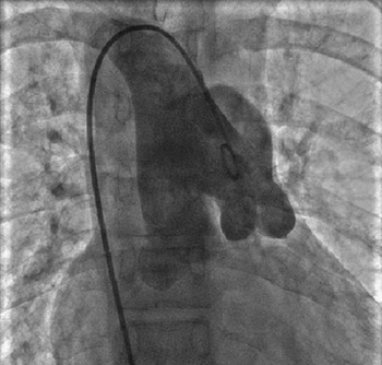
In patients where the primary diagnosis cannot explain the clinical scene, the final diagnosis should be confirmed by cardiac catheterization (Fig. 2).Reference Tirado, de Soto and Montero10 Recent reports on the use of helical computed tomography in accurate delineation of aortopulmonary windows may make angiography redundant in the future.Reference Sridhar, Kalyanpur, Suresh, Sharma and Maheshwari11 The lesion has also been imaged accurately using magnetic resonance imaging, again obviating the need for angiography.Reference Teo, Goldberg, Strouse, Vermilion and Bove12
An angiogram shown in frontal projection, with a pigtail catheter passed retrograde through a right aortic arch across an aortopulmonary window into the pulmonary trunk. Note the two distinct arterial valves.

The aortopulmonary windows are associated with other cardiac defects in more than half of patients, the most frequent being interruption of the aortic arch, usually at the isthmus, persistent patency of the arterial duct, ventricular septal defect, coronary arterial anomalies, and tetralogy of Fallot.Reference Faulkner, Oldham, Atwood and Graham13 We found ventricular septal defect to be the most frequent association, present in one-fifth of our cohort, as did another group,Reference Bertolini, Dalmonte, Bava, Moretti, Cervo and Marasini14 followed in our series by persistent patency of the arterial duct and interrupted aortic arch.
In conclusion, an aortopulmonary window is a rare congenital cardiac malformation, often associated with other significant intracardiac lesions. A high index of suspicion for the lesion is warranted in cases of unexplained severe pulmonary arterial hypertension, and/or unexplained dilation of the left heart. The lesion can accurately be diagnosed at the initial echocardiographic study in more than nine-tenths of cases. Even a meticulous echocardiographic examination, nonetheless, may produce false negative diagnoses. Hence, whenever the clinical situation and the echocardiographic diagnosis do not coincide, additional evaluation is mandated. So-called complex variants can pose a diagnostic challenge, and are easily overlooked, especially when the echocardiographic findings seemingly explain the clinical picture. If the lesion goes undiagnosed until the patient is in the operating room, the surgical plan can be jeopardised, sometimes with ruinous effect if the operating team is not equipped adequately for the altered management. This can be a serious issue at centres that are not used to correcting patients with complex congenitally malformed hearts. Hence, routine evaluation in each and every echocardiographic study to establish the presence of adjacent discrete and separate walls of the intrapericardial components of the aorta and the pulmonary trunk is well worth the effort.