Pseudomonas aeruginosa is a gram-negative nonfermenting bacillus in water and soil that has been implicated in healthcare-associated infections in intensive care settings. 1 , Reference Trautmann, Lepper and Haller 2 Patients in neonatal intensive care units (NICUs) are particularly vulnerable to infection because of underdeveloped immune systems, limited skin barrier functions, and exposure to invasive procedures and devices.Reference Polin, Denson and Brady 3 Multiple P. aeruginosa outbreaks have been documented in NICUs and have been attributed to contaminated medications, respiratory equipment, laryngoscopes, and personnel hand contamination.Reference Crivaro, Di Popolo and Caprio 4 – Reference Davis, Jensen and Van Hal 12 Pseudomonas aeruginosa outbreaks have also been associated with contaminated hospital tap waterReference Molina-Cabrillana, Artiles-Campelo and Dorta-Hung 11 – Reference Rogues, Boulestreau and Lasheras 14 because the organism is hardy and can form biofilms in plumbing fixtures that resist chemical treatment.Reference Williams, CR and MJ 15
In fall 2013, a P. aeruginosa outbreak involving 15 patients and 2 deaths occurred in a 28-bed NICU that admits 370–450 patients/year. The NICU has private patient rooms each with 1 sink, and is located in a newly constructed building that opened to patient care in May 2013. During outbreak response, the hospital notified the county health department, diverted admissions to another NICU, and performed water remediation, including installation of disposable membrane filtration point-of-use (POU) filters on faucets in November 2013. Environmental samples collected during November 2013–May 2014 from prefiltered water in the NICU yielded P. aeruginosa, but no strain types matched patient isolates. During May 21–June 9, 2014, new faucets were installed in the NICU and POU filters were removed. Starting June 22, 2014, a second cluster of cases occurred, including 1 patient death. Again, admissions to NICU were diverted. On September 23, 2014, the state health department invited the Centers for Disease Control and Prevention (CDC) to identify risk factors for transmission and provide recommendations to prevent additional cases. Herein, we present findings and recommendations from the CDC investigation.
METHODS
Case Definition and Case Finding
We defined a case as the first positive P. aeruginosa culture from a clinical or surveillance specimen in a patient admitted to the NICU between June 1, 2013, and September 30, 2014. Cases were identified through hospital microbiology record review. We classified cases as either clinical or surveillance based on the indication for testing. Surveillance specimens were collected during periods when unit policy mandated weekly cultures for all patients. This policy was in effect from November 26, 2013, to January 31, 2014, and from August 29, 2014, to September 30, 2014. Certain surveillance cultures were collected outside these periods at provider discretion.
Infection Control Assessment
We conducted observations of NICU infection control and clinical care. We conducted >10 hours of hand hygiene audits at 30–120 minutes per session (median length, 35 minutes) during all shifts. A hand-hygiene opportunityReference Boyce and Pittet 16 was marked unsuccessful if the hand-hygiene practice observed did not comply with the NICU policy. Hospital occupational health services examined NICU personnel hands and reviewed health histories regarding potential P. aeruginosa colonization (eg, history of otitis externa).
We also conducted audits of contact precautions and central-line insertion and maintenance practices using standardized audit tools. 17 We observed respiratory therapy, feeding practices, equipment reprocessing, pharmacy practices, environmental cleaning, an eye exam, and routine patient care.
Environmental Evaluation
We performed environmental sampling in NICU patient areas with the strongest epidemiologic links to cases (ie, rooms with multiple cases or a recent case) on September 29, 2014. To collect samples, we removed POU filters from faucets and collected 1-liter water samples. POU filters were replaced after sampling. We also collected swab samples from faucets and drains and sponge-stick samples from sink basins. We collected additional sponge-stick and swab samples from ventilator equipment, breast pumps, an incubator humidity outlet, and shelves adjacent to the patient room sinks. We collected water samples from pipes delivering hot and cold water to the floor where the NICU was located. Samples were submitted to CDC for culture by membrane filtration of water and by homogenization and extraction of swabs and sponge sticks. Nonlactose fermenting, pigmented colonies that grew on MacConkey selective agar were identified as P. aeruginosa using an automated biochemical identification system.
To determine relatedness, pulsed-field gel electrophoresis (PFGE) was performed at the CDC on 21 P. aeruginosa environmental isolates and 10 case isolates (5 surveillance and 5 clinical). We selected case isolates from the hospital laboratory that represented the most recent case patients or those with an epidemiologic link to environmental samples.
Risk Factors: Case-Control Study
To assess risk factors for positive P. aeruginosa culture, we conducted a case-control study. Surveillance and clinical cases were combined because the primary study focus was on assessing transmission risk factors. Control patients were deemed eligible if they (1) were admitted to the NICU between June 1, 2013, and September 30, 2014; (2) had a negative surveillance culture for P. aeruginosa; and (3) did not have any cultures positive for P. aeruginosa during the study period. We selected control patients randomly from an eligible patient list. Because case-patient birth weights were markedly lower than those of the general NICU population, we matched case patients 1:1 with a control patient according to the following birth weight categories: ≤1,000 g, 1,001–1,500 g, 1,501–2,500 g, and ≥2,501 g to ensure that control patients received similar exposures as case patients. The index date for controls was the most recent negative surveillance culture date.
We abstracted data from electronic medical records using a standard form that included the following data: demographics, maternal and obstetrical history, medical history and comorbidities, medication and device exposure, nutrition, signs and symptoms before positive culture, laboratory and microbiology findings, environmental exposure (ie, bed type and room number), exposure to NICU personnel, and clinical outcomes. We assessed exposures during the 7 days prior to positive culture or the index date for all variables except antibiotic exposure, which we assessed during the 30 days prior to positive culture.
Statistical Analysis
We performed descriptive analyses of characteristics and exposures of case patients and control patients. We used conditional logistic regression adjusted for gestational age to evaluate the association between case status and each exposure. We reported exact odds ratios and 95% confidence intervals (CIs) due to limited sample size. We pooled matched sets according to the recommendations of Kleinbaum and Klein,Reference Kleinbaum and Klein 18 resulting in models stratified by birth-weight category.
Ethical Review
The CDC National Center for Emerging and Zoonotic Infectious Diseases reviewed this investigation for human subjects’ protection and determined that it constituted an urgent public health response.
RESULTS
Case Finding and Confirmation
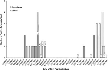
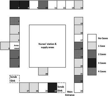
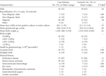
We identified 31 cases that occurred in 2 distinct clusters, September–November 2013 and August–September 2014 (Figure 1) and included 3 deaths. Clinical cases (n=17) consisted of blood, respiratory, urine, and eye cultures; surveillance cases (n=14) included respiratory, groin, and nasal cultures. Most case patients were Hispanic (58%); approximately half (52%) were male (Table 1). Approximately half (52%) had a birth weights of ≤1,000 g. For comparison, <10% of patients admitted to this NICU were extremely low birth weight (≤1,000 g) during the study period. Cases occurred in patients occupying different rooms and areas in the NICU (Figure 2).
Epidemic curve for Pseudomonas aeruginosa cases (n=31) including environmental and engineering interventions in the neonatal intensive care unit, June 2013–September 2014. Environmental and engineering interventions overlaying the epidemiologic curve. A, Aerators removed from faucets hospital wide. B, Laminar flow devices replaced in all neonatal intensive care unit (NICU) rooms. C, Hyperchlorination of NICU and part of labor and delivery. D, Descaling of all sink fixtures building wide. E, Laminar flow devices exchanged with antimicrobial devices. Sink fixtures cleaned building wide. F, Cleaning and disinfection of all faucet fixtures positive for P. aeruginosa. G, Point-of-use (POU) filters installed. H, NICU faucets replaced and POU filters removed. J, Hyperchlorination performed building wide and continuous chlorine dioxide treatment initiated.

Layout of neonatal intensive care unit with number of case patients occupying each room depicted. The number of case patients who occupied each room in the 7 days prior to positive culture is shown according to the legend.

Pseudomonas aeruginosa Outbreak in a Neonatal Intensive Care Unit Attributed to Hospital Tap WaterFootnote a

a Characteristics of case patients (n=31) and controls (n=31) matched by birth weight. Clinical exposures refer to the period 7 days before positive culture or incident date.
b Calculated using exact conditional logistic regression with pooled matched strata.
c Calculated using Fenton 2013 criteria.Reference Fenton and Kim 19
Infection Control Assessment
Before our investigation, the hospital implemented multiple infection control measures, including contact precautions for patients with a positive P. aeruginosa culture. NICU policy required personnel to use gloves during all patient contact.
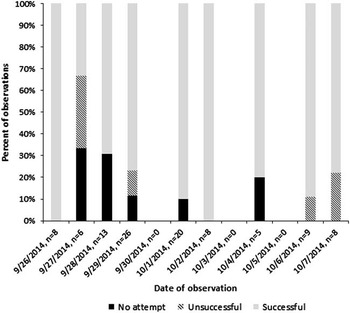
Our investigation revealed inconsistent hand-hygiene practices among direct patient care personnel, including use of soap and water over alcohol-based hand rub (ABHR) when not indicated (Figure 3). NICU policy was changed to specify preferential use of ABHR on October 2, 2014. No risk factors for colonization (eg, hot tub use) or skin breakdown on personnel hands were identified.
Hand-hygiene audit results from observations in the neonatal intensive care unit (NICU), September 26–October 10, 2014. Hand-hygiene audit results by date and success rating as a percentage of successful, unsuccessful, and no-attempt observations per total number of observations per day. A hand-hygiene opportunity was considered unsuccessful if the hand-hygiene practice observed did not comply with the NICU policy at the time of observation. The NICU policy for hand hygiene was to use either soap and water or alcohol-based hand rub (ABHR) until October 2, 2014, when it was changed to reflect a preference for ABHR unless hands are visibly soiled, according to Centers for Disease Control and Prevention guidelines.

Review of feeding practices revealed that breast pump parts were washed in tap water, according to manufacturer instructions; however, the manufacturer recommendation for daily sanitation of breast pump parts was not performed. Other infection control audits by the investigation team did not reveal breaches or other evidence of tap water exposure to NICU patients.
Environmental Evaluation
Our investigation revealed that although NICU patient care had been moved to the new hospital building in May 2013, water treatment, system flushing, and water testing after the construction process was complete were completed in February 2013. From February to May 2013, water in the hospital pipes was idle.
After the first cluster of NICU P. aeruginosa cases was identified in the autumn of 2013, interventions were exercised given suspicion that hospital water supply was the bacteria source. The hospital removed aerators from faucets; cleaned, disinfected, and removed mineral deposits on faucets and sink fixtures; and performed multiple hyperchlorination flushes of the building’s water system (Figure 1). The hospital also installed POU filters on all NICU faucets in December 2013. In May 2014, the hospital removed POU filters when NICU faucets were replaced with a different model thought less likely to become contaminated. After the second cluster of cases began in June 2014, POU filters were reinstalled on NICU faucets September 23, 2014, per our recommendation. A company contracted by the hospital sampled prefiltered water monthly from November 2013 to January 2014, and during May 2014 to detect P. aeruginosa.
Multiple samples grew P. aeruginosa, with high colony counts occurring in May 2014, before faucet replacement; however, no PFGE matches were identified among environmental and patient isolates. In conjunction with hospital engineering staff, we also evaluated the plumbing system for terminal dead ends and areas of poor water flow leading to the NICU; none were found.
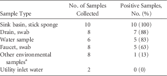
Pseudomonas aeruginosa was isolated from 28 of 42 (67%) environmental samples collected by our CDC team in September 2014. Of 32 samples of tap water and biofilms associated with tap water (eg, faucet, drain, and sink basin), 27 (84%) were positive for P. aeruginosa (Table 2).
Summary of Results of Environmental Sampling for Pseudomonas aeruginosa Culture in a Neonatal Intensive Care Unit

a Swabs of a breast pump (n=1), breast pump kit parts used and washed by a parent (n=1), humidity outlet on an incubator (n=1), mechanical ventilator tubing in use for invasive ventilation (n=1), mechanical ventilator tubing in use for noninvasive continuous positive airway pressure (n=1), and shelves adjacent to the sink (n=3).
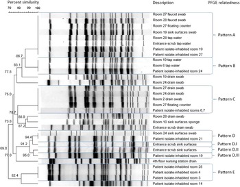
PFGE analysis of CDC environmental samples and patient isolates sent to the CDC laboratory revealed 4 unrelated groups of environmental and patient isolates with indistinguishable PFGE patterns (Figure 4). Isolates from 2 case patients were indistinguishable by PFGE from environmental isolates collected in the rooms occupied by each case patient.
Dendogram for environmental and case-patient isolates collected during a Pseudomonas aeruginosa outbreak in a neonatal intensive care unit. Dendogram displays the pulsed-field gel electrophoresis (PFGE) patterns obtained from select environmental and case-patient isolates. Groupings identified by related PFGE patterns are labeled at the right side of figure.

Risk Factors: Case-Control Study
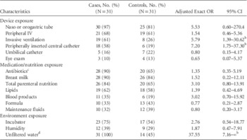
After adjustment for gestational age, case patients had higher odds of exposure to a peripherally inserted central catheter (PICC) (exact odds ratio [eOR], 7.20; 95% CI, 1.75–37.30) or invasive ventilation (eOR, 5.79; 95% CI, 1.39–30.62), compared with controls (Table 3). In addition, case patients had higher odds of having received care in a room with no POU filter installed on the sink faucet during the 7 days before positive culture (eOR, 37.55; 95% CI, 7.16–∞). All 31 case patients were in rooms without POU filters during the 7 days before positive culture, compared with 14 (45%) control patients.
Pseudomonas aeruginosa Outbreak in a Neonatal Intensive Care Unit Attributed to Hospital Tap WaterFootnote a

NOTE. OR, odds ratio; CI, confidence interval.
a Exact conditional logistic regression models with pooled matched strata adjusted for gestational age. Exposures occurred in the 7 days before positive or index culture.
b P<.05.
c Exposure in the 30 days before positive culture or index date.
d Based on the median unbiased estimate.
DISCUSSION
Tap water was implicated as the source of a P. aeruginosa outbreak in an NICU that occurred after a new building was opened. Case clusters occurred before POU filters were installed and after they were removed. Residing in a room without a POU filter was the strongest risk factor for having a positive P. aeruginosa culture. POU filter reinstallation interrupted the outbreak in the short term. Patients with PICCs or invasive ventilation were also at increased risk.
A substantial percentage of environmental samples from water, faucets, drains, sink basins, and shelves near sinks were positive for P. aeruginosa. Although isolates indistinguishable by PFGE were identified from patients and the environment, a high level of genetic diversity existed among environmental and patient isolates. This finding is consistent with a previous NICU outbreak investigationReference Davis, Jensen and Van Hal 12 and with potential biofilm formation in the pipes, faucets, or drains. Biofilm promotes genetic diversityReference Boles, Thoendel and Singh 20 and is known to form on pipes or other fixtures in stagnant water.Reference Williams, CR and MJ 15 , Reference Cooksey, Jhung and Yakrus 21 Water in the hospital remained stagnant for 3 months after completion of hospital construction, allowing ample time for biofilm formation. Flushing or disinfection of water systems might be prudent if water remains stagnant >8 hours.Reference Favero, Petersen, Boyer, Carson and Bond 22 – Reference Bland and Favero 24 Daily flushing of unused taps in NICUs is recommended. 25
Although water was implicated as the source, we did not identify a definitive mechanism of exposure of patients to the water. However, opportunities for tap water exposure might have been related to hand washing and other care practices (eg, cleaning of breast pump parts). We did not identify breaches in infection control related to PICCs or ventilators; however, the presence of these devices might increase exposure to staff hands during manipulation or might be a marker for increased vulnerability to infection. Consistent with other NICU outbreaks of P. aeruginosa, case patients were more likely to be extremely low birth weight compared with the general population of NICU infants.Reference Crivaro, Di Popolo and Caprio 4 , Reference Moolenaar, Crutcher and San Joaquin 7
Before our investigation, staff did not receive consistent guidance emphasizing the preferential use of ABHR over washing with soap and water, according to CDC guidelines.Reference Boyce and Pittet 16 Hand washing with contaminated water by healthcare personnel might have contributed to transmission. We recommended that the hospital revise policies for hand hygiene in accordance with CDC guidelines. In addition, given the evidence of water contamination, the hospital implemented a policy of using ABHR after hand washing with soap and water in the NICU, until water remediation efforts could be ensured. Continuous education of healthcare personnel, auditing, and feedback about adherence to hand-hygiene policies were recommended. Staff hands could have also been indirectly contaminated with P. aeruginosa through touching sink basins or nearby shelves. Although colonized staff have been implicated in transmission of P. aeruginosa in previous outbreaks,Reference Moolenaar, Crutcher and San Joaquin 7 , Reference Mermel, McKay, Dempsey and Parenteau 26 , Reference Zawacki, O’Rourke, Potter-Bynoe, Macone, Harbarth and Goldmann 27 we did not find an association between specific staff members and cases.
Our epidemic curve and case-control study provide evidence that POU filters interrupted this P. aeruginosa outbreak in the short term, consistent with published literature.Reference Trautmann, Halder, Hoegel, Royer and Haller 28 , Reference Vianelli, Giannini and Quarti 29 Although a single study determined that POU filters are the most economical means of protecting vulnerable hospital patients from waterborne pathogens,Reference Hall, Hodgson and Kerr 30 others caution against their use as a long-term solution. 31 We recommended POU filtration continue until P. aeruginosa is no longer detected in the hospital water. We recommended validation of remediation measures through monthly microbiologic testing of prefiltered water and validation of the POU filters by testing postfilter water monthly before the scheduled replacement of the filter. The goal of remediation was total heterotrophic plate counts ≤500 CFU/mL (in accordance with Environmental Protection Agency standards for safe drinking water 32 ) and no Pseudomonas. Cultures of postfilter water should be negative.
During the following months, the hospital followed these recommendations and continued additional water remediation efforts, including decontamination of faucets and drains, routine flushing of faucets, hot water flushing of the hospital water system, hyperchlorination, monitoring temperature and water flow variability, and continuous supplemental chlorine dioxide. Despite remediation, prefiltered water samples continued to grow P. aeruginosa, although levels declined. Postfilter water samples were negative.
From January to November 2015, 8 new cases of P. aeruginosa occurred in NICU patients, representing an increase over baseline, with 1 death. The hospital continued to work closely with the state health department during this time; CDC was periodically consulted. In response to these cases, the hospital hired outside experts to direct water remediation. We continued to recommend regular testing of postfilter water to ensure proper filter function and strict adherence to infection control measures until eradication of P. aeruginosa could be maintained. The reasons for continued cases was unknown, but multiple possibilities were considered, including breaches in infection control or POU filter failure.
The state health department considered the outbreak over in March 2016, after substantial reduction of P. aeruginosa in water samples was achieved and no new cases were reported. The hospital ceased routine patient surveillance cultures at this time. By June 2016, P. aeruginosa was no longer detected in the fixtures in the NICU (<1 CFU/100 mL); the hospital opted to continue using POU filters as an added precaution.
Multiple factors limited our investigation. The case-control study was limited by the size of the NICU population and precluded matching of cases and controls using a ratio greater than 1:1, matching by NICU admission date, or performing multivariable modeling.
Our findings are consistent with the statement made by Williams et alReference Williams, CR and MJ 15 that waterborne healthcare-associated infections occur “at the 3-way intersection of nonsterile potable water, susceptible individuals, and a lapse in infection control practices.” All 3 factors likely contributed to this outbreak. Although interruption of the outbreak with POU filters provided a short-term solution, eradication of P. aeruginosa in the hospital water, faucets, and sinks was necessary to protect patients. This outbreak highlights the importance of addressing and understanding the inherent risks (eg, biofilm formation) in healthcare facilities where water has been stagnant for extended periods.
ACKNOWLEDGMENTS
We acknowledge the substantial contributions of the hospital staff where the outbreak occurred and their commitment to patient safety. The findings and conclusions in this report are those of the authors and do not necessarily represent the official position of the Centers for Disease Control and Prevention.
Financial Support. No financial support was provided relevant to this article.
Potential conflicts of interest. All authors report no conflicts of interest relevant to this article.