INTRODUCTION
The family Thoracostomopsidae Filipjev, Reference Filipjev1927 comprises predatory marine nematodes distinguished by an often large body size, the presence of a cephalic capsule and the buccal cavity with a mandibular complex and onchium (teeth) forming grasping jaws enabling them to capture food items (Filipjev, Reference Filipjev1927; Nicholas, Reference Nicholas2004; Smol & Coomans, Reference Smol, Coomans, Abebe, Traunspurger and Andrássy2006). Molecular data confirm the previous morphological classifications of the superfamily Thoracostmopsidae (Bik et al., Reference Bik, Lambshead, Thomas and Lunt2010a).
Oxyonchus species from this family are commonly found in intertidal and shallow subtidal sandy sediments, but have also been reported from depths of 200 m on the continental shelf from the Sea of Japan. Members of the Thoracostomopsidae have been found in the Atlantic, Indian, and Pacific Oceans and most are described from European waters, whereas for the western coast of the Pacific Ocean there is a shortage of knowledge about the biodiversity of this group. It is known that the seas of the Pacific region are reckoned to be the marine area with greatest biodiversity on Earth (Nicholas, Reference Nicholas2004).
Oxyonchus was erected by Filipjev (Reference Filipjev1927) who established the new subgenus Oxyonchus for Enoplolaimus hamatus Steiner Reference Steiner1916. He also transferred Enoplolaimus acantholaimus Saveljev, Reference Saveljev1912, E. australis de Man Reference De Man1904 and E. dentatus Ditlevsen Reference Ditlevsen1918 to the new genus. De Coninck & Schuurmans Stekhoven (Reference De Coninck and Schuurmans Stekhoven1933) raised subgenus to genus and provided a key to the species of Oxyonchus. They reviewed the genus and considered O. stateni Allgen, Reference Allgen1930 as insufficiently characterized and possibly a synonym of O. australis (De Man, Reference De Man1904) Filipjev, Reference Filipjev1927. However subsequently, Wieser (Reference Wieser1953) revised the genus Oxyonchus, considered O. stateni a synonym of O. australis, re-described the species E. dentatus Ditlevsen Reference Ditlevsen1918 and provided a key to the species. Allgen (Reference Allgen1959) disagreed with the position of Wieser (Reference Wieser1953) regarding the synonymy of O. stateni and O. australis but did not provide additional figures or information to support the validity of O. stateni. Later, more new species were added to the genus: O. subantarcticus (Mawson, Reference Mawson1958); O. brachysetosus, O. crassicolis, O. macrodon, O. notodentatus, O. parastateni (Allgen, Reference Allgen1959), O. culcitatus (Wieser, Reference Wieser1959); O. ditlevseni (Inglis, Reference Inglis1964); O. striatus (Keppner, Reference Keppner1988); O. evelynae, O. longisetosus (Nicholas, Reference Nicholas2004).
At present, Oxyonchus identification is not an easy task and it is often based on minor morphological characteristics, requiring time and resources consuming methods. Many species were discovered and described in the 19th Century and descriptions were often based on a single specimen or on a single sex, sometimes even on a single juvenile.
Keppner (Reference Keppner1988) and Nicholas (Reference Nicholas2004) greatly extended the key of Wieser (Reference Wieser1953) to the species of Oxyonchus and included 14 valid species described by 2004. Because in most of the species the mouth parts are not adequately described, the diagnosis given for the known Oxyonchus species can only be a working diagnosis. A comparison of buccal armature of different thoracostomopsids, in general, can be used as further illustration of this unsatisfactory situation (Fadeeva & Zograf, Reference Fadeeva and Zograf2010). One of the most characteristic peculiarities of the nematode tissue is an ability to form cuticular structures. The organization of the buccal armature is a specific characteristic of the genus Oxyonchus. Wieser (Reference Wieser1953) and Inglis (Reference Inglis1964) made a detailed study of the thoracostomopsid head and proposed specialized terminology for the head structures currently used in the descriptions of representatives of this group. So, the majority of the thoracostomopsids are characterized by the possession of a conical buccal cavity, surrounded by one dorsal lip and two subventral lips, and armed with powerful jaws (Inglis, Reference Inglis1964). The latter are formed by three mandibular plates, each supporting a forwardly directed onchium (tooth). The pharyngeal muscles are attached anteriorly to the cephalic cuticle forming a cephalic capsule (Inglis, Reference Inglis1964). The labial sensilla are setiform. The morphology of the buccal armature in various genera and the taxonomy of the family were reviewed and revised by Greenslade & Nicholas (Reference Greensdale and Nicholas1991) and Nicholas (Reference Nicholas2004) in detail.
The confocal microscopy technique allows description of the peculiarities of certain structures: cephalic capsule, fenestrae, mandibular ring, mandibular plates, spicules and gubernacula. Obtained pictures may be rotated to observe structures from different points of view.
The aim of this study was to describe three new Oxyonchus species found at the Russian coast of the Sea of Japan and the Sea of Okhotsk and stored in the Collection of Marine Nematodes at the Far Eastern Federal University, Russia. Special attention was paid to the head structure as revealed by confocal microscopy.
MATERIALS AND METHODS
Intertidal sediment samples were collected in several locations in the Sea of Japan and the Sea of Okhotsk at a depth of 7–200 m over a period of several years. Samples were collected using a tubular 20 cm2 bottom sampler to a depth of 5 cm. Living nematodes were sorted from the sediment, preserved in 10% hexamethylenetetramine-buffered formalin and transferred to pure glycerin using Seinhorst's (Reference Seinhorst1959) rapid method as modified by De Grisse (Reference De Grisse1969). The nematodes were then mounted on permanent slides.
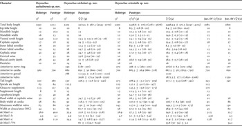
Microscopic examination was done using an Axio Imager A1 light microscope (Сarl Zeiss) and a confocal microscope (Leica LSM SPE). The slides were imaged using confocal laser-scanning microscopy (LSM) for autofluorescence (Fadeeva & Zograf, Reference Fadeeva and Zograf2010). Fluorescence image stacks were registered in the 488 nm (green) channel. The scanning step size was usually 0.5 µm. The number of optical sections in a series ranged from 30 to 70, depending on the size of the specimen. Detailed morphometric data were obtained from camera lucida drawings and are given in Table 1.
Emended diagnosis (after Smol & Coomans (Reference Smol, Coomans, Abebe, Traunspurger and Andrássy2006) and original data): Enoplolaiminae. Lips high or low. Cuticle smooth or striated. Cephalic organ present or absent. Cephalic capsule well developed, broad, with well developed fenestrae and incisions; posteriorly can be divided into six lobes or undulate. The anterior edge of the cephalic capsule is strengthened with wide mandibular ring of specialized cuticle associated with mandibles. Mandibles well developed, arch-shaped, rods connected by broad transverse bar with claws, denticles can be present at the inner surface of the mandibular plate. Teeth (onchium) unequal: two large ventrosublateral teeth (onchium) that extend to the anterior end of the mandibles. Dorsal tooth (onchium) small. Spicules short (1–3 anal body diameters (a.b.d)). Pre-cloacal supplement present or absent. Gubernaculum with or without apophysis. Marine.
Morphometrics of the Oxyonchus species. All measurements are in μm.

Type species O. hamatus (Steiner, Reference Steiner1916) Filipjev, Reference Filipjev1927
Other species:
O. acantholaimus (Saveljev, Reference Saveljev1912)
= Enoplolaimus acantholaimus Saveljev, Reference Saveljev1912
O. australis (de Man, Reference De Man1904)
= E. australis de Man, Reference De Man1904
O. brachysetosus Allgen, Reference Allgen1932
O. campbelli Allgen, Reference Allgen1932
O. crassicollis Allgen, Reference Allgen1932
O. culcitatus (Wieser, Reference Wieser1959)
O. dentatus (Ditlevsen, Reference Ditlevsen1918)
= E. dentatus Ditlevsen, Reference Ditlevsen1918
= E. crassidens Ditlevsen, Reference Ditlevsen1930
= E. elegans Schulz, 1932
O. ditlevseni (Inglis, Reference Inglis1964)
O. dubius (Filipjev, Reference Filipjev1918)
= E. dubius Filipjev, Reference Filipjev1918
O. evelynae (Nicholas, Reference Nicholas2004)
O. longisetosus (Nicholas, Reference Nicholas2004)
O. macrodon Allgen, Reference Allgen1959
O. nicholasi sp. nov.
O. notodentatus Allgen, Reference Allgen1959
O. orientalis sp. nov.
O. pachylabiatus Stekhoven, Reference Stekhoven1946
O. parastateni (Allgen, Reference Allgen1959)
O. polaris (Filipjev, Reference Filipjev1927)
= E. polaris Filipjev, Reference Filipjev1927
O. problematicus Filipjev, Reference Filipjev1946
O. sakhalinensis sp. nov.
O. stateni Allgen, Reference Allgen1930
O. striatus (Keppner, Reference Keppner1988)
O. subantqrcticus Mawson, Reference Mawson1958
For revision see Keppner (Reference Keppner1988) and Nicholas (Reference Nicholas2004).
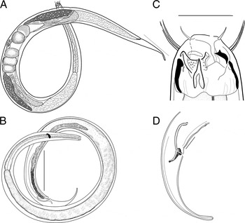
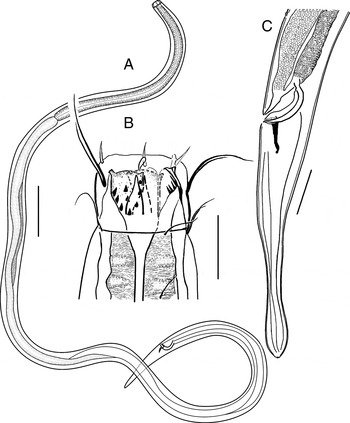
Oxyonchus orientalis sp. nov.: (A) female; (B) male holotype; (C) male head end; (D) male tail end. Scale bars: A = 300 µm; B = 100 µm; C = 25 µm, D = 50 µm.

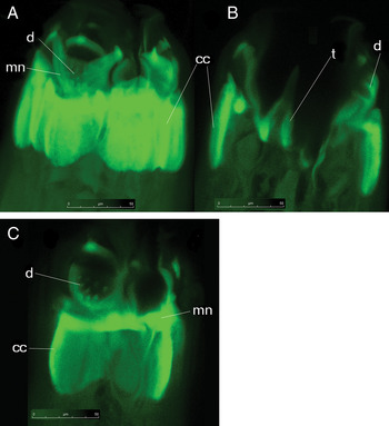
Laser-scanning microscopy pictures of Oxyonchus orientalis sp. nov.: (A) details of the male head skeletal component; (B) ventrosublateral tooth; (C) distribution of small denticles on inner surface of mandible. cc, cephalic capsule; d, denticles; mn, mandible; t, tooth. Scale bars: A, B, C = 50 µm.

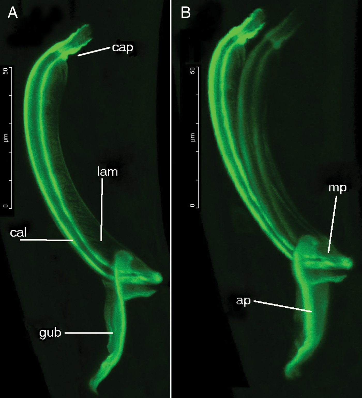
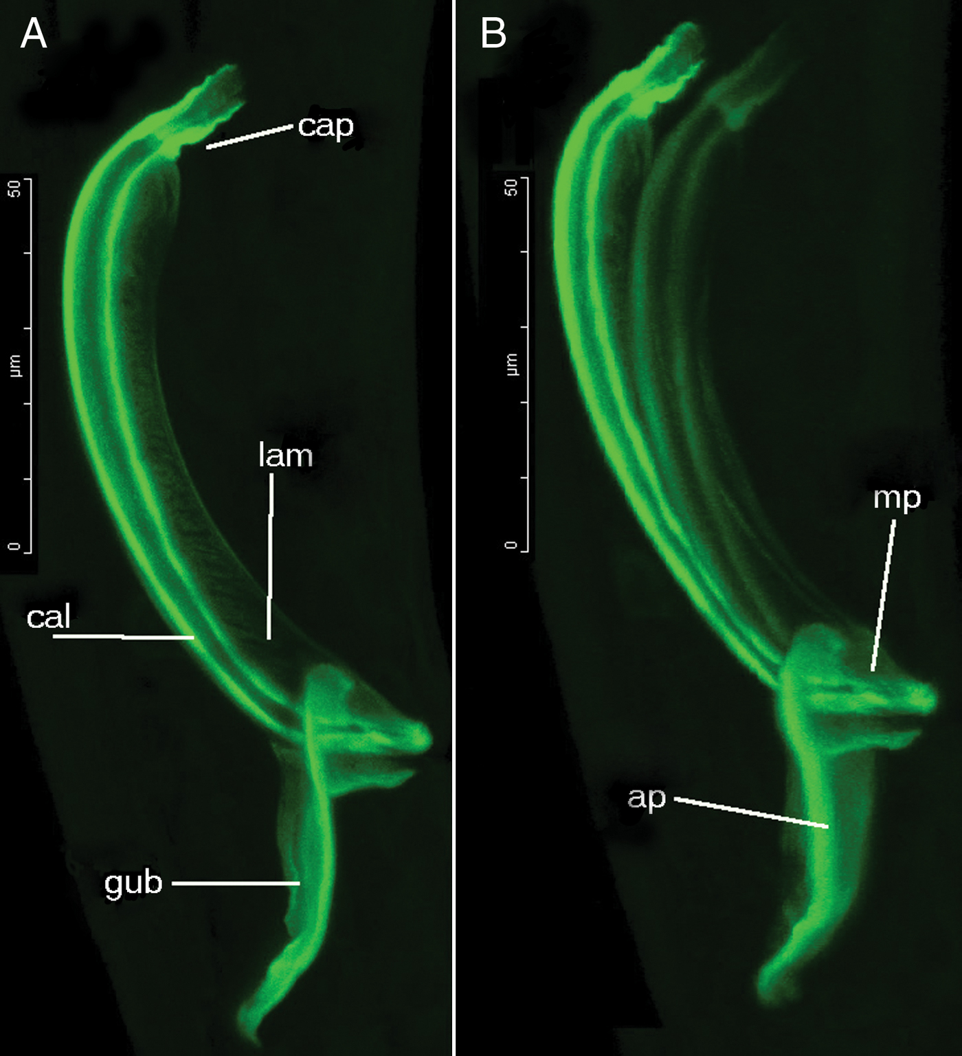
Laser-scanning microscopy pictures of Oxyonchus orientalis sp. nov. Male holotype: (A) lateral view of spicular apparatus; (B) laterodorsal view of spicular apparatus. ap, apophysis; cap, capitalum; cal, calomus; lam; lamina; mp, median part of gubernaculum. Scale bars: A, B = 50 µm.
TYPE MATERIAL
Holotype: adult male, formalin-fixed, mounted on slide (MN 12222), in glycerin. The Sea of Japan, Peter the Great Bay, Island Russkyi coordinates: 43°01′N 131°94′E; depth 25 m, collected by Dr S.I. Maslennikov, 19 May 2007, deposited in the collections of the Division of Biological Sciences, Zoological Museum of Far Eastern Federal University, Vladivostok, Russia.
Paratypes: three males (MN 12000) and three females (MN 12222, MN 2531) same date and site, deposited in the collections of the Division of Biological Sciences, Zoological Museum of Far Eastern Federal University, Vladivostok, Russia. Juvenile male and female, formalin-fixed, mounted on slide MN-3000, 3001 in glycerin. The Sea of Japan, Rudnaya Pristan, coordinates: 44°21′6N 136°12′7E; depth 146.5 m, fine sand. Collected by Dr V.V. Morduchovich, 4 July 2003, deposited in the collections of the Division of Biological Sciences, Zoological Museum of Far Eastern Federal University, Vladivostok, Russia.
ENVIRONMENTAL DATA
Station 12: bottom water temperature 15.8°C, salinity 35‰, sediment mean grain size 259 µm, silt/clay content 11.75%.
ETYMOLOGY
The species name is derived from the Latin orientalis that means ‘eastern'.
MEASUREMENTS
See Table 1.
DESCRIPTION
Body long cylindrical slightly narrowing towards anterior end and sharply narrowing towards tail. The head in the lip region narrower than buccal region. Cuticle thick with distinct transverse striation. Mouth opening surrounded by one dorsal and two ventrosublateral lips separated by deep clefts, each bearing very thin lip-lobe at its apex. The six inner labial sensillae short; outer labial sensillae slightly longer than 4 cephalic setae. However, the setae are often difficult to recognize in this species. Six outer labial setae equal to 0.60 cephalic diameters. Cephalic organ not observed. The cephalic capsule has a distinct junction with the pharyngeal tissue, well developed, as high as 0.8–0.9 diameter at the level of cephalic setae. Anterior margin of the cephalic capsule with long incisions and small fenestrae accommodating bases of cephalic setae. Amphideal fovea indistinct. Very short setae are sparsely distributed elsewhere along the body.
Lips are supported internally by arched mandibular plates with strengthened rims that connect adjacent mandibular plates below the clefts so that the cuticular rims surround the mouth with a cuticular mandibular ring. Mandibular complex well developed. Broad mandibular plate with irregular arrangement of numerous small denticles. Base of each subventral lip with a robust, triangular, outwardly curved onchium, the tip of which does not extend beyond anterior edge of the mandibular arch. The dorsal onchium is reduced to a knob.
Reproductive system diorchic, both anterior and posterior opposed testes are located to the left of intestine. Two spicules (2.1–2.3 anal diameters long), curved into segment of a semicircle with narrow velum, their distal tips are pointed. Distal end of spicules passes through the slit of the gubernaculum supported by long dorsocaudal apophysis (50–56 µm). Tubular precloacal supplement finely sclerotized (15 µm long and 2 µm wide) and situated near the proximal region of the spicules, 137–163 µm (2.3 a.b.d.) from the cloaca. Slender tail 4.6–5.5 a.b.d. long with slightly swollen tip.
Females closely resemble males, but are larger. Reproductive system didelphic, amphidelphic, ovaries reflexed. Length of anterior branch of reproductive system 440 µm, posterior branch 430 µm. Vagina straight with thick walls and well developed sphincter muscles. Tail length 4.1–4.8 a.b.d.
DIFFERENTIAL DIAGNOSIS
Oxyonchus orientalis sp. nov. has short cephalic sensillae (0.6 cephalic diameters), strongly developed broad mandibular plate with irregular arrangement of numerous small denticles. Another species with short cephalic setae, O. brachysetosus from Antarctica is too poorly described and considered species inquirenda (Keppner, Reference Keppner1988; Nicholas, Reference Nicholas2004).
The new species resembles O. dentatus and O. hamatus in the shape of spicules and the head structure. Nevertheless, it differs from each species by well-developed cephalic capsule, the slender spicules and gubernaculum with long dorsal apophyses with characteristic shape. Oxyonchus orientalis sp. nov. can be distinguished from O. dentatus by the longer spicules (122–147 µm versus 84 µm), by a longer tail (4.6–5.5 a.b.d. versus 4–4.5 a.b.d.), from O. hamatus by the smaller body size (3084–4155 µm versus 5623 µm) and by the number of denticles (12–16 versus 6).
Oxyonchus sakhalinensis sp. nov. Male holotype. (A) entire body; (B) head end; (C) tail end. Scale bars: A = 200 µm; B = 25 µm; C = 10 µm.

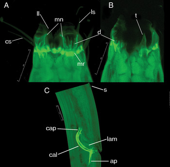
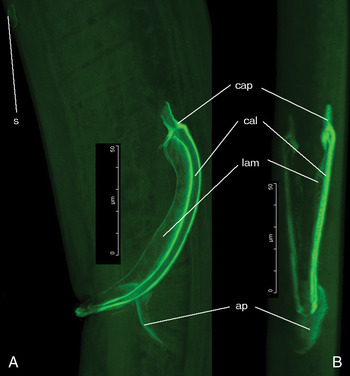
Laser-scanning microscopy pictures of Oxyonchus sakhalinensis sp. nov.: (A) details of the male head skeletal component; (B) lateroventral view of the head; (C) lateral view of spicular apparatus. ap, apophysis; cap, capitalum; cal, calomus; cs, cephalic seta; d, denticles; lam, lamina; ll, lip lobe; ls, labial seta; mn, mandible; mr, mandibular ring; s, supplement; t, tooth. Scale bars: A, B, C = 25.

TYPE MATERIAL
Holotype: adult male, formalin-fixed, mounted on slide MN-3031 in glycerin. The Sea of Okchotsk, the Sakhalin Bay, coordinates: 54°19′3N 140°53′5E, depth: 54 m. Collected by Dr V.V. Morduchovich, 12 July 2003.
Paratype: adult female mounted on slide MN-2931 (coordinates: 54°07′5N 141°02′6E depth: 29.8 m). Collected by V.V. Morduchovich, 12 July 2003. Material deposited in the collections of the Division of Biological Sciences, Zoological Museum of Far Eastern Federal University, Vladivostok, Russia.
ENVIRONMENTAL DATA
Station 29: bottom water temperature –1.2°C, salinity 33.4‰, sediment–gravel.
Station 30: bottom water temperature –0.2°C, salinity 33.3‰, sediment–gravel.
ETYMOLOGY
The name sakhalinensis refers to ‘from the Sakhalin Island'.
MEASUREMENTS
See Table 1.
DESCRIPTION
Body long, cylindrical and slightly narrowing towards anterior end. Cuticle smooth. Head dome-shaped with three lips. Mouth opening surrounded by one dorsal and two ventrosublateral lips separated by deep clefts, each bearing very thin lip-lobe at its apex. Inner labial sensilla long and inserted at the base of lip flaps; outer labial and cephalic sensilla situated at the anterior end of cephalic capsule. Six inner labial sensilla slightly smaller than cephalic setae. Six outer labial setae equal to 1.3 cephalic diameters. Two cirri inserted on outer surface of ventrosublateral lips at the level slightly posterior to the apex of the mandibular plates. Amphideal fovea indistinct. The cephalic capsule is fairly short (Figure 5A).
The mandibular plates are rounded and each contains 10 denticles in the field of inner surface. Two subventral onchia small, triangular, curved and extend anteriorly while the dorsal onchium is very short (Figure 5B). Several rings of short cervical setae present in the neck region.
Diorchic. Anterior outstretched testis extends to cardia, posterior one reflexed. Stout spicules (1 a.b.d. long), arcuate, cephalated (10 µm), with broad alae and sharp distal ends (Figures 4C & 5C). Dorsocaudal apophysis of gubernaculum is 33–40 µm long. Single small supplementary sensory receptor embedded in ventral cuticle situated 1.8–2.1 a.b.d. anterior to cloaca. Tail short, equal to 4.8 a.b.d. long, with slightly swollen tip (Figure 4C).
DIFFERENTIAL DIAGNOSIS AND RELATIONSHIPS
Despite the small number of available adults Oxyonchus sakhalinensis sp. nov. is particularly characterized by the weakly developed short cephalic capsule, and the rounded thin mandibular plates with 10 denticles.
Described species belongs to the group of species with short cephalic sensillae (1–1.2 head diameters) and short spicules: O. dentatus, O. ditlevseni, and O. australis. The new species differs from O. dentatus by a relatively short body length (2392–3212 µm versus 3630 µm), and spicule length (63–64 µm versus 84 µm), by the number of mandibular denticles (10–12 versus 15–20) and weak developed cephalic capsule. Oxyonchus sakhalinensis sp. nov. differs from the O. australis by the number of denticles (10 denticles versus 6). Oxyonchus ditlevseni (described from only one male) is similar to O. sakhalinensis in many measurements but differs from the new species by having ring of thin long setae immediately posterior to the cephalic capsule, by longer body (3500 µm versus 2392–3212 µm), and by the shorter cephalic setae (36/12 µm versus 47/25 µm).
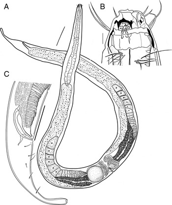
Oxyonchus nocholasi sp. nov.: (A) female; (B) male holotype head end; (C) male holotype tail end. Scale bars: A = 200 µm; B = 10 µm, C = 100 µm.

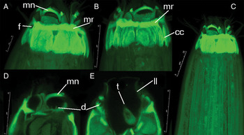
Laser-scanning microscopy pictures of Oxyonchus nicholasi sp. nov.: (A) detail of female head skeletal component; (B) detail of male head skeletal component; (C) pharyngeal region of female; (D) mandibles and denticles of male head; (E) ventrosublateral tooth of male head. cc, cephalic capsule; d, denticles; f, fenestrae; ll, lip lobe; mn, mandible; mr, mandibular ring; t, tooth. Scale bars: A = 10 µm, B, C, D, F = 25.

Laser-scanning microscopy pictures of Oxyonchus nicholasi sp. nov.: (A) lateral view of spicular apparatus; (B) dorsal view of spicular apparatus. ap, apophysis; cap, capitalum; cal, calomus; lam, lamina; s, supplement. Scale bars: A, B = 50.

TYPE MATERIAL
Holotype: adult male, formalin-fixed, mounted on slide MN-4211 in glycerin. The Sea of Okchotsk, the Sakhalin Bay, Baikal Lagoon coordinates: 53°32′5N 142°30′16E, depth: 7 m. Collected by Dr V.V. Morduchovich, 13 June 2006.
Paratype: adult female mounted on slide MN-4211 same date and site. Material deposited in the collections of the Division of Biological Sciences, Zoological Museum of Far Eastern Federal University, Vladivostok, Russia.
ENVIRONMENTAL DATA
Station 12: bottom water temperature 12.39°С, salinity 18.25‰, sediment silty sand.
ETYMOLOGY
The new species is named in honour of Professor Warwick L. Nicholas for his contributions in the field of marine nematology.
MEASUREMENTS
See Table 1.
DESCRIPTION
Body cylindrical; strongly narrowing towards anterior end. Cuticle smooth. Mouth opening surrounded by one dorsal and two ventrosublateral lips separated by deep clefts. Six inner labial setae 0.8 cephalic diameters. Outer labial sensilla and cephalic setae are equal in size and situated in one circle. Numerous long cervical setae present in the neck region. Amphid pore not identified. The cephalic capsule is a prominent thick and dense cuticular structure with well developed fenestrae and incisions. The anterior edge of the cephalic capsule is strengthened with a wide mandibular ring of specialized cuticle associated with mandibles. The numbers of denticles at the mandibular plates are difficult to count because of curvature of the plates, but they are less numerous than in O. orientalis sp. nov, about 14–16 on each mandible. Base of each subventral lip with a strong outwardly curved onchium, the tip of which may or may not extend to or beyond the mandibular arch. Subventral onchia are large, curved and extending anteriorly, the dorsal onchium is very short.
Anterior testis outstretched, posterior one reflexed. Spicules slender, 1.5 a.b.d. long, arcuate, with broad alae and short tube-form capitulum (Figures 6C & 8). Dorsocaudal apophysis short (28 µm). Tubular precloacal supplement finely sclerotized (15 µm long and 2 µm wide) and situated 133 µm (2 a.b.d.) from the cloaca. Tail 3.5 a.b.d. long, conical in its anterior part, cylindrical in its posterior part, with notably swollen tip (Figure 6C).
Female closely resembles male, but is larger. Six outer labial sensilla and four cephalic setae are equal in size, inserted at the anterior margin of cephalic capsule. Few short cervical setae present.
Reproductive system didelphic, amphidelphic, ovaries reflexed, with 3 fully developed eggs in each oviduct. Fully formed intrauterine eggs are 145 × 150 µm and are lying oblique. The length of the anterior ovary 440 µm, posterior–430 µm. Tail 3.5 a.b.d. long, conical in its anterior part, cylindrical in its short posterior part; tail tip notably swollen.
DIFFERENTIAL DIAGNOSIS AND RELATIONSHIPS
Oxyonchus nicholasi sp. nov. is particularly characterized by the short equal in size outer labial sensilla and cephalic setae (0.8 cephalic diameters), subventral large, curved extend anteriorly to onchia, well developed capsule, fenestrae and incisions, by the numerous long cervical setae immediately posterior to the cephalic capsule and caudal setae.
Oxyonchus nicholasi sp. nov. is closely related to the O. sakhalinensis sp. nov. but can be separated by the structure of the cephalic armature (well developed capsule, fenestrae and incisions), by the numerous long cervical setae and tail shape (tip of the tail notably swollen), the absence of cirri, shorter equal in size cephalic sensilae (1.3 versus 0.8 cephalic diameters), and by relatively large number of denticles (14–16 denticles versus 10).
DISCUSSION
In general, the biogeographical distribution of marine nematodes species is poorly known (Gage, Reference Gage1996; Rex, Reference Rex1997; Tchesunov, Reference Tchesunov2006; Fonseca et al., Reference Fonseca, Vanreusel and Decraemer2006). The Oxyonchus nematodes are usually found worldwide at sandy-beach ground water and intertidal and shallow-water sandy sediments to depths of 300 m and feed on microturbellaria and other small animals (Nicholas, Reference Nicholas2004). There are already several publications discussing distribution in free-living marine nematodes (order Enoplida) at large scales (Mando-Ocampo et al., Reference Mundo-Ocampo, Lambshead, Bebenham, King, De Ley, Backdwin, Tandingan De Lay, Rocha-Olivares, Waumann, Thomas, Packer and Boucher2007; Bik et al., Reference Bik, Lambshead, Thomas and Lunt2010a, Reference Bik, Thomas, Lunt and Lambsheadb).
Most species of Oxyonchus have relatively few individuals and rather narrow distributions, being characteristic constituents of distinct habitat types and assemblages (Gerlach & Riemann, Reference Gerlach and Riemann1974). The earlier descriptions of species now placed in Oxyonchus are inadequate and require new taxonomic and ecological studies in adjacent areas. Therefore, it is difficult to obtain information about the distribution patterns of species of this genus.
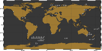
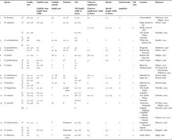
In order to unravel the possible morphological relationships between Oxyonchus species the distribution of species were analysed in relation to environmental and geographical distribution. Therefore, the species were plotted on a world map (Figure 9), and the comparative table of the species was made (Table 2).
Global map of distribution the species of the genus Oxyonchus: 1, O. acantholaimus; 2, O. australis; 3, O. brachysetosus; 4, O. campbelli; 5, O. crassicollis; 6, O. culcitatus; 7, O. dentatus; 8, O. ditlevseni; 9, O. dubius; 10, O. hamatus; 11, O. macrodon; 12, O. notodentatus; 13, O. pachylabiatus; 14, O. parastateni; 15, O. Polaris; 16, O. problematicus; 17, O. stateni; 18, O. subantqrcticus; 19, O. longisetosus; 20, O. evelynae; 21, O. striatus, 22, O. orientalis sp. nov.; 23, O. sakhalinensis sp. nov.; 24, O. nicholasi sp. nov. *, described from juvenile; ^, species inquirenda.

Morphometry of valid Oxyonchus species.

?, denotes missing data. Data were taken from original descriptions.
Until now, Oxyonchus dentatus (Ditlevsen, Reference Ditlevsen1918) Filipjev has been found in many localities alongside coastlines of Europe. This species spread from the Atlantic to the Pacific and Southern Ocean (Gerlach & Riemann, Reference Gerlach and Riemann1974; Pereira et al., Reference Pereira, Fonseca, Mundo-Ocampo, Guilheme and Rocha-Olivares2010). Oxyonchus dentatus dwells in full-salinity waters as well as in brackish bights and estuaries. The species is the most common species in shallow water and usually inhabits dissipative beach with wide tidal range, fine sand with small waves. The distribution of O. dentatus shows a large variability in its habitat. The highest intraspecific variation pertains to the most widely spread cosmopolitan species O. dentatus, suggesting that morphological plasticity enhances adaptation to different environmental conditions. Oxyonchus culcitatus is widespread on the American coast (Puget Sound) and Australian coast of the Pacific Ocean (Nicholas, Reference Nicholas2004).
The rich group of species is provided by the uncommon species in the sense of ‘rare species’ nematodes that only occur near Europe—O. pachylabiatus (the Baltic Sea) and O. acantholaimus (the White Sea), near Asia—O. problematicus (the eastern Siberian Sea). Three species (O. acantholaimus, O. hamatus and O. polaris) were found in the Barents Sea, two species (O. hamatus and O. polaris) were found in the Kara Sea. The distribution of three species is restricted to subtropical and tropical latitudes: O. dubius in the Mediterranean Sea and the Black Sea, O. striatus in the Gulf of Mexico, and O. ditlevseni on the African coast. Oxyonchus evelyanae and O. longisetosus were described from the Australian coast of the Pacific Ocean. Species of O. australis and O. subantarcticus are found mainly in the Antarctic, the Indian Ocean and Pacific Ocean near New Zealand (Nicholas, Reference Nicholas2004).
There are several subgroups within Oxyonchus nematodes. The group of morphologically similar species characterized by short cephalic setae composed of European species. The differences between species appear as the difference in the number of denticles (O. acantholaimus possess 14–15 denticles while O. hamatus and O. polaris have 4–6 denticles on the mandibular plates), tail lengths (O. dubius and O. problematicus distinguished from other European species by the tail up to 7–10 a.b.d long). Therefore, it is possible that shallow-water European species are close relatives.
Three species: O. nicholasi sp. nov., O. orientalis sp. nov. and O. sakhalinensis sp. nov. were described in adjacent areas of Russian Far Eastern Seas based on recently collected specimens. These species have been recorded in two sites (the Sea of Japan and the Sea of Okhotsk) widely separated by hundreds of kilometres and differing in depth, bottom temperature and salinity preference. Habitat transitions are relatively complicated amongst nematode species of this region.
The Sea of Japan is considered to have been formed sometime between 10 and 20 million years ago, following large scale crustal movement (Isozaki et al., Reference Isozaki, Kazumasa, Nakama and Yanai2010). This basin is bathymetrically and oceanographically isolated, due to the shallowness of the Nevelscoi Strait from the Amur River estuary, lengthy and shallow water. The Amur River estuary is geographically located between the Sea of Japan (Nevelskoi Strait) and the Sea of Okhotsk (part of the Sakhalin Bay). Due to desalination of the Amur River estuary waters (to 5–8‰) cold-water nematodes are not able to move into the Sea of Japan from the Sea of Okhotsk using the Nevelskoi Strait. Oxyonchus orientalis sp. nov. and O. sakhalinensis are marine geographically restricted morphospecies and their differences may be explained by the isolation (the presence of the shallow Nevelskoi Strait with low salinity). Salinity also seems to play a major role in the occurrence of two species—O. nicholasi sp. nov. and O. sakhalinensis sp. nov., and determined their niche separation. This species needs simple physiological adaptations to move between different physical environments. The Peter the Great Bay of the Sea of Japan has an unusual mix of subtropical and temperate taxa (Adrianov, Reference Adrianov2004). It has abundant endemic species among invertebrates and is considered to be among the most biologically diverse regions.
New species share many morphological similarities with European specimens of Oxyonchus. Using diagnostic morphological characters in a traditional taxonomic approach O. nicholasi sp. nov. and O. orientalis sp. nov. may be placed into a species group with short cephalic setae (less than 1 head width) according to the key of Nicholas (Reference Nicholas2004) and are similar to each other but can be separated by the size and shape of the tail and other features. Based on the present LSM observation, we suggest that all these differences may be considered as main taxonomic characters of this species separated geographically.
The species group (O. longisetosus, O. evelynae and O. culcitatus) includes species with the long cephalic setae which are found mainly in the Pacific Ocean. Oxyonchus australis and O. subantarcticus having a circumpolar distribution differentiated by the short cephalic setae and spicules from other species. Oxyonchus striatus from the Gulf of Mexico differs from all other species of the genus in the presence of the distinctly striated cuticle, long spicules, and by the gubernaculum without apophysis. Oxyonchus ditlevseni on the African coast differs from other species by the shape of spicules and gubernaculum.
Evolutionary histories perhaps have strongly influenced nematodes species composition at ocean scale as considered some authors (Fonseca et al., Reference Fonseca, Vanreusel and Decraemer2006, Reference Fonseca, Muthumbi and Vanreusel2007). It is possible, that the distribution of Oxyonchus species can be explained on a global scale by means of palaeogeographical events as was shown for many other species of nematodes (Fonseca et al., Reference Fonseca, Vanreusel and Decraemer2006). Additional molecular studies are needed in order to assess the distribution of species of Oxyonchus. Finally, we consider that to advance taxonomy of marine nematodes an integrative approach is required encompassing morphological and molecular analyses during biodiversity surveys.
ACKNOWLEDGEMENTS
The authors are grateful to Professor Etiene Geraert for help with English writing. We are grateful for the constructive comments and suggestions of the anonymous referee, which have improved the manuscript. The study was financially supported by the Russian Foundation for Fundamental Researches through grant awards N 09-04-00271-a, Grant of the Far Eastern Branch of the Russian Academy of Sciences N 11-III-В-06-109 and Grant of the Government of the Russian Federation No. 11.G34.31.0010