Introduction
Fructose-1,6-bisphosphate aldolase (FBPA) is an ubiquitous enzyme essential for glycolysis, gluconeogenesis and the Calvin cycle in living organisms. It can be divided into two distinct classes based on the mechanism of catalysis: class I FBPA (FBPA I) utilizes a Schiff base reaction mechanism and class II FBPA (FBPA II) uses divalent metal ions (Rutter, Reference Rutter1964; Marsh & Lebherz, Reference Marsh and Lebherz1992). Besides catalytic activities, FBPA is immunogenic and can induce immunity in many infectious diseases (Srivastava et al., Reference Srivastava, Schmidt, Certa, Dobeli and Perrin1990; Wanidworanun et al., Reference Wanidworanun, Nagel and Shear1999; McCarthy et al., Reference McCarthy, Wieseman, Tropea, Kaslow, Abraham, Lustigman, Tuan, Guderian and Nutman2002; Palm et al., Reference Palm, Weiland, Griffiths, Ljungstrom and Svard2003). Antibodies to FBPA have been successfully found in humans infected with Plasmodium falciparum (Srivastava et al., Reference Srivastava, Schmidt, Certa, Dobeli and Perrin1990; Genrich et al., Reference Genrich, Guarner, Paddock, Shieh, Greer, Barnwell and Zaki2007). This protein has also been used as a target for rapid diagnosis of malarial infection (Tjitra et al., Reference Tjitra, Suprianto, Dyer, Currie and Anstey1999; Gatti et al., Reference Gatti, Bernuzzi, Bisoffi, Raglio, Gulletta and Scaglia2002).
Schistosomiasis continues to be an important parasitic disease of humans worldwide, with an estimated more than 200,000 deaths occurring per year (Chitsulo et al., Reference Chitsulo, Loverde and Engels2004). Cases of human schistosomiasis have been reported in Africa, South America, the Caribbean, South-East Asia and China (Chitsulo et al., Reference Chitsulo, Loverde and Engels2004). Infection can be determined by either recovering ova from stools or detecting specific antibodies in serum. The circumoval precipitin test (COPT), indirect haemagglutination assay (IHA) and indirect fluorescence assay (IFA) were the serological methods used for detection, and antigens from adult worms or eggs were widely used for these assays (De Jonge et al., Reference De Jonge, Rabello, Krijger, Kremsner, Rocha, Katz and Deelder1991; Wu, Reference Wu2002; Zhu, Reference Zhu2005); however, the sensitivity and specificity of these tests were unsatisfactory at low levels of infection (Guan & Shi, Reference Guan and Shi1996). Therefore, enzyme-linked immunosorbent assays (ELISAs) have been developed and many antigens have been evaluated with this test (Hamilton et al., Reference Hamilton, Klinkert and Doenhoff1998).
Various strains of Schistosoma japonicum have been described in China, the Philippines (Hsu & Hsu, Reference Hsu and Hsu1960), Japan (Moose et al., Reference Moose, Williams and Ota1963), Indonesia (Cross, Reference Cross1976) and Taiwan (Hsu et al., Reference Hsu, Li, Wang, Fan and Huang1952). Of these, the Formosan strain is morphologically, ecologically and physiologically similar to the other strains (Hsu et al., Reference Hsu, Li, Wang, Fan and Huang1952; Hsu & Hsu Li, Reference Hsu and Hsu Li1956; Hsu et al., Reference Hsu, Davis and Hsu1962). It is also closely related to the Chinese strain in the genetic analysis of mitochondrial deoxyribonucleic acid (DNA) (Guo & Niu, Reference Guo and Niu2004) and in protein patterning by sodium dodecyl sulphate–polyacrylamide gel electrophoresis (SDS–PAGE) (Qiu et al., Reference Qiu, Liu, Xue, Zhang, Li, Hu and He1999). However, the non-human or zoophilic nature of the Formosan strain has been strongly supported in data from epidemiological studies and experiments on human volunteers (Hsu et al., Reference Hsu, Li, Wang, Fan and Huang1952; Hsu & Hsu Li, Reference Hsu and Hsu Li1956). It has also been reported that the pathology in Formosan strain-infected Rhesus monkeys is milder than that caused by the Chinese, Japanese or Philippine strains (Hsu & Li Hsu, Reference Hsu and Li Hsu1960). Therefore, it is safer to use the Formosan strain for manipulation.
FBPA has been recovered from S. mansoni and was able to induce a strong immune response in mice (Harrop et al., Reference Harrop, Coulson and Wilson1999). It is most likely that this protein will also be present in S. japonicum. Therefore, the objectives of this study were to recover FBPA from the Formosan strain of S. japonicum and to evaluate its diagnostic value.
Materials and methods
Participants
Ninety-one persons (70 men and 21 women) were recruited from Yueyang, Hunan province, an area endemic for schistosomiasis in China, and 29 individuals (26 men and 3 women) were recruited from the Department of Allergy, Immunology, and Rheumatology, Taichung Veterans General Hospital, Taiwan. After written consent, blood and faecal samples were collected from each participant. The stool specimen of each enrollee was examined for ova and parasites according to the method described by Katz et al. (Reference Katz, Chaves and Pellegrino1972). Sera obtained from the enrollees were divided into two groups and stored at − 80°C for further investigation.
Schistosoma japonicum Formosan strain
The Formosan strain of S. japonicum was originally collected from infected buffalos at the Hanpo Farm in Fengyuan District, Changhua County, Central-West Taiwan and had been maintained in our laboratory since 1987. Oncomelania hupensis chiui snails were used as the intermediate hosts and 8- to 10-week-old male C57BL/6J mice served as the final hosts. Adult worms were obtained from mesenteric vasculature by the perfusion technique (Duvall & DeWitt, Reference Duvall and DeWitt1967) and were washed three times with normal saline. They were then snap-frozen in liquid nitrogen for further experimentation.
Preparation of recombinant FBPA of Schistosoma japonicum Formosan strain
Preparation of cDNA from Schistosoma japonicum Formosan strain
Total ribonucleic acid (RNA) was extracted from 100 adult worms using the TRizol reagent (Invitrogen, Carlsbad, California, USA) according to the manufacturer's instructions, and cDNA was prepared according to the method described by Nagano et al. (Reference Nagano, Pei, Wu, Wu, Cui, Boonmars and Takahashi2004) with a slight modification. Briefly, 20 μl reaction buffer containing 5 μg extracted RNA, 2 μl of 10 mm dNTP, 1 μl oligo(dT)18, 1 μl RiboLock ribonuclease inhibitor (Fermentas International Inc., Ontario, Canada), and 1 μl reverse transcriptase (Fermentas) was added to a microcentrifuge tube and incubated at 42°C for 60 min followed by incubation at 70°C for 10 min. The synthesized cDNA was stored at − 20°C until experimentation.
Preparation of gene encoding FBPA
The gene encoding full-length putative FBPA was amplified by polymerase chain reaction (PCR) from the synthesized cDNA using oligonucleotide primers with BamHI restriction sites. Based on the nucleotide sequences of the putative FBPA gene (GenBank accession number AY813526), the primers designed for PCR were:
5′-CGGGATCCAAAATGCCACGATTCC-3′ and
5′-GCCCTAGGTTCAGTTGGTGTGATGC-3′
(BamHI restriction sites underlined).
The reaction mixture (50 μl) consisted of 1 μl synthesized cDNA (100 ng), 5 μl 10 × PCR buffer, 4 μl dNTP mix (2.5 mm), 2.5 units of SuperTaq XL DNA Polymerase (Protech Technology Enterprise Co., Taipei, Taiwan), and 1 μl each of 10 μm primer. After amplifying for 35 cycles (denaturation at 94°C for 30 s, annealing at 58°C for 30 s, and extension at 68°C for 60 s), the PCR products were digested with BamHI (New England BioLabs, Beverly, Massachusetts, USA) and the nucleotide sequence was determined using an ABI DNA automatic sequencer (Model 3730; Applied Biosystems, Foster City, California, USA) followed by analysis with BioEdit software (Hitachi Software Engineering, Tokyo, Japan).
Expression and purification of recombinant protein
The purified gene encoding FBPA was cloned into a pQE-30 vector (Qiagen Co., Victoria, Australia) and inserted into Escherichia coli JM-109 for expression. The bacteria were cultivated in lysogeny broth (LB) and production of recombinant protein was induced by adding 1 mm isopropyl-β-d-thiogalactopyranoside (IPTG) at 37°C for 4 h. The induced cells were collected by centrifugation at 4000 g for 20 min, resuspended in lysis buffer (50 mm NaH2PO3, 300 mm NaCl, 1 mm phenylmethylsulphonyl fluoride, 0.05% Triton X-100; pH 8.0) containing 10 mm imidazole, and then sonicated for 10 min in an ice bath. The supernatant was obtained by centrifugation at 8000 g for 20 min at 4°C, gently mixed with nickel–nitrilotriacetic acid (Ni–NTA) resin (Qiagen) in a disposable column at 4°C for 30 min, and then washed with wash buffer (50 mm NaH2PO3, 300 mm NaCl containing 20 mm imidazole) twice. The recombinant protein was eluted with elution buffer (50 mm NaH2PO3, 300 mm NaCl containing 250 mm imidazole) and stored at − 80°C for further experimentation. Purity of the eluted protein was analysed by PAGE with 10% SDS and Coomassie blue R 250 stain (United States Biological, Swampscott, Massachusetts, USA).
Western blotting
The ability of purified FBPA to react with antibodies in sera of schistosome-infected patients was demonstrated by Western blotting. Purified FBPA was transferred to a polyvinylidene difluoride (PVDF) membrane (MSI, Westborough, South Dakota, USA) with a mini transblot cell (Bio-Rad Laboratories, Inc., Richmond, Virginia, USA) and the transferred membrane was first blocked by 5% bovine serum albumin (BSA) at 37°C for 30 min. After decanting the blocking solutions, sera from the ova-positive participants diluted to 1:200 with phosphate buffered saline (PBS) were added to each well of an incubation tray (Bio-Rad Laboratories) at 37°C with shaking for 1 h and then washed three times with 5 ml PBS containing 0.05% Tween-20 (PBST) for 10 min. The membranes were allowed to react with goat anti-human immunoglobulin G (IgG; H+L) (Zymed Laboratories, San Francisco, California, USA) (1:5000 dilution) at 37°C for 30 min and then washed three times with 5 ml PBST (PBS with 0.05% Tween-20) for 10 min. Colour formation was developed after adding 3,3′,5,5′-tetramethylbenzidine (TMB) single solution (Zymed Laboratories).
ELISA-analysed infected sera
Total IgG ELISA was performed as described previously (Nagano et al., Reference Nagano, Pei, Wu, Wu, Cui, Boonmars and Takahashi2004). Each well of the ELISA plates (Falcon, Franklin Lakes, New Jersey, USA) was coated with 1 μg of recombinant FBPA in carbonate–bicarbonate buffer (pH 9.6) and incubated overnight at 4°C. After blocking with 0.5% skimmed milk diluted in 0.15 m PBS (pH 7.2), 100 μl of the diluted sample (1:200 in PBS) was added to each well and incubated at 37°C for 1 h. The plates were washed three times with PBST for 5 min, then 100 μl diluted secondary antibody (goat anti-human IgG (H+L); Zymed Laboratories) (1:2000 in PBS) was added to each well and incubated for 30 min. After washing, 100 μl 2,2′-azino-bis 3-ethylbenzothiazoline-6-sulphonic acid (ABTS) substrate buffer solution (Zymed Laboratories) was added to develop the colour for 30 min and the absorbance was measured at a wavelength of 405 nm using an automatic ELISA reader (Power WaveX 340; Bio-TEK Instruments Inc., Winooski, Vermont, USA).
Statistical analysis
The optical densities (ODs) of participants without schistosome ova were recorded, and the mean of the ODs as well as the standard deviation (SD) were calculated. The cut-off value for the ELISA was determined as the mean of the ODs plus 2SD (Saliki & Lehenbauer, Reference Saliki and Lehenbauer2001). Specificity and sensitivity of the ELISA were defined as described by Galen (Reference Galen1980): sensitivity was determined as the number of positives in the ELISA divided by the number of positives in the stool examination, and specificity was determined as the number of negatives in the ELISA divided by the number of positives in the stool examination.
Results
Stool examination
Of the 91 participants recruited from China, 34 had ova of S. japonicum in their stool samples and 57 had ova of other parasites besides S. japonicum – 16 had ova of Ascaris lumbricoides, 15 of hookworm, 13 of Paragonimus westermani and 13 of Clonorchis sinensis. On the other hand, none of the participants recruited from Taiwan had parasites or ova. The participants were divided into two groups based on the presence of schistosome ova.
Molecular characterization of FBPA from S. japonicum Formosan strain
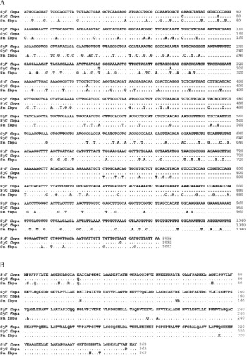
The gene encoding FBPA from the Formosan strain was successfully amplified by PCR using cDNA obtained from adult worms of S. japonicum. The nucleotide sequence has been submitted to the GenBank database under the accession number DQ497790. As shown in fig. 1A, the DNA sequence of this gene was 1092 bp in length and was 99% similar to the Chinese strain (GenBank accession number DQ497791), with differences at six locations: 61, 75, 270, 297, 345 and 1078 bp. The cDNA sequence of the Formosan strain was also 84% similar (920/1092 bp) to S. mansoni FBPA (GenBank accession number L38658) (fig. 1A) (El-Dabaa et al., Reference El-Dabaa, Mei, El-Sayed, Karim, Eldesoky, Fahim, LoVerde and Saber1998).
Sequences of nucleotides (A) and amino acids (B) of fructose-1,6-bisphosphate aldolase (FBPA) from Schistosoma japonicum (Formosan strain, SjF; Chinese strain, SjC) and S. mansoni (Sm). The estimated molecular mass is 39.6 kDa.

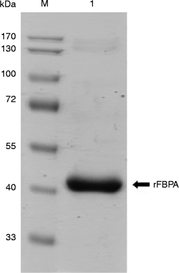
This gene encoded a protein with 363 amino acids at a molecular mass of 39.6 kDa, and the amino acid sequence had a similarity of 99% and 95% with the proteins from the Chinese strain of S. japonicum and S. mansoni, respectively (fig. 1B). This recombinant protein was a 6 × His-tagged fusion protein, and the molecular mass was 39.6 kDa +6 × His (fig. 2).
Sodium dodecyl sulphate–polyacrylamide gel electrophoresis (SDS–PAGE) of recombinant fructose-1,6-bisphosphate aldolase (FBPA) from Schistosoma japonicum Formosan strain. The molecular mass was 39.6 kDa +6 × His. Lanes: 1, purified recombinant FBPA; M, molecular mass standard.

Western blot analysis
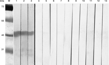
The ability of purified FBPA to react with the sera of Schistosoma-infected participants was demonstrated by Western blotting, the results of which are shown in fig. 3. A strong reaction band near 40 kDa was seen in lanes 1 to 3, in which recombinant FBPA was reacted with the sera of participants with schistosome ova. On the other hand, no band or weakly stained bands were seen in lanes 4–13 (sera from participants in whom no schistosome ova were found).
Western blot analysis of recombinant fructose-1,6-bisphosphate aldolase (FBPA) from Schistosoma japonicum Formosan strain reacted with sera of patients with and without ova of S. japonicum (determined by stool examination). Lanes: 1–3, FBPA reacted with sera of four patients with S. japonicum ova; 4 and 5, reacted with sera of two patients containing no parasites; 6 and 7, reacted with sera of two patients with Ascaris lumbricoides ova; 8 and 9, reacted with sera of patients with hookworm ova; 10 and 11, reacted with sera of patients with Paragonimus westermani ova; 12 and 13, reacted with sera of patients with Clonorchis sinensis ova; M, molecular mass standard.

ELISA on the sera of participants
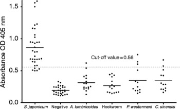
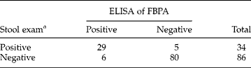
The ODs of the ELISA using purified FBPA from the Formosan strain on 120 participants are shown in fig. 4. The mean OD of 86 schistosome ova-negative participants was 0.270 and the SD of this group was 0.145. The cut-off point was selected as the mean plus 2SD, which was 0.56 in this study. A total of 35 sera samples were found to be positive by this assay, of which 29 were from participants who had ova of S. japonicum in their stool samples and the other six were from participants with ova of A. lumbricoides (1), P. westermani (3) and C. sinensis (2) (fig. 4).
Enzyme-linked immunosorbent assay (ELISA) using recombinant fructose-1,6-bisphosphate aldolase (FBPA) from Schistosoma japonicum Formosan strain on the sera of 120 participants. The cut-off values are indicated by the long dotted lines, and was 0.56 for this study. The sensitivity and specificity were 85.3% and 93.0%, respectively.

On comparison of these results with that of the faecal examination, the sensitivity and specificity of this ELISA were calculated as 85.3% and 93.0%, respectively (table 1).
Correlation of results of the ELISA assay of FBPA with the finding of ova of S. japonicum by faecal examination.
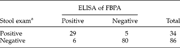
Sensitivity: number of positives by ELISA/number of positives by stool exam = 85.3% (29/34).Specificity: number of negatives by ELISA/number of negatives by stool exam = 93.0% (80/86).
a Based on the finding of S. japonicum ova in a stool exam.
Discussion
Egg microscopy is a traditional and widely used method to diagnose schistosome infection. This method is the least costly and technical assistance is available (Doenhoff et al., Reference Doenhoff, Chiodini and Hamilton2004). However, it is considered to be labour intensive and time consuming (Zhu, Reference Zhu2005); therefore, serological tests for large-scale and easier operation have been developed. ELISA for schistosomiasis was developed by Schinski et al. (Reference Schinski, Clutter and Murrell1976); peptides, protein, antibodies or hormones of schistosomes were used for this assay and FBPA was selected as the antigen.
Aldolase in Plasmodium species is an important and well-characterized enzyme of the glycolytic pathway (Srivastava et al., Reference Srivastava, Schmidt, Certa, Dobeli and Perrin1990), and it is also considered to be a potential target for antimalarial drug design (Wanidworanun et al., Reference Wanidworanun, Nagel and Shear1999). Tjitra et al. (Reference Tjitra, Suprianto, Dyer, Currie and Anstey1999) have used the combined P. falciparum–P. vivax immunochromatographic test (ICT malaria P.f/P.v) with aldolase (panmalarial antigen) and histidine-rich protein 2 (HRP-2) to diagnose malaria in eastern Indonesia; the sensitivity for P. vivax malaria was 96% for parasitaemias of more than 500 parasites/μl. Gatti et al. (Reference Gatti, Bernuzzi, Bisoffi, Raglio, Gulletta and Scaglia2002) have also used the ICT malaria P.f/P.v. with panmalarial antigen and HRP-2 to detect malaria in 241 travellers and immigrants in Italy; the sensitivity and specificity for P. falciparum infection were 94.4% and 94.5%, respectively. Furthermore, aldolase is an immunoreactive protein in human giardiasis (Palm et al., Reference Palm, Weiland, Griffiths, Ljungstrom and Svard2003) and in Candida albicans infection in murine models (Pitarch et al., Reference Pitarch, Diez-Orejas, Molero, Pardo, Sanchez, Gil and Nombela2001). In helminthic parasites, FBPA of Onchocerca volvulus (McCarthy et al., Reference McCarthy, Wieseman, Tropea, Kaslow, Abraham, Lustigman, Tuan, Guderian and Nutman2002) has been identified by immunoscreening with sera of infected patients. In bacteria, FBPA is also a secreted antigenic protein; it reacts with serum antibodies from broiler chickens immunized with virulent Clostridium perfringens (Kulkarni et al., Reference Kulkarni, Parreira, Sharif and Prescott2006). All of these studies indicate that FBPA can be an important antigen in the development of new diagnostic tools.
FPBA of S. mansoni has been cloned from cercariae (El-Dabaa et al., Reference El-Dabaa, Mei, El-Sayed, Karim, Eldesoky, Fahim, LoVerde and Saber1998) and lung-stage larvae (Harrop et al., Reference Harrop, Coulson and Wilson1999), and could induce a strong immune response (Harrop et al., Reference Harrop, Coulson and Wilson1999). In the present study, we cloned and expressed FBPA from adult worms of S. japonicum Formosan strain. This FBPA has 99% and 95% amino acid sequence similarity, as well as 99% and 84% cDNA sequence similarity, with S. japonicum Chinese strain (GenBank accession number DQ497791) and S. mansoni (GenBank accession number L38658), respectively (El-Dabaa et al., Reference El-Dabaa, Mei, El-Sayed, Karim, Eldesoky, Fahim, LoVerde and Saber1998). In the Western blotting, we confirmed that recombinant FBPA from S. japonicum Formosan strain reacts with antibodies in the sera of patients infected with S. japonicum Chinese strain. These results show that FBPA from S. japonicum Formosan strain can be used as a diagnostic antigen to detect schistosome infection.
The diagnostic serology of Schistosoma infection has used antigens that the parasite possesses, excretes or secretes to detect antibodies in circulating blood of infected patients. Crude extracts from schistosome ova and adult worms that are composed of multiple proteins, glycoproteins and lipoproteins have been important immunodiagnostic tools for the diagnosis of schistosome infection (Zhu, Reference Zhu2005). However, these antigens do not have satisfactory specificities and sensitivities, especially in individuals with mild infection (Guan & Shi, Reference Guan and Shi1996). Purified and recombinant proteins have been used in the diagnosis of schistosome infection and are more specific and sensitive than the crude extracts in ELISA (Doenhoff et al., Reference Doenhoff, Chiodini and Hamilton2004). Recently, recombinant peptide antigens such as rSj26GST, rSj23HD, rSj32 and Sj31-b have been developed for immunodiagnosis in China (Zhu, Reference Zhu2005).
In the present study, recombinant FBPA cloned from the Formosan strain was used as an antigen for ELISA in the detection of human schistosomiasis. By comparing the results obtained with those of stool examination, the sensitivity of this assay was calculated to be 85.3%. In the schistosome ova-negative individuals, six were positive for other ova, and all of them were recruited from the Hunan Province of China, an area endemic for schistosomiasis. The mean OD of the schistosome ova-negative individuals from Taiwan (0.191 ± 0.062) was lower than that of the other schistosome ova-negative individuals from China (0.315 ± 0.154). This could be due to a previous infection of schistosomiasis or a higher antibody level in individuals from the endemic area (fig. 4).
In conclusion, recombinant FBPA from S. japonicum Formosan strain can be a useful diagnostic tool for the detection of antibodies against S. japonicum in humans.
Acknowledgements
We thank the Tongji Medical College, Huazhong University of Science and Technology in the People's Republic of China (Hubei Province), and the Institute of Parasite Disease Control and Prevention, Chinese Center for Disease Control, for providing the sera for this study.