Introduction
Intractable aspiration leads to central nervous system (CNS) problems in patients of all ages, and the number of cases requiring surgical treatment has been increasing recently. Several surgical techniques are used for prevention of aspiration. Total laryngectomy is one of the most reliable methods, but it is irreversible. Lindeman first reported a diversion surgery for intractable aspiration in 1975.Reference Lindeman1 This method differs from laryngectomy in that it is reversible. Several modifications to Lindeman's method have been reported since then.Reference Krespi, Quatela, Sisson and Som2–Reference Baron and Dedo4 Patients lacking speech communication who are at high risk of aspiration are good candidates for this surgery. Laryngotracheal separation and tracheoesophageal diversion are currently the most commonly used surgical methods for complete prevention of aspiration. After these surgeries, the amount of saliva suctioning is reduced considerably, which improves the quality of life (QoL) of patients as well as caregivers.
In this study, we focus on tracheoesophageal diversion. Although this is relatively easy,invasive surgical relatively easy, although invasive surgical technique is associated with few complications. Moreover, it completely prevents aspiration, although oral food intake depends on the status of swallowing function. The purpose of this study was to describe our experience with this technique in treating intractable aspiration and report the peri-operative outcomes. The advantages of this method are also discussed.
Materials and methods
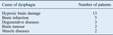
We retrospectively reviewed 25 patients (17 males and 8 females; median age, 25 years; range, 0–78 years; (Figure 1)) who underwent tracheoesophageal diversion for intractable aspiration between 2003 and 2009 at our hospital. Criteria for diversion included repeated pneumonia due to intractable aspiration, lack of speech communication and low probability of recovery estimated by the neurological findings. Of 25 patients,13 patients had experienced hypoxic brain damage and five brain infarction (Table I). All patients were medically managed for the prevention of post-operative aspiration pneumonia. All surgeries were performed under general anaesthesia. The observation period was 37 ± 20 months (range, 2–65 months). Four patients died after surgery.
Lateral view of the tracheoesophageal diversion procedures: (a) end-to-side anastomosis and (b) side-to-side anastomosis.

Causes of dysphagia
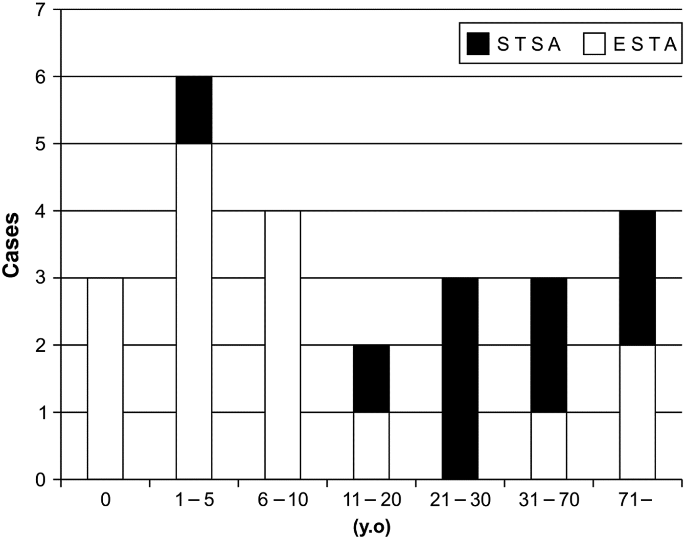
Two methods of tracheoesophageal diversion were used: end-to-side anastomosis mainly for patients who were not already tracheostomised and side-to-side anastomosis only for those who were pre-tracheostomised (Figure 1). Unlike the method used by Krespi et al.,Reference Krespi, Quatela, Sisson and Som2 the side-to-side anastomosis method we used only involved anastomosis of the back side of the tracheal rings and cricoid cartilage to the front side of the esophagus. End-to-side anastomosis was performed in 16 patients and side-to-side anastomosis in 9. The age distributions of the patients in the end-to-side anastomosis and side-to-side anastomosis treatment groups were different, with end-to-side anastomosis performed in younger patients (Figure 2).
Age distribution according to surgical method: (a) end-to-side anastomosis (ESTA) and (b) side-to-side anastomosis (STSA).
We investigated the cutting level of the trachea. We used the unpaired t-test to statistically analyse the differences in operative time and bleeding amount between the side-to-side anastomosis and end-to-side anastomosis groups, as well as between children and adults. We investigated the complications associated with the surgery and the feeding routes of patients before and after surgery. We examined the short-term and long-term complications of surgery. We also examined differences in nutrition route after surgery between non-adults and adults.
Results
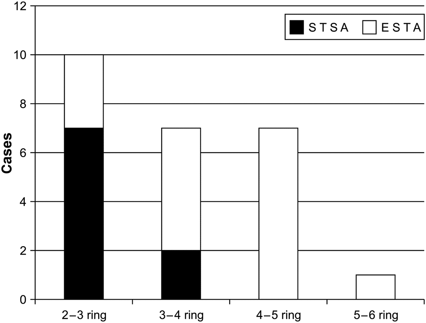
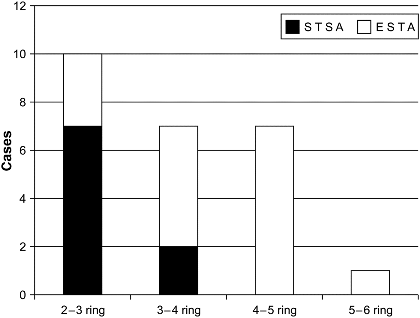
The cutting level of the tracheal ring depended on the tracheotomy position, as seen in Figure 3. Most patients undergoing side-to-side anastomosis were tracheostomised at higher tracheal ring levels, indicating a tendency towards a high cutting level. Cutting between the fourth and fifth rings and the fifth and sixth rings was only performed in non-adults (<20 years old). The average operative time for side-to-side anastomosis (191 minutes) was significantly longer than that for end-to-side anastomosis (141 minutes). There was greater bleeding among the side-to-side anastomosis group than the end-to-side anastomosis group, but the difference was not significant (Table II). Cutting levels were significantly lower, operative times were significantly shorter, and bleeding was significantly less (Table II) in non-adults (<20 years old) than in adults (>20 years old).
Cutting level of the trachea: (a) end-to-side anastomosis (ETSA) and (b) side-to-side anastomosis (STSA).
Operation time and amount of bleeding during operation

*p < 0.05 (unpaired t-test). ESTA = End-to-side anastomosis; STSA = side-to-side anastomosis.
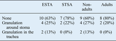
For the present study, we defined peri-operative complications as those occurring within one month of surgery and long-term complications as those occurring at a later time. Peri-operative complications were observed only in two (8 per cent) patients: one developed infection and one developed hematoma (Table III). All long-term complications were associated with tracheostomy tube insertion. Granulation formation around the stoma or within the tracheal mucosa was observed in 32 per cent of patients (Table IV). End-to-side anastomosis cases were observed to have more long-term complications. No long-term complications were observed among the ventilator-supported patients. Fewer complications were experienced by adults than non-adults, and granulation formation within the trachea was not observed in adults (Table IV). Fistula formation was not observed in any patients regardless of age.

Long-term complications*
* More than one month after surgery. ESTA = End-to-side anastomosis; STSA = side-to-side anastomosis.
After surgery, 18 patients (72 per cent) could be fed orally; however, only four patients (16 per cent) were principally fed orally. Fourteen patients took food through a combination of oral and other routes. Of the 21 patients for whom the oral route was not the primary nutritional route, nasogastric (NG) tube feeding was required in 12 (48 per cent) and feeding through gastrostomy was necessary in 7 (28 per cent). The remaining two patients were fed via an enteric fistula. Oral intake was possible only for adult cases. No difference in nutritional status was observed between the end-to-side anastomosis and side-to-side anastomosis groups (Table V).
Feeding route: pre- and post-operations
NG = nasogastric; ope = operations; ESTA = End-to-side anastomosis; STSA = side-to-side anastomosis.
Discussion
Patients with intractable aspiration resulting from central nervous system damage caused by brain infarction or hypoxia, as well as those with recurrent aspiration pneumonia, require surgery to prevent aspiration. Various surgical methods have been developed for this purpose. Laryngotracheal diversion was first reported by Lindeman,Reference Lindeman1 and we used this method for non-tracheostomised patients in the present study. This surgical technique is generally suitable for end-to-side anastomosis, which cannot be easily performed in tracheostomised patients, because the tissue around the tracheostomy often becomes scarred. Krespi et al. Reference Krespi, Quatela, Sisson and Som2 reported a method of tracheoesophageal anastomosis for pre-tracheostomised patients. This method involves partial removal of tracheal cartilage and anastomosis of the oesophagus. The methods of LindemanReference Lindeman1 and Krespi et al. Reference Krespi, Quatela, Sisson and Som2 both allow misdirected food to enter the oesophagus.
Later, Yarington and SuttonReference Yarington and Sutton3 and Baron and DedoReference Baron and Dedo4 described a laryngotracheal separation procedure without diversion. MontgomeryReference Montgomery5 performed glottic closure for 12 patients, and aspiration was prevented in all 12 patients. Sasaki et al. Reference Sasaki, Milmoe, Yanagisawa, Berry and Kirchner6 reported glottic closure with muscle flaps, and Habal and MurrayReference Habal and Murray7 used an epiglottic–arytenoid flap to close a larynx in a nine-year-old girl who did not experience any recurrent episodes of pneumonitis after surgery. Biller et al. Reference Biller, Lawson and Beak8 reported laryngoplasty for total glossectomy, involving closure of the epiglottis and the aryepiglottic fold. Since then, their method has been used not only for prevention of aspiration, but also for retention of phonatory function.
The aforementioned techniques were aimed at preventing flow of food into the trachea and preserving the laryngeal framework. Although they can completely prevent aspiration, none strike a balance between both vocal function and prevention of aspiration. In our study, tracheoesophageal diversion completely prevented pneumonia caused by aspiration. After surgery, there was no salivary flow into the trachea, and therefore there was a considerable decrease in the amount of saliva suctioning. This has the potential to improve the QOL of patients, parents and caregivers.
The cutting level of the trachea was most often the third–fourth or fourth–fifth tracheal ring gap and did not affect prognosis or the probability of complications. In pre-tracheostomised patients, the cutting level depended on the location of the tracheostomy before operation. It is easier to perform an anastomosis of the trachea and oesophagus when the cutting level is lower, because the trachea is easier to bend. We performed end-to-side anastomosis at lower cutting levels, so we could anastomose the trachea and oesophagus end-to-side. Furthermore, because the tracheal cartilage is softer in young patients, we cut the trachea as low as possible in the children. In the pre-tracheostomised cases, the stoma was often infected and granuloma formation observed; so the fresh tissue was limited and it was difficult to bend and anastomose end-to-side.
Laryngotracheal separationReference Yarington and Sutton3, Reference Baron and Dedo4 is the most common surgery used to prevent intractable aspiration; however, it is associated with the occurrence of tracheocutaneous fistula. In a study by Zocratto et al.,Reference Zocratto, Savassi-Rocha, Paixão and Salles9 fistulae occurred in 14 (23 per cent) of 60 patients. Eibling et al. Reference Eibling, Snyderman and Eibling10 reported that fistulae occurred in 13 (38 per cent) of 34 patients, but the length of hospital stay was not affected. Manrique et al. Reference Manrique, Settanni and Camponês do Brasil Ode11 found that fistulae occurred in 4 (17 per cent) of 23 paediatric patients, all of all of which were involved in laryngotracheal separation. In contrast to these studies, no fistulae were observed in our study. This indicates that, even though anastomosis of the trachea to the oesophagus requires more effort on the part of the surgeon, it may reduce the probability of tracheocutaneous fistula.
During swallowing, the pressure generated by the oropharynx and mesopharynx and the saliva that enters the larynx can escape through laryngoesophageal or tracheoesophageal fistulae. When anastomosis is not performed, the swallowing pressure increases in the larynx and directly exerts pressure on the laryngeal or tracheal suture. Eisele et al. Reference Eisele, Yarington, Lindeman and Larrabee12 compared tracheoesophageal diversion with laryngotracheal separation and reported that fistulae occurred in 3 (18 per cent) of 17 separation cases and in none of the 14 diversion cases. This suggests that, compared with diversion, separation increases the risk of developing tracheocutaneous fistula.
The alimentary route after surgery primarily depends on the swallowing function and the general condition of the patient. In the study by Zocratto et al.,Reference Zocratto, Savassi-Rocha, Paixão and Salles9 62 per cent of the patients were able to accept oral feeding after surgery. Eibling et al. Reference Eibling, Snyderman and Eibling10 reported that 11 (32 per cent) and 3 (9 per cent) of 34 patients could accept regular or only liquid diets, respectively. Eisele et al. Reference Eisele, Yarington, Lindeman and Larrabee12 found that 9 (60 per cent) of 15 laryngotracheal separation patients and 8 (73 per cent) of 11 tracheoesophageal diversion patients could accept oral feeding. In our study, only 16 per cent of the patients could accept oral feeding. This worse result was likely because the neurological and nutritional statuses of patients in our study were more severe than those of patients in other studies. The neurological status was worse in non-adult cases; as the feeding status. The nutrition status after operation depends on the neurological status of the patient; so the operation does not ensure oral intake. After surgery, patients can learn safe oral intake; so the operation is beneficial for those patients.
The reversibility of the end-to-side anastomosis and side-to-side anastomosis methods is speculative. Yarington and SuttonReference Yarington and Sutton3 performed reconstruction in two of six cases after tracheoesophageal diversion. Eibling et al. Reference Eibling, Snyderman and Eibling10 reported that 2 of 34 cases in their study underwent reconstructive surgery. Eisele et al. Reference Eisele, Yarington, Lindeman and Larrabee12 reported that 5 of 31 patients underwent reconstructive surgery, but one of these underwent additional laryngectomy. Pletcher et al. Reference Pletcher, Mandpe, Block and Cheung13 reported a case of reversal after laryngotracheal separation. Swallowing function can recover if the cause is neural in nature. Many studies have shown small percentages of reconstruction/reversals.Reference Yarington and Sutton3, Reference Eibling, Snyderman and Eibling10–Reference Pletcher, Mandpe, Block and Cheung13 In our study, none of the patients could undergo tracheal reconstruction. Two possible reasons for this were our strict criteria for surgery and the lack of accurate evaluation of swallowing function after surgery. However, the most likely reason was that we often performed other surgeries (such as myotomy of the cricopharyngeal muscle and laryngeal elevation) for borderline cases.
• The results of the present study indicate that tracheoesophageal diversion is a safe and feasible procedure for patients with intractable aspiration
• This method differs from laryngectomy in that it is potentially reversible, and it reduces the probability of fistula formation compared with separation
• It is suitable for patients who lack speech communication and are at high risk of aspiration
• However, potential candidates should be carefully evaluated before this surgery is undertaken
The operative time for side-to-side anastomosis was significantly longer than that for end-to-side anastomosis. This indicates that if the swallowing function of the patient has a low probability of recovery, then diversion or separation should be performed instead of tracheostomy in the first place. Age was also found to influence outcome. The tracheal cartilage of children is softer than that of adults. Therefore end-to-side anastomosis was easier to perform in children, and the resulting operative time and occurrence of bleeding were less than in adults. Tomita et al. Reference Tomita, Tanaka, Shinden and Ogawa14 compared laryngectomy and tracheoesophageal diversion cases. They found operative time for diversion to be shorter than that for total laryngectomy, but the effects on swallowing dysfunction were almost similar. In that study, the time required to perform total laryngectomy was 2 or 3 hours, which is similar to our study. In our case, the operative time was not so long,bleeding was little and no fistula was observed;hence tracheoesophageal diversion is one of reasonable method for intractable aspiration.