Introduction
This third instalment in the series of magnetic resonance imaging (MRI) physics discusses the area of pulse sequences. This topic receives little attention within the general literature not only because of its specialisations but also because the underlying concepts are quite complex. However, as neuroimaging techniques continue to experience growth and often become the focus of neuropsychiatric research, it is important to have an understanding of what pulse sequences are and their associated terminology. In basic terms, the pulse sequences define the manner in which the radiofrequency pulses, which generate the detectable signals, and magnetic field gradients, which provide the spatial encoding of the signals, are applied Reference Jackson, Ginsberg and Schomer(1). They are computer programs that control the MRI hardware to go through the MRI signal measurement process. Often a pulse sequence diagram is used that shows the sequence of events that will occur during the MR imaging. There are hundreds of pulse sequences available for imaging and spectroscopy and each is specific to creating images of the required properties. Some of the commonly used sequences that are most often encountered are described below.
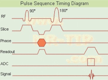
The spin echo sequence is the most commonly used pulse sequence in neuroradiology (Fig. 1). It uses a 90° slice selective radio frequency (RF) pulse for excitation that dephases the spins and then an 180° RF pulse to rephase the spins to form an echo. This echo is the MR signal that is collected by the acquisition system and is used to fill in one line of k-space. However, to obtain a complete image, we need to fill in the entire k-space and this is achieved by using phase encoding gradients. For example, if we have 256 steps of phase encoding we would get 256 echoes to fill entire k-space. The Fourier transform of the k-space then gives us an image. To calculate how much time it would take to get an image, we can use formula-A. If we have a repetition time (TR) of 3 s, with a single averaging and 256 phase encoding steps, the scan time would be 12.8 min. However, it needs to be borne in mind that the formula below represents the time for a single slice and thus to obtain an MRI of the entire brain would take more than one slice and as such would increase the scan time considerably. For example, if we were to acquire 200 slices so as to acquire an entire brain volume, we would be looking at a scan time of several hours.
Spin echo sequence (courtesy mr-tip.com).


where TR, repetition time; ø, number of phase encodes; Av, number of averages.
To reduce the scan times,we use the fast spin echo (FSE) sequences. In FSE sequence, we apply the 180° RF pulse multiple times within the same TR to collect multiple echoes. For example, if we collect eight echoes instead of one, the scan time above will be reduced to 1.6 min.
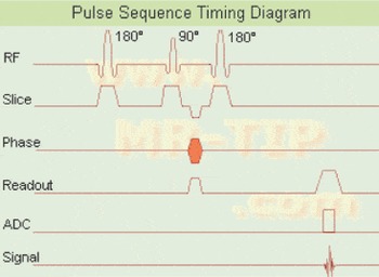
Another sequence used commonly is the inversion recovery sequence, which is a spin echo sequence with an additional 180° inversion pulse (Fig. 2). This pulse precedes the 90° excitation pulse and 180° rephasing pulse of a spin echo sequence. The time between the inversion pulse and the excitation pulse is called the inversion time (IR). Image contrast can be manipulated by changing the inversion time Reference Weishaupt(2), and these sequences are typically used for T1-weighting or fat suppression but can also be used for T2-weighting. Two commonly used inversion recovery sequences are short TI inversion recovery (STIR) and fluid-attenuated inversion recovery (FLAIR). STIR is used for fat suppression because it eliminates the signal from fat by using shorter inversion times. FLAIR uses longer inversion times and is used to suppress signals from fluids such as cerebrospinal fluid, while detecting signals from brain tissue, tumours, fat and oedema.
Inversion recovery sequence (courtesy mr-tip.com).

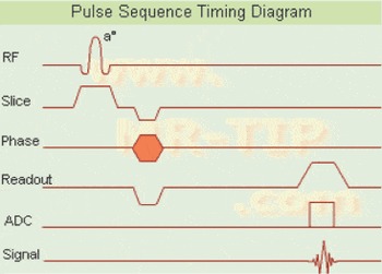
The gradient echo (Fig. 3) sequences are different again in that they use a pair of bipolar magnetic field gradients to produce an echo after the 90° excitation pulse instead of using the 180° refocusing pulse. Because no 180° pulse is needed to generate the echoes, very short TR can be achieved that make the sequences significantly faster than the spin echo sequences. This reduction in scan time however comes with a compromise of reduced signal-to-noise ratio and increased artefacts because of changes in magnetic susceptibility between different tissues. In the case of gradient echo sequences, because short repetition times are used, there is part of the signal that is left over after each cycle. This signal must be destroyed when T1-weighted images are acquired and applying ‘spoiler’ gradients does this. The spoiled gradient echo (sometimes referred to as SPGR) sequences are widely used in the clinical settings and are often used as the structural sequence preceding fMRI protocols. Along with SPGR, FLASH (fast low angle shot) is another popular spoiled gradient sequence.
Gradient echo sequence (courtesy mr-tip.com).

In MRI, we also have hybrid sequences. They use a series of spin echoes and gradient echoes to generate the signal. An example of such a hybrid sequence is GRASE (gradient and spin echo). In this instance, a series of 180° RF pulses is applied to generate several spin echoes; in addition several gradient echoes are produced for each spin echo by rapidly switching the readout gradient polarity. This makes the GRASE sequence faster than FSE.
The above pulse sequences and techniques form the foundation for routine clinical MRI and from these specialized applications have been developed for the research community. In particular, for neuropsychiatry applications such as fMRI, spectroscopy and diffusion tensor imaging are all now possible.
The byte
The above foundation sequences used in neuroimaging can be further modified to create more sophisticated techniques. For example, modifying the spin echo or the gradient echo sequence by the addition of a magnetization transfer (MT) pulse adds a new mode of image contrast. This MT pulse selectively saturates protons that have restricted motion typically those associated with macromolecules such as proteins and membrane phospholipids Reference Balaban and Ceckler(3). This magnetization is transferred from the macromolecular protons to the free water protons. The image contrast also depends on the concentration of these macromolecules. MT pulses have been used to improve sensitivity of post-contrast T1-weighted imaging of brain lesions and multiple sclerosis plaques. Adding the appropriate gradient field pulses during the echo time, a conventional spin echo sequence can be used to emphasize effects of diffusion Reference Jackson, Ginsberg and Schomer(1). T2-weighted images can be acquired with and without the diffusion gradient pulses. Post-processing of the images can generate an apparent diffusion coefficient image in which pixel intensity is directly proportional to the diffusion coefficient. To this end diffusion weighted imaging has been used to evaluate damage to white matter tracts. An inversion pulse can also be used with gradient echo technique to acquire fast three-dimensional T1-weighted images. These sequences are commonly known as MPRAGE sequences and are widely used for structural imaging. Finally, echoplanar and spiral sequences are other sequences that can be used to further reduce the acquisition times in MR imaging which makes them suitable for fMRI applications.
Conclusion
The continual development of pulse sequences has lead to an increasing range of available imaging modalities for the both clinical and scientific communities. Desirable properties from image contrast to the acquisition time are controlled by the pulse sequence. To this end, it is important to have an understanding (albeit rudimentary) of the basic as well as the more sophisticated pulse sequences (as described above) that are routinely used in the neuropsychiatry.