INTRODUCTION
Schistosomiasis causes severe morbidity and mortality in over 200 million people worldwide, with a further 600 million currently at risk of infection (King et al. Reference King, Dickman and Tisch2005; Gryseels et al. Reference Gryseels, Polman, Clerinx and Kestens2006). All schistosomes are transmitted between mammalian definitive hosts and fresh water snail intermediate hosts. Adult worms occur as worm pairs in specific venous sites of the mammalian host. Females lay hundreds to several thousands of eggs daily, many of which are trapped in host tissues or, alternatively, are released in the faeces or urine. The eggs hatch in fresh water to release free-swimming miracidia which infect snails. Inside the snail the miracidia transform into mother sporocysts that produce daughter sporocysts by asexual reproduction, which in turn release cercariae into the external, aquatic environment. The cercariae penetrate mammalian skin and migrate via the lungs to the venous system where they eventually develop into adult worms. These developmental phases are accompanied by remarkable morphological, biochemical and molecular changes in the various life-cycle stages (Vermeire et al. Reference Vermeire, Taft, Hoffmann, Fitzpatrick and Yoshino2006).
The major pathological effects of schistosomiasis result from the deposition of eggs within human liver and other organs. This deposition is followed by a subsequent intense granulomatous response induced by these eggs. With continuing questions concerning the long-term efficacy of the therapeutic drug, praziquantel, to control schistosomiasis (King, Reference King2007), and the current lack of effective anti-schistosome vaccines, there is considerable interest in utilizing post-genomic technology to develop new drugs or subunit vaccines against schistosomes, by exploiting parasite molecules pivotally involved in essential metabolic processes and/or critical phases of development. These approaches have been aided by valuable resources available in the expressed sequence tag (EST) databases for schistosomes (Hu et al. Reference Hu, Yan, Shen, Liu, Zhu, Song, Xu, Wang, Rong, Zeng, Wu, Zhang, Wang, Xu, Wang, Fu, Zhang, Wang, Brindley, McManus, Xue, Feng, Chen and Han2003; Verjovski-Almeida et al. Reference Verjovski-Almeida, DeMarco, Martins, Guimaraes, Ojopi, Paquola, Piazza, Nishiyama, Kitajima, Adamson, Ashton, Bonaldo, Coulson, Dillon, Farias, Gregorio, Ho, Leite, Malaquis, Marques, Miyasato, Nascimento, Ohlweiler, Reis, Ribeiro, Sa, Stukart, Soares, Gargioni, Kawano, Rodrigues, Maderia, Wilson, Menck, Setubal, Leite and Dias-Neto2003; Liu et al. Reference Liu, Lu, Hu, Wang, Cui, Chi, Yan, Wang, Song, Xu, Wang, Zhang, Zhang, Wang, Xue, Brindley, McManus, Yang, Feng, Chen and Han2006). These public datasets have been utilized to design and construct microarrays, with the resultant transcriptional data highlighting considerable numbers of genes from Schistosoma species for study (Fitzpatrick and Hoffmann, Reference Fitzpatrick and Hoffmann2006; Gobert et al. Reference Gobert, McInnes, Moertel, Nelson, Jones, Hu and McManus2006; Moertel et al. Reference Moertel, McManus, Piva, Young, McInnes and Gobert2006; Vermeire et al. Reference Vermeire, Taft, Hoffmann, Fitzpatrick and Yoshino2006; Jolly et al. Reference Jolly, Chin, Miller, Bahgat, Lim, DeRisi and McKerrow2007). Recently, we described the fabrication and use of a 22 575 feature 60-mer microarray to investigate transcription patterns between and within discrete Chinese (SJC) and Philippine (SJP) strains of Schistosoma japonicum (Moertel et al. Reference Moertel, McManus, Piva, Young, McInnes and Gobert2006), and to show stage-associated transcription between mature adult worms and lung schistosomula from amplified S. japonicum mRNAs (Chai et al. Reference Chai, McManus, McInnes, Moertel, Tran, Loukas, Jones and Gobert2006). In order to further address transcriptional differences in S. japonicum, 8 gender- or strain-associated molecules identified by microarray analysis (Moertel et al. Reference Moertel, McManus, Piva, Young, McInnes and Gobert2006) were selected for the present study. These molecules were first analysed in key life-cycle stages (SJC and SJP male/female adult worms; SJC eggs, miracidia and cercariae) using real-time PCR to gain a further appreciation of their differential expression pattern during development. Then, functional analysis of a subset of 4 of the genes encoding metabolic enzymes was undertaken in order to correlate gender-associated transcript levels with enzyme activity in protein extracts from adult S. japonicum (SJC strain).
MATERIALS AND METHODS
Chemicals and biochemicals
All chemicals and biochemicals, including DNase- and RNase-free water used in isolation and storage of total RNA samples, were supplied by Sigma-Aldrich (Sydney, Australia), unless stated otherwise.
Selection of S. japonicum transcripts
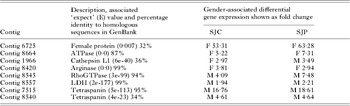
Probes associated with differential transcription (P-value ⩽0·001) in previous microarray analyses (Moertel et al. Reference Moertel, McManus, Piva, Young, McInnes and Gobert2006) were further interrogated to identify particular S. japonicum genes of interest. Male- and female-associated genes from SJC and SJP were selected initially by microarray analysis of male and female adult worms (Moertel et al. Reference Moertel, McManus, Piva, Young, McInnes and Gobert2006) and secondarily by in silico analysis (using 6 frame translation and BLAST analysis http://www.ncbi.nlm.nih.gov/) which included description, protein prediction and a detailed investigation of the probe and assembled sequence. Then, the genes were further screened for biologically relevant ontology, including association with cellular metabolic processes (GO:0044237), catalytic activity (GO:0003824), biological regulation (GO:0065007), or characterized as novel genes, exemplified by the tetraspanins (Table 1).
Schistosome genes selected for study with their identified function and strain-or gender-associated microarray fold expressionFootnote *
(SJC, Chinese S. japonicum; SJP, Philippine S. japonicum; M, Male adult; F, Female adult. Expect (E) values were obtained by BLAST (NCBI 2006).)

* Data from Moertel et al. (Reference Moertel, McManus, Piva, Young, McInnes and Gobert2006) where all probes were differentially expressed at a P-value ⩽0·001.
Isolation of adult worms, eggs and miracidia of S. japonicum
The maintenance of snails, the collection and storage of S. japonicum were carried out as described previously (Moertel et al. Reference Moertel, McManus, Piva, Young, McInnes and Gobert2006). Eggs of SJC were isolated and purified using modifications of a previously described protocol, whereby ethylenediaminetetraacetic acid (EDTA) and ethylene glycol tetraacetic acid (EGTA) were omitted from the wash buffers (Dalton et al. Reference Dalton, Day, Drew and Brindley1997). Eggs were used either for total RNA extraction using TRIzol (Invitrogen, Mount Waverley, Australia) or the production of miracidia. The miracidia were hatched and isolated using a previously described protocol (Dalton et al. Reference Dalton, Day, Drew and Brindley1997). Finally, the miracidial pellet was resuspended in 1 ml of TRIzol (Invitrogen) and stored at −20°C until the total RNA was isolated.
Isolation of SJC cercariae
Snails were exposed to a bright light at room temperature for 3 h to encourage cercariae to emerge. Cercariae attracted to the water surface were transferred to 50 ml Falcon tubes. The cercariae were sedimented by centrifugation (3000 g, 4°C for 20 min), transferred to 10 ml tubes, pelleted again and re-suspended in 500 μl of TRIzol reagent.
Isolation of total S. japonicum RNA and complementary DNA synthesis for real-time PCR analysis
Total RNA was isolated from adult worm pairs or adult males or females, eggs, miracidia or cercariae using published procedures (Hoffmann et al. Reference Hoffmann, Johnston and Dunne2002). Considerable care was taken to ensure that all total RNA samples used for real-time PCR were of high quality (A260/A280 nm ⩾1·7 in nuclease-free water) assessed using a Bioanalyzer RNA Pico LabChip (Bioanalyzer), with minimal degradation, as recommended (Bustin and Nolan, Reference Bustin and Nolan2004). Samples were treated with DNase (Promega) before complementary DNA (cDNA) was synthesized from total RNA using a modified SuperScript™ III protocol (Invitrogen, Melbourne, Australia) with p(dT)15 primers (Roche, Sydney, Australia). The amplified samples were quantified by a ND-1000 spectrophotometer and stored in microfuge tubes at −20°C until used.
Real-time PCR
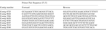
Transcription patterns determined previously by microarray analysis (Moertel et al. Reference Moertel, McManus, Piva, Young, McInnes and Gobert2006) were validated using real-time PCR. Forward and reverse primers (Sigma-Aldrich) were designed from S. japonicum transcripts (Table 2). The PCR products from these transcripts were amplified and sequenced to confirm sequence identity. All cDNA samples synthesized from aliquots of the same total RNA used for the microarray experiments were adjusted to 50 ng/μl, being quantified by a ND-1000 spectrophotometer (Nano Drop, Wilmington, USA). Then, 5 μl aliquots were combined with 10 μl of SYBER® Green (Applied Biosystems), 3 μl of water and 2 μl (5 pmol) of each forward and reverse primers in a 0·1 ml tube (Corbett Research, Sydney, Australia). All reactions were performed in a Rotor-Gene (3000) thermal cycler (Corbett Research) and the data analysed using Rotor Gene 6 software (Corbett Research). In order to minimize indiscriminate binding of double-stranded DNA, separate reverse transcription and PCR steps were used (Bustin and Nolan, Reference Bustin and Nolan2004). Melting curves were used to optimize the cycling conditions and to verify the specificity of the PCR. Primary normalization was calculated by quantification of sample cDNA templates. All real-time PCR experiments were undertaken in duplicate, the confidence threshold (CT) of the second set being normalized to the first set before evaluation. This was done using the Rotor Gene 6 software, importing the standard curve of the first set to that of the second. The mean copies per reaction and standard deviation values shown in Table 3 were calculated from the mean of 4 or more original and normalized CT values.
Primers used for real-time PCR to investigate gene expression in the different Schistosoma japonicum life-cycle stages

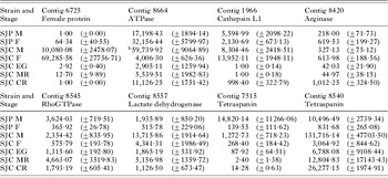
Real-time PCR analysis of selected Schistosoma japonicum contigs in copies per reactionFootnote *
(SJP, S. japonicum Philippine (Sorsogon, Luzon) strain; SJC, S. japonicum Chinese (Anhui) strain; M, adult male; F, adult female; EG, eggs; MR, miracidia; CR, cercariae.)
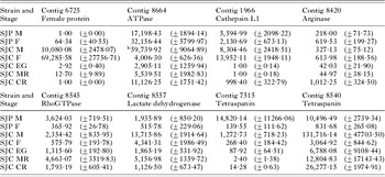
* The results shown are the mean values of duplicate sets of real-time PCR data comprising 4 or more replicates. The mean values for each contig are in copies/reaction calculated from the standard curve for each primary data set (±S.D.). bContig 8664 (ATPase) in SJC M was the only gender-associated probe revealed by microarray analysis not validated by real-time PCR.
Enzyme assays
For these assays, adult males and females of the SJC strain were homogenized in 0·1% Triton X-114 or 100 mM K2PO4 pH 7, 2 mM EDTA in phosphate-buffered saline (PBS). All enzyme assays were undertaken at room temperature (RT) (23–25°C) in triplicate and blanks and positive controls included, according to manufacturers' instructions. The total protein concentrations of the homogenates were determined using the Bradford dye-binding procedure (Bio-Rad) according to the manufacturer's protocol.
ATPase activity was determined by nicotinamide adenine dinucleotide (NADH) oxidation where 1 molecule of NADH is converted to NAD+ by the enzyme, which corresponds to the production of 1 molecule of ADP (Moller et al. Reference Moller, le Maire and Andersen1988). Pooled adult male or female worms of the SJC strain were homogenized in 0·1% Triton X-114 and centrifuged at 8000 g, 4°C for 5 min. The total protein content in the supernatant was quantified and stored at −70°C until used. For each sample, 30 μl of 0·1 M MgATP (Tris base, Sigma), 6 μl of 15 mM NADH, 10 μl of Mg-phosphoenolpyruvate (50 mM MgCl2 and 50 mM phosphoenolpyruvate) and 6 μl of lactate dehydrogenase (800–1200 units/mg protein) (LDH) were added to 500 μl of medium [0·1% Triton X-114, 0·1 mM CaCl2, 0·1 M KCl, 0·01 M TES in PBS]. To each of the tubes, 10 μl of pyruvate kinase (0·01 M TES and 0·1 M KCl at a protein concentration of 5 mg/ml) were added and left to incubate for 2 min at RT. Finally, 400 μg (total volume of 600 μl) of adult SJC male or SJC female protein sample were added to individual tubes and the rate of NADH oxidization was measured (after 1 min) at 340 nm for 4 min, reading the absorbance every 30 sec in a UVette (Crown Scientific). The rate of NADH oxidation in international units (IU) was calculated by the Beer Lambert law, using the molar extinction coefficient of 6220 for NADH (McMillan et al. Reference McMillan, Stimmler, Foth, McFadden and Muller2005).
Arginase activity, measuring the conversion of arginine to ornithine and urea, was determined using a quantitative colorimetric assay at 494 nm, employing a QuantiChrom arginase assay kit (Bioassay Systems). An aliquot (40 μl) of protein extract (40 μg) from either adult male or female SJC worms in 0·1% Triton X-114 was combined with 10 μl of the substrate buffer and incubated at RT for 2 h. Samples were then added to a 96-well flat-bottomed plate (Corning Inc) with the appropriate blank controls. Urea reagent (supplied with kit) was then added to the individual wells to stop the arginase reaction and incubated at RT for 15 min. The absorbance was then measured at 493 nm and enzyme activity (in IU) calculated according to the kit instructions.
GTPase activity was measured in adult male and female SJC worms using a GTPase colorimetric assay kit (Innova Biosciences). Male and female worms were homogenized separately in 0·5 M Tris (Invitrogen), pH 7·5, before removal of free inorganic phosphate (Pi) by dialysis (molecular weight cut off: 10 000). Aliquots (100 μl) containing 5 μg of protein were then combined with the substrate buffer mix in individual wells of a 96-well plate, after which gold mix (malachite green formulation) was added to all samples; blanks and the plates incubated at RT for 2 min. Then, the stabilizer (Innova Biosciences) was added to all samples prior to another incubation at RT for 30 min. The optical density of each well was read at 650 nm to determine inorganic phosphate (Pi) production. A standard phosphate curve was produced to calculate specific GTPase enzyme activity (in IU) in each sample by measurement of Pi (μM) released over time.
Adult male and female SJC worms were separately homogenized in 500 μl of 100 mM K2PO4 pH 7, 2 mM EDTA in PBS, and the homogenates centrifuged at 10 000 g for 15 min at 4°C. The supernatants were removed, their protein concentration determined, and stored at −70°C. A QuantiChrom assay kit (Bioassay Systems) was used to quantify the activity of lactate dehydrogenase (LDH) in each sample at 565 nm. A 10 μl aliquot of sample, for both male and female worms containing 28·5 μg of protein was transferred into separate wells of a 96-well plate. These samples were then combined with colour reagent and substrate buffer (Bioassay Systems) before reading the absorbance at 565 nm within 1 min and again after 25 min, following the manufacturer's instructions. All absorbance values were measured, taking into account blank and calibrator (supplied with the QuantiChrom assay kit) readings, to calculate the amount of LDH enzyme activity (in IU).
RESULTS
Real-time PCR was used to validate previous microarray data obtained for adult male and female SJC and SJP (Moertel et al. Reference Moertel, McManus, Piva, Young, McInnes and Gobert2006) and to expand the gene expression profile in miracidia, eggs and cercariae for 8 selected putative gender- or strain-associated gene transcripts (Table 1). We selected these genes from our previous identification of a panel of 1163 male and 1016 female probes differentially expressed in adult SJP, and 1047 male and 897 female probes differentially transcribed between adult SJC (P-value ⩽0·001) (Moertel et al. Reference Moertel, McManus, Piva, Young, McInnes and Gobert2006). These probes were screened further based on meaningful annotation, which included 6-frame characterization and protein BLAST analysis. The gene sequences had 85–99% identity to expected contiguous products (data not shown). Additionally, a subset of 4 of the cDNA transcripts was selected to correlate transcript level with enzyme activity in adult male and female SJC worms.
Contig 6725 was shown previously in microarray analysis (Moertel et al. Reference Moertel, McManus, Piva, Young, McInnes and Gobert2006) to be strongly up-regulated in adult female worms of both SJC and SJP (Table 1). The real-time PCR analysis confirmed this finding, showing a significantly higher signal in both the SJC and SJP female worms compared with males (Table 3). No significant variation (within 1 standard deviation) could be detected for this transcript between pools of SJC eggs or miracidia. The expression levels in adult male and female SJC were substantially higher than in their SJP counter parts (Table 3). Investigation of the complete assembled sequence for Contig 6725, including a 6-frame translation, identified similarity (E-value 0·007) with Fs800, a previously described female-associated protein from S. mansoni (Reis et al. Reference Reis, Kuhns, Blanton and Davis1989).
Earlier microarray analysis (Moertel et al. Reference Moertel, McManus, Piva, Young, McInnes and Gobert2006) indicated that Contig 8664 was female-associated in both SJC and SJP (Table 1). This was verified by real-time PCR analysis of adult worms of SJP in which there was an up-regulation in females compared with males, but this was not the case for SJC in which the transcription level was 15-fold less in females (Table 3). BLAST analysis of the translated amino acid sequence of Contig 8664 showed the protein contained a conserved protein adenosine 5′-triphosphatase (ATPase) subunit region. Accordingly, we measured ATPase enzyme activity in homogenates of male and female SJC; the specific activity in adult females (0·115±0·005 IU/μg protein) was more than 9-fold greater than in adult males (0·012±0·000 IU/μg protein) (Table 4), which again correlated well with the cDNA microarray but not with the real-time PCR findings.
Enzyme activities in extracts of SJC adult worms and relative up-regulated fold changes in expression levels in males or females determined by enzymatic assay, microarray analysis and real-time PCR
(One IU represents the conversion of 1 μmole of substrate to product per min.)

BLAST analysis of the complete translated amino acid sequence of Contig 1966 indicated high sequence identity (60%) to the protease enzyme, cathepsin L1. Real-time PCR analysis of Contig 1966 (Table 3) confirmed the microarray result (Table 1), including higher levels of transcription in SJP males compared with females and the reverse with SJC worms. A significant decrease in primary signal from SJC adult worms to eggs and miracidia was also shown by real-time PCR for Contig 1966 (Table 3).
Real-time PCR (Table 3) confirmed the up-regulation of Contig 8420 in SJP and SJC females shown by the microarray analysis (Table 1). There was also a significantly increased transcription in cercariae (Table 3) compared with the other developmental stages of the SJC strain. A 6-frame translation of Contig 8420 and subsequent protein BLAST analysis indicated a conserved arginase sequence in the protein. The up-regulation in females, as shown by real-time PCR, did not correlate with the direct enzymatic measurement of arginase activity using SJC worm extracts. The arginase activity in males was 0·408±0·014 U/μg protein, which was 1·3 times higher than the corresponding activity (0·310±0·016 U/μg protein) in females (Table 4).
BLAST analysis of protein inferred from Contig 8545 confirmed its putative identification as a GTPase. The real-time PCR gene expression of Contig 8545 confirmed the microarray results (Table 1) with substantially higher expression in SJC and SJP males (Table 3). Enzymatic assays also confirmed that GTPase was expressed at a higher level in males of SJC (Table 4).
BLAST analysis of protein inferred from Contig 8557 (lactate dehydrogenase-like protein of S. japonicum; complete sequence) confirmed its identity as a lactate dehydrogenase. The microarray analysis (Table 1) showing an up-regulation in SJC and SJP males was confirmed by real-time PCR analysis, showing a 3·75- and a 3·15-fold increase in SJP and SJC males, respectively (Table 3). This pattern was also reflected in the enzyme activity of LDH in SJC adults where the mean activity was 0·435±0·052 U/μg protein in males, 1·23-fold greater than that in females (Table 4).
Previous microarray analysis (Moertel et al. Reference Moertel, McManus, Piva, Young, McInnes and Gobert2006) showed an up-regulation of transcription linked to contigs 7515 and 8540 in males compared with females for both strains SJC and SJP (Table 1). BLAST analysis indicated that both probes encoded putatively conserved tetraspanin regions. Real-time PCR confirmed the microarray results for contigs 8540 and 7515 between adult males and females but not the higher expression of Contig 7515 in SJC males compared with those of SJP, or Contig 8540 in SJP males compared with those of SJC (Table 3).
DISCUSSION
Investigations of the different transcriptional profiles of male and female schistosomes are central to an increased understanding of their sexual and developmental biology. In the present study, we used real-time PCR analysis as a tool to investigate differences in transcription between the adult male and female worms of the Chinese (SJC) and Philippine (SJP) strains of S. japonicum revealed previously by microarray analysis (Moertel et al. Reference Moertel, McManus, Piva, Young, McInnes and Gobert2006). Additionally, real-time PCR was utilized to expand the transcriptional profiles of selected genes to other life-cycle stages of the SJC strain. While it is recognized that real-time PCR may not always provide independent verification of transcription levels obtained by microarray analysis (Anfosso et al. Reference Anfosso, Efferth, Albini and Pfeffer2006), the majority of data obtained herein indicated similar patterns using the two approaches (see Tables 1 and 3). The present analysis also included enzyme activity assays of extracts of male and female SJC worms, in order to correlate transcript levels with enzyme levels for a subset of the differentially expressed molecules.
A series of previous comparative transcriptomic studies have indicated major differences in the transcription profiles between male and female schistosomes (Moertel et al. Reference Moertel, McManus, Piva, Young, McInnes and Gobert2006; Wuhrer et al. Reference Wuhrer, Koeleman, Fitzpatrick, Hoffmann, Deelder and Hokke2006). Major gender-associated differences were also presented in the current study, an example being Contig 6725 representing a molecule with sequence identity to the female-specific fs800 protein of S. mansoni (Reis et al. Reference Reis, Kuhns, Blanton and Davis1989). This molecule was substantially up-regulated in females of both SJC and SJP (cf. Tables 1 and 3). Reis et al. (Reference Reis, Kuhns, Blanton and Davis1989) showed that fs800 is expressed in the vitellaria of females and that fs800 expression ceased when egg production stopped in senescent worms (Reis et al. Reference Reis, Kuhns, Blanton and Davis1989). The function of this molecule appears unrelated to eggshell formation but to other aspects of embryogenesis (Reis et al. Reference Reis, Kuhns, Blanton and Davis1989). We noted very low levels of transcription in both eggs and miracidia (Table 3) but no detectable levels in cercariae. Further characterization of this molecule may reveal a specific functional role in the sexual maturation of females of both S. mansoni and S. japonicum.
Previous studies have shown that ATPases play an important role in calcium homeostasis in schistosomes through transferring Ca2+ from the cytosol to the sarcoplasmic reticulum (Talla et al. Reference Talla, de Mendonca, Degand, Goffeau and Ghislain1998; Noel et al. Reference Noel, Cunha, Silva and Mendonca-Silva2001; Xiao, Reference Xiao2005). The transcription of the gene corresponding to Contig 8664, whose translated protein sequence indicated sequence similarity to Ca2+ ATPases was, apart from SJC females, significantly up-regulated in adults compared with eggs, miracidia and cercariae as shown by real-time PCR (Table 3). The up-regulation of this contig in SJC males, as determined by real-time PCR, was unexpected and probably due to unidentified homology differences between the strain (Moertel et al. Reference Moertel, McManus, Piva, Young, McInnes and Gobert2006).
Schistosomes express at least 2 cathepsin L proteinases, SmCL1 and SmCL2 (both identified in S. mansoni) (Brady et al. Reference Brady, Dowd, Brindley, Ryan, Day and Dalton1999). Cathepsin L1 is used in the metabolism of haemoglobin by adult females, whereas L2 is located around the uterus and the gynaecophoric canal (Michel et al. Reference Michel, Ghoneim, Resto, Klinkert and Kunz1995). The male association shown by the putative cathepsin inferred from Contig 1966 in SJP reported here is unusual, as previous studies have shown that cathepsin L1 is more highly expressed in female schistosomes and is not associated with a copy number polymorphism (Hoffmann, Reference Hoffmann2004; Fitzpatrick et al. Reference Fitzpatrick, Johansen, Johnston, Dunne and Hoffmann2004, Reference Fitzpatrick, Johnston, Williams, Williams, Freeman, Dunne and Hoffmann2005; Linzmeier and Ganz, Reference Linzmeier and Ganz2006). In order to further elucidate the possible function of the gene linked to Contig 1966, the primer product and probe section of the completely assembled sequence were translated and subjected to BLAST. This analysis revealed that this soluble protein contains a peptidase co-activator independent of the af-2 (CIA) conserved region C1, and is a member of a subfamily comprising cysteine peptidases, similar to papain and including the mammalian cathepsins B, C, F, H, L, K, O, S, V, X and W. The unexpectedly high signal for cathepsin L1 detected in cercariae (Table 3) may reflect additional function(s) of the enzyme in this life-cycle stage.
Arginase belongs to a family of metallo-proteins that occur ubiquitously in prokaryotes and eukaryotes (Muller et al. Reference Muller, Walter and Wrenger2005). In mammals, 2 arginase genes, types I and II, have been identified; type I occurs in liver cells and type II has a wide tissue distribution. Although the SjARG gene has been identified in adult S. japonicum (Li et al. Reference Li, Yu, Wu and Hu2006) and ornithine production has been reported in this species (Kawanaka et al. Reference Kawanaka, Hayashi and Carter1986; Fan et al. Reference Fan, Huang, Wang and Li2002) and S. mansoni (Cesari et al. Reference Cesari, Ballen, Perrone, Oriol, Hoebeke and Bout2000), the arginase enzyme has not been studied in schistosomes in any detail. The up-regulation of Contig 8420 (arginase) in females compared with males by microarray analysis (Table 1) and real-time PCR (Table 3) shown here was not confirmed by enzyme activity measurements of SJC extracts, which suggests the presence and activity of multiple types of the gene. The presence of multiple forms of this gene is also supported by the low copy/reaction number shown in SJC eggs (Table 3), as previous studies have suggested that ornithine is a nitrogenous excretory product of this life-cycle stage (Kawanaka et al. Reference Kawanaka, Hayashi and Carter1986). Further protein sequence analysis and expression data together with a detailed study of this enzyme in S. japonicum may provide an explanation of the observed differential expression among the males, females and eggs of S. japonicum.
The enzyme Rho GTPase is involved in the regulation of several important processes within schistosomes, including organization of the actin cytoskeleton, gene transcription, cell cycle progression and membrane trafficking (Vermeire et al. Reference Vermeire, Osman, LoVerde and Williams2003). Vermeire et al. (Reference Vermeire, Osman, LoVerde and Williams2003) showed that Rho GTPase was present at the highest level in adult females of S. mansoni, when compared with males, schistosomula (3-h-old) and cercariae. The present data suggest a substantially higher level of transcription for the gene linked to Contig 8545 in adult males of S. japonicum (Tables 1, 3 and 4), which suggests more extensive requirements in the male for the production of tegument, relative to the female. The tegument includes cytoskeletal components and requires extensive membrane turnover and remodulation (Gobert et al. Reference Gobert, Stenzel, McManus and Jones2003; Jones et al. Reference Jones, Gobert, Zhang, Sunderland and McManus2004). Why this requirement is not mirrored in male S. mansoni is not immediately apparent, but it may reflect a discrete species-specific difference. The role of Rho GTPase in other life-cycle stages has been reported (Vermeire et al. Reference Vermeire, Osman, LoVerde and Williams2003); these authors showed that the Rho1 was not detectable or not expressed at the protein level in cercariae. Similarly, SJC exhibited low levels of mRNA in cercariae as well as in eggs and miracidia. The GTPase we report is likely to be a different isoform of the enzyme (94% in the conserved region) to that described by Vermeire et al. (Reference Vermeire, Osman, LoVerde and Williams2003) who suggested the presence of multiple members of the Rho GTPase family in S. mansoni. Indeed, with the exception of 1 molecule (GenBank Accession number AY158217), all of the paralogues suggested (Vermeire et al. Reference Vermeire, Osman, LoVerde and Williams2003) are represented on the microarray used in the present study, and were found to be up-regulated in adult male compared with female worms for both strain SJC and SJP (Moertel et al. Reference Moertel, McManus, Piva, Young, McInnes and Gobert2006).
LDH is an ubiquitous enzyme (Guerra-Sa et al. Reference Guerra-Sa, Franco, Pena and Rodrigues1998; Lu et al. Reference Lu, Hu, Peng, Xie, Li, Wu and Yu2006; Wiwanitkit, Reference Wiwanitkit2007) that catalyses the conversion of pyruvate to lactate with the concomitant interconversion of NADH and NAD as the terminal reaction in glycolysis. Guerra-Sa et al. (Reference Guerra-Sa, Franco, Pena and Rodrigues1998) reported the putative preferential transcription for LDH in S. mansoni males compared with females; further, they showed that LDH was expressed in S. mansoni eggs, miracidia, cercariae and schistosomula, with higher levels in adults (Guerra-Sa et al. Reference Guerra-Sa, Franco, Pena and Rodrigues1998). Similarily, the current study showed a higher level of transcription representing Contig 8557 (LDH) in adult S. japonicum, highest levels being in males compared with females (Tables 1, 3 and 4). These findings correlate with the uptake of glucose by schistosomes; it is the dorsal tegument of the male which is the major absorptive surface for the pair (Camacho and Agnew, Reference Camacho and Agnew1995).
The presence of tetraspanins on the surface of S. mansoni was reported recently, and 2 recombinant tetraspanins (Sm-tsp-1 and Sm-tsp-2) were able to produce significant protection against a challenge infection in immunized mice (Tran et al. Reference Tran, Pearson, Bethony, Smyth, Jones, Duke, Don, McManus, Correa-Oliveira and Loukas2006). The 2 putative S. japonicum tetraspanins identified here, represented by contigs 7515 and 8540, have not been studied previously. Their transcription pattern was similar to Contig 5872 (CD63-like protein Sm-TSP-2 mRNA, complete sequence) being up-regulated in males compared with females; 2·97- and 4·27-fold for SJP and SJC, respectively (Moertel et al. Reference Moertel, McManus, Piva, Young, McInnes and Gobert2006).
In summary, this study is the first undertaken on schistosomes involving real-time PCR and enzymatic analysis to investigate differentially expressed transcripts identified by microarray experimentation. The results indicated variable transcription and expression patterns throughout the S. japonicum life-cycle. The 8 target contigs described here serve as a basis for further investigation of differential gene expression during the schistosome development and for studying the sexual dimorphism of adult worms. Continual refinement and annotation of the microarray used in the current study should support future work in this area.
The authors wish to thank Mary Duke (QIMR) for maintaining the S. japonicum life-cycles and for the provision of parasite materials for analysis, Dr Terrance Piva (RMIT) for advice with some of the enzyme analysis, and Dr Malcolm Jones for commenting on the manuscript. The financial support of the Sandler Centre for Basic Parasitic Research (USA), the Wellcome Trust (UK)/NHMRC (Australia) (ICRG Award; WT071657MA) and the National Health and Medical Research Council of Australia is acknowledged.