Introduction
Fibrovascular polyps of the hypopharynx and oesophagus are rare, benign tumours arising from the upper part of the gastrointestinal tract. Polyps arising from the oesophagus generally originate from the cervical oesophagus and upper oesophageal sphincter. Often, they arise at the level of the cricopharyngeal muscle. These tumours can attain very large sizes after slow growth over a long period.Reference Zevallos, Shah and Baredes1 It is not easy to determine the prevalence of these tumours from the literature; however, according to autopsy studies conducted since 1968, the prevalence of benign oesophageal tumours is known to be less than 1 per cent.Reference Attah and Hajdu2 Mortality and morbidity can be reduced by timely diagnosis and treatment.
Patients commonly present with nonspecific symptoms associated with oesophageal or airway obstruction. However, advanced cases may present with severe respiratory distress.Reference Owens, Donovan, Alford, McKechnie, Franklin and Stewart3 In such cases, surgical removal is indicated.
Case report
A 49-year-old man presented to the emergency department with increasing difficulty swallowing, advanced respiratory distress and weight loss, as well as a hypopharyngeal mass protruding from his mouth.
One year previously, the patient had undergone endoscopy in the gastroenterology department, and had been recommended to undergo biopsy for a 1 × 1 cm hypopharyngeal mass. However, he had rejected any intervention at that time.
During the current presentation, endoscopical examination showed a mass with a wide pedicle covered with smooth mucosa, arising from the right lateral aspect of the hypopharynx and having two heads extending to the oropharynx (Figure 1).
Giant fibrovascular polyp arising from the right lateral aspect of the hypopharynx, and having two heads visualised in the oropharynx. Part (b) shows pre-operative view, as seen standing at the patient's head. T = tongue; HP = hard palate

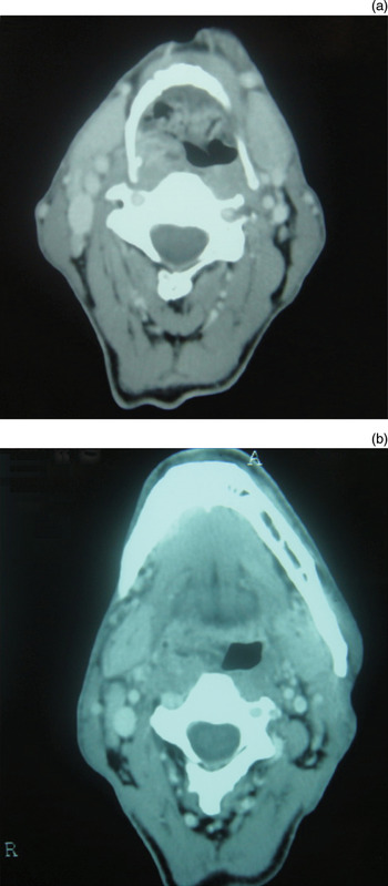
Pre-operative computed tomography (CT) showed that the origin of the tumour was the right lateral aspect of the hypopharynx (Figure 2).
Pre-operative, axial computed tomography scan of the neck, showing the tumour originating from the right lateral aspect of the hypopharynx. A = anterior; R = right

Because of intubation difficulty, a tracheostomy was performed. With the aid of a mouth gag and retractor, the pedicle of the mass was identified endoscopically, and the 11 × 5 × 3 cm mass was completely resected via the per-oral route, assisted by bipolar cautery (Figure 3).
Gross appearance of the giant fibrovascular polyp after per-oral endoscopic removal. The polyp measured approximately 11 × 5 × 3 cm.

In the first post-operative week, the patient was restricted to a liquid diet. The tracheostomy was closed on post-operative day three.
No recurrence was detected over two years of follow up.
Histopathological examination of the surgical specimen revealed tissue surrounded by hyperplastic squamous epithelium, and containing many congested vascular structures, chronic inflammatory cells and focal, mature adipose tissue, within an oedematous subepithelial fibrous stroma (Figure 4).
Photomicrographs showing the polyp tissue surrounded by hyperplastic squamous epithelium, and containing many congested vascular structures, chronic inflammatory cells and focal, mature adipose tissue, within an oedematous subepithelial fibrous stroma. (H&E; ×40)

Discussion
Fibrovascular polyps of the oesophagus and hypopharynx are rare tumours, and their exact incidence is unknown. Malignant transformation of those tumours is reportedly rare.Reference Owens, Donovan, Alford, McKechnie, Franklin and Stewart3, Reference Petry and Shapshay4
Fibrovascular polyps have been reported to frequently arise from two specific regions in the posterior hypopharyngeal wall.Reference Owens, Donovan, Alford, McKechnie, Franklin and Stewart3 One of those sites is the area between the superior and inferior cricopharyngeal muscles (known as the Killian dehiscence), and the other is the area between the inferior cricopharyngeal muscle and the proximal end of the oesophagus (known as the Laimer–Haeckermann area or Laimer triangle).
The underlying pathophysiological mechanism of polyp formation is not known. In the present case, the polypoid formation detected on earlier endoscopy probably enlarged progressively, due particularly to pressure changes during eating.
The symptoms of fibrovascular polyps vary according to their size, location and complications. Fibrovascular polyps of the oesophagus can remain asymptomatic for long periods. The most common complaints are dysphagia associated with oesophageal obstruction, regurgitation and aspiration during eating. However, patients may experience nonspecific respiratory symptoms such as chronic cough, dyspnoea and recurrent pneumonia, as well as clinical emergencies such as complete airway obstruction.Reference Watson-Williams5
The diagnosis is established by endoscopic and radiological evaluation. One study found that 25 per cent of intramural and intraluminal oesophageal tumours can be overlooked at endoscopy.Reference Whitman and Borkowski6 Moreover, polyp mucosa can often be mistaken for normal oesophageal or pharyngeal mucosa.Reference Owens, Donovan, Alford, McKechnie, Franklin and Stewart3, Reference Whitman and Borkowski6 Thus, radiological study is required, not just to detect the mass but also to determine its origin and extension.Reference Borges, Bikhazi and Wensel7 Whitman and Borkowski have defined the computed tomography and magnetic resonance imaging findings for fibrovascular polyps.Reference Whitman and Borkowski6
The differential diagnosis of fibrovascular polyp includes hamartoma, inflammatory polyp, lipoma, haemangioma, lymphangioma and schwannoma, as well as several rare neoplasms such as carcinoid tumour and chemodectoma.Reference Eberlein, Hannan, Josa and Sugarbaker8
Surgical excision is the definitive treatment.Reference Watson-Williams5 The surgical approach is chosen based on the size and location of the mass. If surgical resection cannot be performed rapidly, then the patency of the airway should be ensured by pre-operative intubation or tracheostomy.Reference Owens, Donovan, Alford, McKechnie, Franklin and Stewart3, Reference Nuyens, Bhatti and Flint9
Tumours with an appropriate pedicle can be excised endoscopically. This method is noted to be more suitable for smaller tumours, for which haemorrhage control and complete visualisation are reportedly more difficult.Reference Owens, Donovan, Alford, McKechnie, Franklin and Stewart3, Reference Eliashar, Saah, Sichel and Elidan10
Owens et al. reported performing lateral pharyngotomy in two patients with fibrovascular polyps located close to the cricopharyngeal muscle.Reference Owens, Donovan, Alford, McKechnie, Franklin and Stewart3 They were able to gain complete control of the tumour while ligating the polyp vessels with sutures, and recommended this method as it facilitated the identification of any mucosal abnormality, particularly in the dehiscence area.
Pham et al. described the endoscopic removal of a giant fibrovascular polyp in the oesophagus.Reference Pham, Rees and Belafsky11
• Fibrovascular polyps occur most commonly in the cervical oesophagus, and are extremely rare in the hypopharynx
• Endoscopic excision is favoured for smaller lesions when possible; open surgery via lateral pharyngotomy is an alternative
• Securing the airway is the most important part of surgical management
In our case, a per-oral endoscopic approach enabled us to gain complete control over the polyp pedicle, even while the polyp itself protruded from the mouth. Since intubation of our patient may have proved difficult, a tracheostomy was performed to secure the airway. The 11 × 5 × 3 cm mass was then excised via the per-oral route, with the help of a mouth gag and retractor, while controlling haemorrhage with bipolar cautery.
Conclusion
In patients presenting with nonspecific symptoms of the oesophagus and upper airway, particularly difficulty swallowing, a comprehensive examination is essential before considering nonorganic causes. Fibrovascular polyps should be considered in the differential diagnosis of obstructive lesions in this region. It is essential to instigate appropriate pre-diagnosis measures to prevent possible complications, such as airway obstruction. Endoscopy and radiological studies are the main means of diagnosis. Securing the airway pre-operatively is crucial. If the posterior hypopharyngeal wall dehiscence area can be accessed adequately by endoscopy, then endoscopic excision should be preferred, in order to avoid the potential morbidity of open surgery.