Double outlet right ventricle is a rare form of CHD, where both of the arterial roots are supported in their greater part by the right ventricle. Reference Karmegaraj, Kumar, Srimurugan, Sudhakar, Simpson and Vaidyanathan1 A vast majority of the hearts with double outlet right ventricle have an interventricular communication. Reference Ebadi, Spicer, Backer, Fricker and Anderson2 However, it has been recognised that a double outlet right ventricle can exist in the absence of an interventricular communication. Reference Menon, Kumar, Mathew, Venkateshwarn, Jayakumar and Dharan3
Case 1
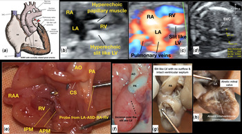
A 34-year-old second gravida mother of South Asian ethnicity was referred to our foetal cardiology clinic at 19 weeks 5 days of gestation because of an abnormal four-chamber view in an obstetric scan. Foetal echocardiography showed usual atrial arrangement with the heart in the left hemithorax. The systemic and pulmonary venous drainages were normal. There was a large secundum atrial septal defect. The left atrioventricular valve was atretic and left ventricle was slit-like (Fig 1b and c). The great arteries were normally related and committed to the right ventricle. The ascending aorta [0.24 cm (z score −2.45)] and aortic isthmus [1.2 mm (z score −3.49)] appeared small (Fig 1d). The foetus was in sinus rhythm with no evidence of hydrops fetalis. There were no other extra-cardiac anomalies. A staged single-ventricle palliation with the possible need for a Norwood-type operation in the newborn period was proposed. The parents opted for termination of pregnancy because of the uncertain prognosis of this condition.
(a) A schematic cartoon of DORV, mitral valve atresia, slit-like left ventricle, intact ventricular septum, and normally related great arteries. (b) Four-chamber view showing mitral valve atresia, hyperechoic papillary muscle, and slit-like left ventricle; (c) Four-chamber view showing normal drainage of two pulmonary veins into left atrium and slit-like LV; (d) Three-vessel view showing normally related great vessels with smaller ascending aorta. (e) Dissection of the right ventricle showing normal tricuspid valve morphology with thickened anterior papillary muscle, intact interventricular septum, and both great vessels were normally related and committed to the right ventricle. A probe passed from left atrium coarsed through the atrial septum and right atrium into the right ventricle, (f): Lateral view of the heart showing the slit-like LV; (g and h) Dissection of the LV showing a blind pouch with no inflow, outflow, and intact ventricular septum.

A foetal autopsy was performed after parental consent. The weight of the foetus was 210 g. External examination showed a male foetus with no obvious congenital anomalies. The heart was dissected using the approach described by Erickson and colleagues, Reference Erickson4 and described according to the sequential segmental analysis proposed by Anderson and colleagues. Reference Anderson and Shirali5
There was a usual arrangement of the abdominal and thoracic organs. The heart was in the left hemithorax. The inferior caval vein was intact and drained into the right-sided atrium. There were bilateral superior caval veins with no bridging vein. The pulmonary veins drained normally into the left-sided atrium. The right-sided atrial appendage was larger and more pyramidal with pectinate muscles extending beyond the appendage. The left-sided atrial appendage was “finger like” with no pectinate muscle extending into the atrium. The right atrium opened into the anterior ventricle through a morphologically normal tricuspid valve (Fig 1e). The left atrioventricular connection was absent and the only egress to the left atrium was a large atrial communication. The dominant anterior ventricle appeared to be of right ventricular morphology with a moderator band. The ventricular septum was intact. Both of the great arteries arose from this ventricle and were normally related with the aorta posterior and to the right and the pulmonary artery anterior and to the left. Both arteries were of normal size. The aortic arch was left sided with a normal branching pattern and a narrow isthmus. The branch pulmonary arteries were of normal calibre and the arterial duct was normal. Dissection in the postero-inferior aspect of the ventricular mass revealed an incomplete left ventricle (Fig 1f–h). The sequential analysis was situs solitus, absent left atrioventricular connection (mitral atresia), double outlet right ventricle with an intact ventricular septum, and normally related great arteries. A schematic cartoon of the lesion is shown in Fig 1a.
Case 2
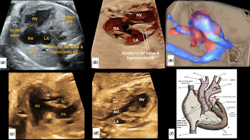
A 28-year-old primigravid mother of South Asian ethnicity was referred to our foetal cardiology clinic at 29 weeks of gestation in the view of hypoplastic left ventricle in an obstetric scan. Foetal echocardiography showed usual atrial arrangement with the heart in the left hemithorax. The systemic and pulmonary venous drainages were normal. There was a large atrial septal defect. The left atrioventricular valve was atretic and left ventricle was hypoplastic (Fig 2a and b). The great arteries were committed to the right ventricle and unobstructed. The aorta arose rightward and anterior to the pulmonary artery. The aorta appeared smaller than the pulmonary artery and the sagittal view of the aortic arch failed to show the typical candy cane curvature, but rather a straight course of the aorta suggestive of an interrupted aortic arch (Fig 2c–e). The foetus was in sinus rhythm with no evidence of hydrops fetalis. There was no patient follow-up. The sequential analysis was situs solitus, absent left atrioventricular connection (mitral atresia), double outlet right ventricle with an intact ventricular septum, aorta arose right and anterior to the pulmonary artery, and an interruption of aortic arch. A schematic cartoon of the lesion is shown in Fig 2f.
(a) Four-chamber view showing hypoplastic left atrioventricular valve and left ventricle. ( b) High-definition live surface rendering of the four-chamber view showing hypoplastic left atrioventricular valve and small pouch-like left ventricle. (c) Right ventricular outflow tract view showing the commitment of both vessels to RV and aorta arising right to the pulmonary artery and prominent bilateral coni (arrows). (d) Sagittal view of the aortic arch failed to show the typical candy cane curvature, but rather a straight course of the aorta suggestive of IAA. (e) High-definition colour rendering of sagittal view of the aortic arch showing discontinuity in the arch after the ascending aorta gives off the innominate artery. (f) A schematic cartoon of DORV, mitral valve atresia, hypoplastic LV, intact ventricular septum, parallel outflows with an interrupted aortic arch. AO=ascending aorta; APM=anterior papillary muscle; ASD=atrial septal defect; CS=conal septum; IPM=inferior papillary muscle; IVC=inferior caval vein; LA=left atrium; LV=left ventricle; LSA=Left subclavian artery; LSVC=left superior caval vein; Lt=left; LAA=left atrial appendage; LCC=left common carotid artery; LSCA=left subclavian artery; PDA=patent ductus arteriosus; PA=main pulmonary artery; Rt=right; RA=right atrium; RAA=right atrial appendage; RV=right ventricle; RSVC=right superior caval vein; RSCA=right subclavian artery; RCC=right common carotid artery; SVC=superior caval vein.

Discussion
Double outlet right ventricle with intact ventricular septum was described in 3 of the 100 morphological specimens. Reference Erickson4 The exact embryological mechanism of this anomaly is not yet understood, although there is some evidence in support of aetiological factors that are important in the development of a double outlet right ventricle associated with a ventricular septal defect in a genetically modified mouse model. Reference Aiello, Spicer, Anderson, Brown and Mohun6 An association with deletion of 22q12 and trisomy 18 has also been reported. Reference L’herminé-Coulomb, Houyel, Aboura, Audibert, Dal Soglio and Tachdjian7,Reference Patel, Muise and Redline11
The very first reported heart with double outlet right ventricle with intact ventricular anomaly was that of a similar anomaly. This was in a pre-surgical era and the diagnosis was established at autopsy. Reference Macmahon and Lipa8 Though double outlet right ventricle, hypoplastic mitral valve, and left ventricle, intact ventricular septum, transposed great arteries had been reported in the literature, its association with an interrupted aortic arch in foetus has never been reported in the literature so far. Patients with double outlet right ventricle with intact ventricular septum will need to undergo staged single-ventricle palliation given that they all have a hypoplastic or atretic or unguarded mitral valve and hypoplastic left ventricle.
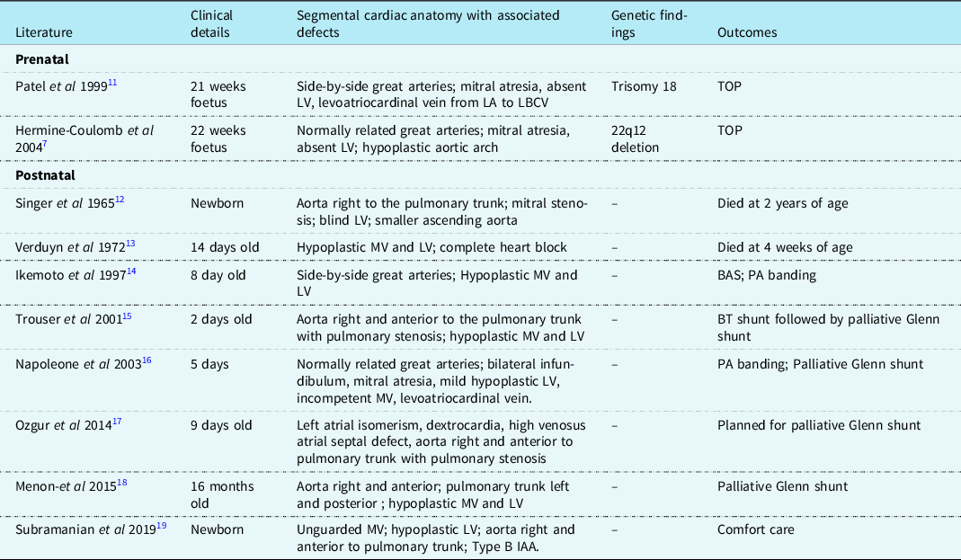
In the current era, a staged single-ventricle palliation can result in survival and a reasonable quality of life although concerns exist about the performance of the right ventricle supporting the systemic circulation in the longer term. Reference Erikssen, Aboulhosn and Lin9 The long-term survival after single-ventricle palliation is only 76% at 25 years. Reference d’Udekem, Iyengar and Galati10 A summary of previously published prenatal and postnatal case reports were shown in Table 1.
Summary of previously published reports of double outlet right ventricle with intact ventricular septum.

BAS = balloon atrial septostomy; BT = Blalock–Taussig; IAA = interrupted aortic arch; LA = left atrium; LBCV = left brachiocephalic vein; LV = left ventricle; MV = mitral valve; PA = pulmonary artery; TOP = termination of pregnancy.
Conclusions
We report two different morphologies of the double outlet right ventricle with an intact ventricular septum. The foetal autopsy in an unusual variant of double outlet right ventricle with an intact ventricular septum, slit-like left ventricle, and normally related great arteries without outflow tract obstruction enabled us to confirm the foetal echocardiographic diagnosis of this rare anomaly.
Acknowledgements
We would like to thank Dr. Mani Ram Krishna DNB, FNB , Assistant Professor, Department of Paediatric Cardiology, Amrita institute of medical sciences, Kochi, Kerala, for helping us during the fetal autopsy.
Financial support
This report received no specific grant from any funding agency, commercial, or not-for-profit sectors.
Conflict of interest
None.
Ethical standards
This article does not contain any studies with human participants or animals performed by any of the authors.