Introduction
Nematodes of the family Rhabdiasidae are commonly found in association with the lungs of amphibians and reptiles worldwide. They are found in Australian, Ethiopian, Palaearctic, Oriental and Neotropical regions (Kuzmin, Reference Kuzmin2013; Tkach et al., Reference Tkach, Kuzmin and Snyder2014, Kuzmin & Tkach, Reference Kuzmin and Tkach2015). Recently, the genus Serpentirhabdias was proposed by Tkach et al. (Reference Tkach, Kuzmin and Snyder2014) in addition to the genera Acanthorhabdias Pereira, 1927, Rhabdias Stiles & Hassall, 1905, Kurilonema Szczerbak & Shapilo, 1969, Neoentomelas Hasegawa, 1989, Pneumonema Johnston, 1916, and Entomelas Travassos, 1930. Given their particular morphological and biological characters (comparatively thin body cuticle, arrangement of lips in two lateral groups, smaller number of eggs, presence of homogony in the life cycles and restricted specificity to snakes) and molecular studies, 14 species of Rhabdias were transferred to Serpentirhabdias (Tkach et al., Reference Tkach, Kuzmin and Snyder2014).
Seven species of Serpentirhabdias are known from the Neotropical region, of which six are parasites of colubroidean snakes, as follows: S. cf. fuscovenosa (Railliet, 1899), S. labiata (Pereira, 1927), S. vellardi (Pereira, 1928), S. lamothei (Martínez-Salazar & León-Régagnon, 2006), S. filicaudalis (Barrella, Santos & Silva, 2009) and S. atracti Kuzmin, Melo & Santos, Reference Kuzmin, Melo and Santos2014 (Barella et al., Reference Barrella, Santos and Silva2010; Kuzmin et al., Reference Kuzmin, Melo and Santos2014); and only S. atroxi Kuzmin, Giese, Melo, Costa, Maschio & Santos, 2016 has been reported in viperidian snakes. Among these nematodes, five have been recorded in snakes from Brazil, S. vellardi in Philodrias patagoniensis (=Philodrias schotti) (Colubridae) and Oxyrhopus trigeminus (Dipsadidae), S. labiata in Xenodon merremi (=Rhadinea merremi) (Dipsadidae), S. filicaudalis in Spilotes pullatus (Colubridae), S. atracti in Atractus major (Dipsadidae) and S. atroxi in Bothrops atrox (Viperidae) (Pereira, Reference Pereira1927, Reference Pereira1928; Barella et al., Reference Barrella, Santos and Silva2010; Kuzmin et al., Reference Kuzmin, Melo and Santos2014, Reference Kuzmin, Giese, Melo, Costa, Maschio and Santos2016).
Pit vipers of the genus Bothrops are among the best-studied most wide-ranging Neotropical snake clades (Carrasco et al., Reference Carrasco, Mattoni, Leynaud and Scrocchi2012). Even with this extensive accumulated knowledge on natural history, taxonomy and phylogenetic relationships (Fenker et al., Reference Fenker, Tedeschi, Pyron and Nogueira2014), only S. atroxi of the Rhabdiasidae was reported parasitizing a snake species of the Bothrops genus (B. atrox) (Kuzmin et al., Reference Kuzmin, Giese, Melo, Costa, Maschio and Santos2016). In addition, there is no study on the nematode fauna of Bothrops moojeni, a large pit viper from riparian areas in central and south-eastern Brazil, throughout the Cerrado morphoclimatic domain (Leloup, Reference Leloup1984; Campbell & Lamar, Reference Campbell and Lamar1989; Borges & Araújo, Reference Borges and Araújo1998; Nogueira et al., Reference Nogueira, Sawaya and Martins2003; Fenker et al., Reference Fenker, Tedeschi, Pyron and Nogueira2014).
During a survey of parasites from B. moojeni snakes in São Paulo State, Brazil, nematodes of the genus Serpentirhabdias were located in the lungs and identified as Serpentirhabdias viperidicus n. sp. using morphological and molecular phylogenetic analyses.
Materials and methods
Collection and examination of nematodes
During a helminthological survey in the period between July 2012 and February 2013, 12 specimens of B. moojeni were collected at ‘Reserva Particular do Patrimônio Natural Foz do Rio Aguapeí’, municipality of Castilho, São Paulo State, Brazil (license permission SISBIO 30772-4). The snakes were euthanized by injection of sodium thiopental and necropsied for parasite survey under a stereomicroscope. A total of 115 nematodes were found in the lungs of six snakes. The parasites were fixed in hot 70% ethanol. For identification, the specimens were cleared with lactophenol on temporary slides. The morphology was analysed using a computerized system of image analysis (LAS DIC, Leica Microsystems, Wetzlar, Germany).
The morphologies of tail, cuticle and oral structures were also evaluated by scanning electron microscopy (SEM). Nematodes were fixed in 70% ethanol, dehydrated in a graded alcohol series and critically point dried. Then they were mounted on an aluminium stub using conductive double-sided tape, coated with gold–palladium and examined with the use of a Quanta 200 scanning electron microscope (FEI Company; from Centro de Microscopia Eletrônica de Botucatu, São Paulo, Brazil) (adapted from Allison et al., Reference Allison, Ubelaker, Webster and Riddle1972).
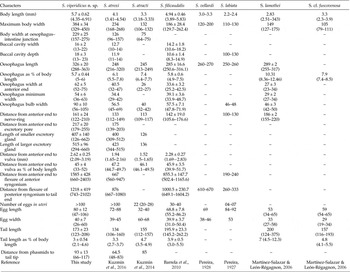
All measurements are reported in micrometres (μm) unless otherwise indicated. Data of the holotype are presented in the text and those of the 33 paratypes and the morphometry of some Serpentirhabdias spp. from the Neotropical region are given in table 1.
A comparison of the morphometrics of Serpentirhabdias viperidicus n. sp. with other Serpentirhabdias spp. from the Neotropical region. Ranges given in brackets.

Molecular analysis
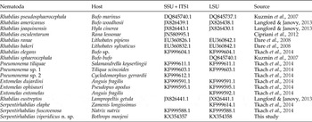
For molecular analysis, some specimens were fixed in absolute ethanol (Merck®) and cut in pieces. Part was taken for scanning electron microscopy and part for DNA extraction. Genomic DNA was extracted from parts of single specimens of worms using DNeasy® Blood and Tissue Kit (Qiagen, California, USA) according to the manufacturer's protocol, with a final volume of 50 μl. DNA fragments were amplified using 28S (partial) containing D2/D3 divergent domains, internal transcribed spacer (ITS; complete) and cytochrome c oxidase I (COI) genes. Primers and cycling conditions are presented in table 2.
The primers, sequences and cycling conditions described in the present study.

Amplification reactions consisted of 25 μl primary polymerase chain reaction (PCR) amplifications, with 3 μl DNA extract, 0.5 or 1.0 μl of each primer and Ready-to-Go PCR beads (Pure Taq™ Ready-to-Go™ PCR beads, GE Healthcare, Chicago, USA). The solution consisted of stabilizers, bovine serum albumin (BSA), deoxyadenosine triphosphate (dATP), deoxycytidine triphosphate (dCTP), deoxyguanosine triphosphate (dGTP), deoxythymidine triphosphate (dTTP), 2.5 units of puReTaq DNA polymerase and reaction buffer. With the reconstituted bead at a final volume of 25 μl, the concentration of each deoxynucleoside triphosphate (dNTP) was 200 μm in 10 mm Tris–HCl (pH 9.0 at room temperature), 50 mm KCl and 1.5 mm MgCl2.
PCR products (2 μl) were run on agarose gel (1%) using gel red and loading buffer to confirm amplicon size and yield. PCR products were purified using QIAquick PCR Purification Kit (Qiagen) and automated sequencing was performed directly on purified PCR products from specimens using BigDye v.3.1 Terminator Cycle Sequencing Ready Reaction kit (Applied Biosystems, Foster City, California, USA) for cycle sequencing. Sequences were run on an Applied Biosystems ABI 3500 DNA genetic analyser. The sequence identity was verified using the Basic Local Alignment Search Tool (BLAST) and contiguous sequences were assembled and edited using Sequencher v. 5.2.4 (Gene Codes, Ann Arbor, Michigan, USA).
Sequences of 28S and ITS genes of Rhabdiasidae were retrieved from GenBank (table 3). As an outgroup the taxon chosen was Rhabditoides regina (EF990726) for partial 28S. Sequences were aligned in ClustalX (Larkin et al., Reference Larkin, Blackshields, Brown, Chenna, McGettigan, McWilliam, Valentin, Wallace, Wilm, Lopez, Thompson, Gibson and Higgins2007) using default settings and saved in FASTA format, Phylip format and NEXUS format. Two alignments were made, one with ITS (complete) and other with 28S (partial). The COI genes were not used, due to the small number of Rhabdiasidae sequences available in GenBank. The numbers of base substitutions per site between sequences were calculated. Standard error estimates were obtained by a bootstrap procedure (2000 replicates). Analyses were conducted using the Kimura 2-parameter model; evolutionary analyses were conducted in MEGA5 (Kimura, Reference Kimura1980; Tamura et al., Reference Tamura, Peterson, Peterson, Stecher, Nei and Kumar2011).
A comparison of host species and GenBank accession numbers of Rhabdiasidae used in the present study. LSU, large small subunit rDNA; ITS, internal transcribed spacer; SSU, small subunit rDNA.

The best-fit model for nucleotide substitution in the resulting matrix was TVM + I, determined by the Akaike information criterion (AIC) in a jModelTest (Posada, Reference Posada2008). Phylogenetic analyses were performed using PhyML v.3.0 (Guindon et al., Reference Guindon, Dufayard, Lefort, Anisimova, Hosdijk and Gascuel2010) for maximum likelihood (ML) and Bayesian Inference (BI) in the BEAST program (Drummond et al., Reference Drummond, Suchard, Xie and Rambaut2012). The Bayesian analyses were run with the following nucleotide substitution model settings: lset nst = 6, rates = invgamma, ncat = 4, shape = estimate, inferrates = yes and basefreq = empirical, which correspond to a general time reversible (GTR) model including estimates of proportion of invariant sites (I) and gamma (G) distributed among-site rate variation. Supports for ML were determined by performing 100 bootstrap replicates and Markov chain Monte Carlo (MCMC) chains were run for 50 million generations, log-likelihood scores were plotted and only the final 75% of trees were used to produce the consensus trees, by setting the ‘burn-in’ parameter at 50 million generations. The trees were generated and edited in FigTree v.1.3.1 (Rambaut, Reference Rambaut2009).
Results
Serpentirhabdias viperidicus n. sp.
Description
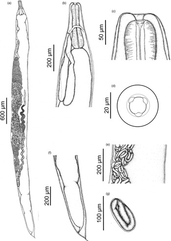
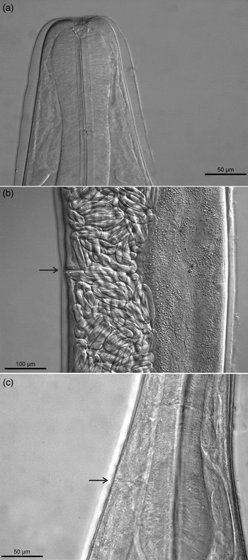
Description based on 34 (holotype and 33 paratypes) gravid females. Head end rounded. Thin and elongate body with thin cuticle, slightly thicker in posterior end of tail (fig. 1f). Cuticular surface regularly transversely striated in anterior region. Body length 6.26 mm and body width at the level of vulva 337. Body width of oesophageal–intestinal junction 179. Oral opening wide, rounded, with six lips slightly noticeable arranged parallel in two lateral groups. Lateral lip slightly smaller than submedian lip. Six small internal labial papillae. Amphidial openings conspicuous. Buccal capsule absent, buccal cavity depth 21 and width 15, formed by very short vestibulum (fig. 1c). Six minute onchia located inconspicuously anterior to oesophastome mid length. Two onchia on each sector of oesophagus (figs 1d and 2a). Oesophagus club-shaped. Posterior dilatation wider than anterior end of oesophagus (fig. 1b). Oesophagus length 316, representing 5% of body length. Width of oesophagus 55 in initial part; width of middle part 39, and 72 at maximum width. Epithelium of oesophagus with fringes (fig. 1c). Nerve-ring surrounding oesophagus at approximately its mid-length, 155 from anterior end of body (fig. 1b).
The morphology of the holotype (a, e, f) and paratypes (b, c, d, g) of Serpentirhabdias viperidicus n. sp. to show the (a) entire body, (b) anterior and lateral view, (c) anterior end ventral view, (d) anterior end apical view, (e) mid-body region with vulva, (f) posterior end and (g) larval egg.

Serpentirhabdias viperidicus n. sp. to show lateral views of (a) anterior end of the onchias, (b) vulva (arrow) and (c) ventral view of excretory pore (arrow).

Excretory pore situated immediately posterior to level of nerve-ring 195 from anterior end (figs 1b and 2c). Larger excretory gland 655 long, its posterior portion located posterior to oesophageal–intestinal junction (fig. 1b). Smaller gland not observed in holotype, but in paratypes is of length 407 ± 140. Intestine thick-walled. Rectum short, funnel-shaped. Contents of intestine black or brown for all extension (fig. 1a).
Genital system typical of the family Rhabdiasidae, amphidelphic with anterior and posterior ovaries. Vulva pre-equatorial and small; vulval lips not salient. Vulval aperture short and transverse. Vagina short, transverse and cuticularized (figs 1e and 2b). Distance from anterior end to vulva 2.86 mm, representing 45% of body length. Uterus large, sac-like, filled with numerous eggs (>100). Eggs containing embryos at various stage of development, wherein developed embryos were located near to the vulva (figs 1g and 2b). Egg size 80.9 ± 11.67 (47–106) × 45.2 ± 5.49 (30–60) (ten eggs measured in uteri close to vulva). Oviducts long, straight, thick-walled, slightly smaller than uterus. Ovaries elongated (fig. 1a).
Tail narrow, conical, tapering in extremity, and there is a little dilatation near to anal aperture (fig. 1f). Laterally, there are two parallel lines but their pores are intercalated, and the latter pore is situated approximately 33 before the anal aperture. A dorsal pore is situated near and before the anal region. Tail length (201), representing 3.2% of body length. A pore-shaped phasmid situated at level of anal aperture and another two in the posterior half of tail, and a pore-shaped phasmid situated in dorsal region of the tail. Distance of pore-shaped phasmid to tail tip, 106. Tail with longitudinal fine striations in the cuticle that begin before the anal opening and extend through the tail.
Taxonomic summary
Order Rhabditida Chitwood, 1933; family Rhabdiasidae Railliet, 1915; Serpentirhabdias viperidicus n. sp. ZooBank number for species: urn:lsid:zoobank.org:pub:FF1E024A-6483-42F6-935E-72F0903C2E1B.
Type host
Bothrops moojeni (Reptilia: Squamata: Serpentes: Viperidae).
Type locality
‘Reserva Particular do Patrimônio Natural Foz do Rio Aguapeí’ Castilho Municipality of São Paulo State, Brazil (21°03′04.9ʺS; 51°52′52.6ʺW).
Site of infection
Lungs.
Prevalence and intensity of infection and range
P = 50%; MII = 19.2 ± 8.4 (2–54).
Type-material
Holotype and five paratypes, deposited at the Coleção Helmintológica do Instituto Oswaldo Cruz (CHIOC), Rio de Janeiro, Rio de Janeiro State, Brazil, under the numbers Holotype 38318 a and Paratypes 38318 b, and 28 paratypes deposited at the Coleção Helmintológica do Instituto de Biociências de Botucatu (CHIBB), Botucatu, São Paulo State, Brazil under the numbers 7280, 7276 and 7304.
Etymology
The specific epithet is derivative from the family of the type host (Viperidae) added to the suffix ‘icus’ (=‘belonging to’).
Remarks
The new species was assigned to the genus Serpentirhabdias based on the following characters: body cuticle thin, cuticle inflation absent, cuticular surface regularly transversely striated in anterior region, circumodal lips arranged in two lateral groups, buccal capsule absent and vulva pre-equatorial (similar to some other species of the genus) (see Tkach et al., Reference Tkach, Kuzmin and Snyder2014).
Serpentirhabdias viperidicus n. sp. differs from all other species of this genus by a combination of characters, such as: (1) its size is the largest and widest of the genus in the Neotropical region; (2) lips slightly notable; and (3) amphidial openings conspicuous. Furthermore, the most striking feature of the new species are: tail length as proportion of body length (3.2%), presence of fine striation at posterior ends, presence of two parallel lines with intercalated pores, a dorsal pore situated near and before the anal region, a phasmid pore situated at the level of the anal aperture and another two in the latter half of the tail, and a phasmid pore situated in the dorsal region of the tail. Distance from a pore-shaped phasmid to tail tip 106.
The new Serpentirhabdias is substantially closely related to S. atroxi due to the absence of a buccal capsule, presence of onchia and hosts that are from the same snake genera in the Neotropical region. However, there are some differences in general size; in the new species the body length is larger than S. atroxi (5.09–6.91 vs. 3.28–5.00 mm); the lips are slightly noticeable in the new species; the excretory pore is situated immediately posterior to the level of the nerve-ring in the new species; the presence of two parallel lines with intercalated pores, an anal phasmid pore situated at the level of the aperture and another two in the latter half of tail in the new species. Finally, S. atroxi is distributed in the Amazon (northern region), while this new Serpentirhabdias is described from the south-eastern region.
Serpentirhabdias viperidicus n. sp. resembles S. vellardi by the absence of distinct lips, absence of a buccal capsule and the size of eggs (69 × 38–46) (see Pereira, Reference Pereira1928). However, S. viperidicus n. sp. differed from this species mainly by body dimension, with almost double the body length. Serpentirhabdias labiata differs from the new species by the presence of distinct lips, a small body and few large eggs in the uterus (84–92 × 53) (see Pereira, Reference Pereira1927).
Serpentirhabdias filicaudalis resembles the new species with regard to body dimensions and oesophagus length/total body length ratio of 5.8% (4.9–7.5%) versus 5.7% (5–6%) in S. viperidicus n. sp. Also, there is no buccal capsule in S. filicaudalis, both species present fine striations at posterior ends and the nerve-ring position is similar. Serpentirhabdias viperidicus n. sp. is also distributed in the south-east of Brazil (see Barrella et al., Reference Barrella, Santos and Silva2010). However, S. filicaudalis is slightly smaller than S. viperidicus n. sp. (3.89–5.83 vs. 4.35–6.91, respectively), with a truncated anterior end, while the new species has a rounded anterior end. In S. filicaudalis the lips are weakly developed (lips slightly noticed in the new species) and the tail is long with a filiform terminal prolongation of the cuticle (Barrella et al., Reference Barrella, Santos and Silva2010), unlike S. viperidicus n. sp., which has a narrow tail that is conical and slightly tapering in the extremity.
Serpentirhabdias atracti resembles the new species by the absence of distinct lips and buccal capsule, but differs from it by having a triangular oral opening, excretory glands that are shorter than the oesophagus (longer than the oesophagus in the new species), number of eggs (20–28 vs. >100) and the shape of the tail (see Kuzmin et al., Reference Kuzmin, Melo and Santos2014).
In addition to these species, two other species of Serpentirhabdias are known from the Neotropical region: S. lamothei and S. cf. fuscovenosa. Serpentirhabdias viperidicus n. sp. differs from these species mainly by the absence of a buccal capsule and by its large body size. Serpentirhabdias lamothei has a triangular oral shape, while the new species presents a rounded oral shape; and the anterior end of this species is truncated differently from that of the new species, which is rounded. They are also different in respect of tail shape (see Martínez-Salazar & León-Régagnon, Reference Martínez-Salazar and León-Régagnon2006). The new species is similar to S. cf. fuscovenosa in its oral shape (rounded), but differs from this species in body size and relative lengths of oesophagus, tail and lips. Finally, all these species are parasites of colubroidean snakes (Pereira, Reference Pereira1927; Pereira, Reference Pereira1928; Martínez-Salazar & León-Régagnon, Reference Martínez-Salazar and León-Régagnon2006; Barrella et al., Reference Barrella, Santos and Silva2010; Kuzmin et al., Reference Kuzmin, Melo and Santos2014), except S. atroxi, which has been considered the only record of this genus in viperidian snakes.
Phylogenetic analysis
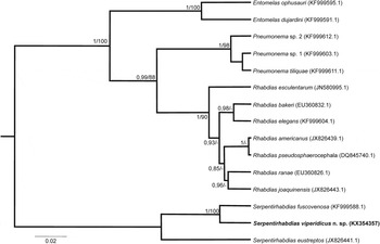
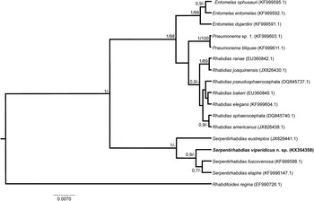
The 28S rDNA alignment consisted of 17 sequences from Rhabdiasidae retrieved from GenBank and a 681 bp alignment with 92 variable and informative sites. The ITS rDNA alignment consisted of 15 sequences from Rhabdiasidae retrieved from GenBank and a 718 bp alignment with 331 variable and informative sites. The sequences were deposited in GenBank. Only the 28S rDNA alignment was used as an outgroup; for the ITS rDNA alignment the Serpentirhabdias clade was the outgroup. Both genes supported topologies with high bootstrap values and similar results (figs 3 and 4). Serpentirhabdias viperidicus n. sp. is in the Serpentirhabdias clade, sister taxon to Serpentirhabdias fuscovenosa (KF999588) for ITS topology (fig. 3) and sister taxon to the clade Serpentirhabdias elaphe (KF999614) and S. fuscovenosa (KF999588) for 28S topology (fig. 4). The ITS topology was best resolved, satisfactorily separated the four genera and added Serpentirhabdias eustreptos (JX826441) to the Serpentirhabdias clade. The 28S topology also separated the four genera as expected, but with lower bootstrap values. The genetic distance analysis involved 17 nucleotide sequences. All positions containing gaps and missing data were eliminated. There was a total of 456 positions in the final dataset. Serpentirhabdias viperidicus n. sp. showed a 7% difference from S. fuscovenosa for the 28S gene, and the ITS gene was more variable, showing a 36% genetic distance difference in the same analysis (not shown).
The phylogenetic relationship of Rhabdiasidae using the 18S + ITS partial gene sequence, with numbers above nodes indicating posterior probabilities (Bayesian analysis) and bootstrap (maximum likelihood analysis), respectively; bootstrap values below 70% are not expressed.

The phylogenetic relationship of Rhabdiasidae using the 28S partial gene sequence, with numbers above nodes indicating posterior probabilities (Bayesian analysis) and bootstrap (maximum likelihood analysis), respectively; bootstrap values below 70% are not expressed.

Discussion
Both molecular topologies showed a distinct lineage composed of three clades: Rhabdias spp. from amphibians closely related to Pneumonema spp. and Entomelas spp. The Serpentirhabdias species clade is distinct and basal (fig. 3). Corroborating previous studies show Rhabdias as a polyphyletic group, Entomelas and Pneumonema as monophyletic taxa and Serpentirhabdias as a separate, monophyletic and basal group among Rhabdiasidae (Langford & Janovy Reference Langford and Janovy2013; Tkach et al., Reference Tkach, Kuzmin and Snyder2014).
The uncertain diagnostic value of various morphological characters has caused instability in the determination of the systematics of Rhabdiasidae (Kuzmin et al., Reference Kuzmin, Tkach and Brooks2007; Tkach et al., Reference Tkach, Kuzmin and Snyder2014). Currently, the morphological traits and molecular tools using different genes are necessary methodological approaches to describe new species (Cipriani et al., Reference Cipriani, Mattiucci, Paoletti, Santoro and Nascetti2012). Serpentirhabdias viperidicus n. sp. was characterized both morphologically and genetically using different and concatenated ribosomal and mitochondrial genes. Both ribosomal genes and different types of phylogeny reconstruction approaches (BI and ML) were used and presented the same results, which gives more confidence to the data. In relation to the COI gene, we present data from the sequence (KX350054) but no phylogenetic reconstruction was made due to the low number of sequences available in GenBank for comparisons.
In the context of the phylogenetic relationships of Serpentirhabdias species, more questions need to be answered regarding life cycles, host–parasite relationships and geographical distribution (Tkach et al., Reference Tkach, Kuzmin and Snyder2014), especially for Neotropical species. As mentioned before, there were seven morphologically described species from snakes in the Neotropical region but no molecular work has been done so far. Possibly the species S. viperidicus n. sp. would be apart from S. fuscovenosa and/or would cluster differently with closely related species from the Neotropical region. To achieve better resolution in the phylogeny, it is important to carry out further sampling and sequencing for a wider range of taxa from the Neotropics, and perhaps more DNA markers should be considered to display better understanding of this group.
The presence or absence of various structures at the anterior end, as well as their variations in shape and position, among nematodes from Rhabdias, have been widely used for species differentiation in this genus (Tkach et al., Reference Tkach, Kuzmin and Snyder2014) and could also be valid for the genus Serpentirhabdias. The absence of the buccal capsule, shared by several species, could form a separate group inside the Serpentirhabdias, especially for Neotropical species, which share this characteristic (Kuzmin et al., Reference Kuzmin, Melo and Santos2014, Reference Kuzmin, Giese, Melo, Costa, Maschio and Santos2016). Seemingly, S. filicaudalis, described with a buccal capsule by Barrella et al. (Reference Barrella, Santos and Silva2010), would be the only separate species from this group (Kuzmin et al., Reference Kuzmin, Melo and Santos2014). However, analysing specimens deposited in the ‘Coleção Helmintológica do Instituto de Biociências de Botucatu’ (CHIBB), more than once, we could observe that Barrella et al. (Reference Barrella, Santos and Silva2010), used morphological characters described from Rhabdias by Vicente et al. (Reference Vicente, Rodrigues, Gomes and Pinto1991), and that these authors made a mistake when they called the ‘buccal cavity’ a ‘buccal capsule’. Therefore, the absence of a buccal capsule is a morphological character shared by known Serpentirhabdias species from the Neotropical region.
The presence of onchia in species of Serpentirhabidias parasitic in viperidian snakes, such as S. atroxi (Kuzmin et al., Reference Kuzmin, Giese, Melo, Costa, Maschio and Santos2016) and S. viperidicus n. sp., allows us to consider them close to each other but separate from their congeneric species, mostly because both are recorded in the same country and family host. Pyron et al. (Reference Pyron, Burbrink and Wiens2013), studying the phylogenetic relationships of the hosts B. atrox and B. moojeni, strongly support these species as sister taxa and such information suggests a potential co-evolutionary hypothesis. We suggest that further studies are necessary, especially because no molecular data are available for other species of Serpentirhabdias.
The snake fauna of the Neotropical region is characterized by high species richness, with 365 species only from Brazil (Costa & Bernis, Reference Costa and Bernis2014), and they present a complexity of ecological relationships among species (Duellman, Reference Duellman1978; Henderson et al., Reference Henderson, Dixon and Soini1979; Vitt, Reference Vitt, Seigel, Collins and Novak1987). This whole snake species diversity is distributed in six different biomes, and they could be potential hosts for Serpentirhabdias, providing a hugely complex community structure that drives species diversification, suggesting a possibility that this region could be the centre of this parasite diversity with high species richness.
The new species is the seventh species of the genus Serpentirhabidas found in Brazil and the eighth in the Neotropical region, evidencing the low number of inventory studies performed in parasite diversity in poorly studied regions, with just a few experts interested in the group.
In addition, this study represents the first description of Serpentirhabdias from Brazil using molecular approaches, demonstrating the scarcity of accurate studies for this genus in the Neotropical region, and it confirms the monophyly of this recent genus proposed by Tkach et al. (Reference Tkach, Kuzmin and Snyder2014).
Acknowledgements
We would like to acknowledge colleagues from the laboratory who helped us in fieldwork and Companhia Energética de São Paulo – CESP, for the logistical support and authorization to work in the area of ‘RPPN Foz do Rio Aguapeí’.
Financial support
Financial support for this study was provided by Fundação de Amparo a Pesquisa do Estado de São Paulo – FAPESP (2011/20186-6 and 2012/24945-1), Coordenação de Aperfeiçoamento de Pessoal de Nível Superior – CAPES (A.A and L.A.F.S grant, M.I.M. grant number AUX-PE-PNPD 3005/2010), and Conselho Nacional de Desenvolvimento Científico e Tecnológico – CNPQ (D.H.M grant number 154739/2015-4).
Conflict of interest
None
Ethical standards
The collection of specimens of B. moojeni was authorized by the ethics committee of University Estadual Paulista (no. 348/2012).