Implications
In this study, the β-3 adrenergic receptor (ADRB3) expression in various tissues of sheep was detected by immunohistochemical technique and by mRNA abundance via real-time PCR. Despite its low expression, the presence of ADRB3 in the spleen and kidney was found in livestock for the first time. Generally, ADRB3 expressed higher in adipose than in non-adipose tissues, and for the former, it was higher in deep fat deposits than in subcutaneous fat. The distributions are consistent with the known functions of ADRB3 in mediating lipolysis and homeostasis in adipose tissues, and relaxation of vascular smooth muscle in the cardiovascular system. The relation between low expression and its function in some viscera, for example, lung, spleen and kidney of sheep deserves further study.
Introduction
β-Adrenergic receptors (ADRBs) are G-protein coupled receptors that mediate effects of the endogenous catecholamines adrenaline and noradrenaline. Three subtypes of ADRBs have been identified, named ADRB1, ADRB2 and ADRB3, respectively. These receptors are present in most mammalian tissues, but the distribution of each subtype varies among tissues in a given species. Among the various physiological roles of ADRBs, the ADRB3 subtype mediates lipolysis and stimulates thermogenesis in adipose tissues (Cannon and Nedergaard, Reference Cannon and Nedergaard2004) by activating uncoupling protein in mitochondria.
In early studies, ADRB3 was identified mainly on the surface of white and brown adipocytes as reviewed by Strosberg (Reference Strosberg1997). In recent years, expression of the protein was also found in the human heart (Rozec and Gauthier, Reference Rozec and Gauthier2006), mammary artery (Rozec et al., Reference Rozec, Serpillon, Toumaniantz, Sèze, Rautureau, Baron, Noireaud and Gauthier2005), placental artery (Rouget et al., Reference Rouget, Barthez, Goirand, Leroy, Breuiller-Fouché, Rakotoniaina, Guérard, Morcillo, Advenier, Sagot, Cabrol, Dumas and Bardou2006), gastrointestinal tract (Anthony et al., Reference Anthony, Sim, Guillaume, Strosberg, Dhillon, Pounder and Wakefield2000), prostate and gall bladder (de Matteis et al., Reference De Matteis, Arch, Petroni, Ferrari, Cinti and Stock2002), urinary bladder detrusor (Igawa et al., Reference Igawa, Yamazaki, Takeda, Hayakawa, Akahane, Ajisawa, Yoneyama, Nishizawa and Andersson1999) and brain (Rodriguez et al., Reference Rodriguez, Carillon, Coquerel, Le Fur, Ferrara, Caput and Shire1995).
It has been confirmed that the mRNA of the ADRB3 gene is expressed in various tissues of domestic animals. The 2.0 kb ADRB3 transcript in the bovine brown adipose tissue (Pietri-Rouxel et al., Reference Pietri-Rouxel, Lenzen, Kapoor, Drumare, Archimbault, Strosberg and Manning1995), and two transcripts of 2.2 and 1.9 kb in porcine subcutaneous fat (McNeel and Mersmann, Reference McNeel and Mersmann1995) were detected by Northern blotting. The mRNA expressed in the adipose tissue (Sumner and McNamara, Reference Sumner and McNamara2007), mammary gland (Inderwies et al., Reference Inderwies, Pfaffl, Meyer, Blum and Bruckmaier2003), gastrointestinal tracts including the cecum, ileum, colon and abomasum (Meylan et al., Reference Meylan, Georgieva, Reist, Blum, Martig, Georgiev and Steiner2004; Kobel et al., Reference Kobel, Engel, Ontsouka, Graber, Blum, Steiner and Meylan2006) and in the liver (Carron et al., Reference Carron, Morel, Hammon and Blum2005) of cattle through real-time polymerase chain reaction (RT-PCR) or ligand binding assay. The ADRB3 mRNA was also present in subcutaneous fat and visceral adipose tissues in dogs (Sasaki et al., Reference Sasaki, Uchida, Niiyama, Yoshida and Saito1998a) and in the ventricle and lungs in pigs (McNeel and Mersmann, Reference McNeel and Mersmann1999).
However, in contrast to studies in humans and the domestic species, the identification of the ADRB3 in ovine tissues has not been reported. In this study, we detect and quantify the expression of ovine ADRB3 in various tissues by immunohistochemistry and by RT-PCR at the mRNA level, aiming at understanding the function of the protein in sheep.
Material and methods
Animal tissues
All experimental animals in this study were raised in Qinshui Demonstration Farm, Shanxi province, China (35°24′N, 115°55′E). South African Mutton Merino (SAMM) was a dual-purpose breed of mutton and wool imported from Australia. Shanxi Dam Line (SXDL) was a newly developed composite sheep line made up of several component breeds (SAMM, Charollais, Corriedale, Small Tailed Han sheep and the native breed in Shanxi province). Breeding rams and ewes were selected and mated from the end of October to early November. Lambing commenced in March and ended in April with weaning at 100 days. After weaning, all animals were managed by indoor feeding during dry seasons in winter and spring, and by grazing on pasture in grass seasons. The ration for indoor feeding consisted of alfalfa hay, grass hay, silage, carrots and mixed concentrates. The pasture consisted of the native grassland of bunch grass and bush-wood dominated by Andropogon saccharoides.
Four 20-month-old virgin rams were randomly selected from each flock and slaughtered. Adipose tissues in subcutaneous, omental, retroperitoneal, mesenteric and perirenal fat and non-adipose tissues in the heart, liver, spleen, lung and kidney were taken immediately after slaughter, snap frozen in liquid nitrogen and then stored at −80°C.
Total RNA extraction and cDNA synthesis
Total RNA from each tissue was extracted using TRIzol Reagent (Invitrogen, Carlsbad, CA, USA) according to the manufacturer’s instructions. About 100 mg of tissue was homogenized with 1 ml TRIzol Reagent using a homogenizer (Ultra-Turrax T18; IKA, Germany) followed by 5 min of incubation at room temperature. After 0.2 ml chloroform was added, the mixture was incubated at room temperature for 10 min and then centrifuged at 12 000 r.p.m. at 4°C for 15 min. The RNA (in the upper aqueous phase) was carefully pipetted to a new tube and precipitated by adding 0.5 ml isopropanol. The precipitate was incubated for 10 min at room temperature, and then centrifuged for 10 min at 12 000 r.p.m. at 4°C. The RNA pellets were washed with 75% ethanol and again centrifuged at 4000 r.p.m. for 5 min at 4°C. The upper aqueous phase was decanted completely, and after drying for 10 min at room temperature the remaining pellets were dissolved in 30 μl RNase-free water. The integrity of RNA was verified by agarose gel electrophoresis and by an optical density (OD) absorption ratio (OD 260 nm/OD 280 nm) between 1.8 and 2.0. RNA was reverse transcribed into cDNA using a PrimeScriptTM RT reagent kit (Takara Bio Inc., Takara code: DRR037A, Japan) and a thermal cycler according to the manufacturer’s instructions. The RT reaction was conducted in 20 μl of reaction mixture (including 4 μl 5 × primescript buffer, 1 μl RT enzyme mix I, 1 μl oligo dT primer, 1 μl random 6 mers, 4 μl total RNA and 9 μl Rnase free H2O) at 37°C for 15 min, and terminated by heating at 85°C for 5 s followed by cooling at 4°C.
ADRB3 gene expression analysis
PCR primers for ADRB3 and GAPDH (Glyceraldehyde-3-phosphate dehydrogenase) were designed using Primer 3.0 plus software based on ovine mRNA sequences found in Genbank. The primers for ADRB3 were forward, 5′-GGCTTCCTGCGGACTTTCT-3′, and reverse, 5′-GGGGAAGGGCAGAGTTTT-3′; and for GAPDH forward, 5′-TGACCCCTTCATTGACCTTC-3′, and reverse, 5′-GATCTCGCTCCTGGAAGATG-3′. GAPDH was amplified as a housekeeping gene.
All PCR reactions were performed in triplicate in an Mx3000P real-time thermal cycler (Stratagene, La Jolla, CA, USA) using the SYBR PrimeScriptTM RT-PCR Kit (Takara Bio Inc., Takara code: DRR041A) following the manufacturer’s instructions. The 25 μl PCR reactions included 12.5 μl SYBR Premix Ex TaqTM II, 0.5 μl forward primer (10 pM), 0.5 μl reverse primer (10pM), 0.5 μl ROX reference dye, 2 μl template and 9 μl water. The reactions were incubated in a 96-well plate at 95°C for 30 s, followed by 45 cycles with each cycle at 95°C for 10 s and at 61°C for 30 s. Following this, the dissociation curves of PCR products were obtained by a cycle of 95°C for 15 s, 61°C for 30 s and 95°C for 15 s, and reaction specificity was determined when there was only one specific peak in the dissociation curve. PCR efficiencies were determined using a relative standard curve derived from a diluted cDNA reaction mixture (a 2-fold dilution series with five measuring points). The R 2 estimates for two standard curves were 0.998 and 0.997, and PCR efficiencies were between 90% and 110%. The CT (cycle threshold: cycle number at which there is a linear increase in fluorescence in the PCR) values were used to quantify the PCR product, that is, the relative expression level of the target gene was expressed as 2−ΔΔCT, where ΔΔCT represents the difference between the CT of the housekeeping gene (GAPDH) and that of the target gene (ADRB3), each being corrected by a control consisting of a sample that was the calibrator.
Immunohistochemical test
Adipose tissue samples were fixed for 24 h with bouins solution, then embedded in paraffin and cut into sections of 5 μm thickness. The StreptAvidin-Biotin-enzyme Complex (SABC) technique of immunohistochemistry was employed using the affinity-purified rabbit polyclonal antibody (Boster, China). In brief, after deparaffinization and rehydration, the activity of endogenous peroxidase was blocked with 0.3% hydrogen peroxide (H2O2), and then antigen retrieved by incubating the sections with trypsinase for 30 min at 37°C. After rinsing with phosphate-buffered saline (PBS, 0.01 mol/l, pH 7.2), the specimens were reacted with 5% bovine serum albumin for 30 min at room temperature, and then incubated with primary antibody (1 : 100) for 24 h at 4°C. After a new washing with PBS, the sections were incubated in SABC (Boster, China) for 30 min. Finally, the sections were reacted with 3,3′-diaminobenzidine (DAB kit; Boster, China) solution for 5 to 10 min until a clearly visible color was developed. The reaction was stopped by extensive washing in double-distilled water and counterstaining of the sections with hematoxylin. Finally, the sections were dehydrated with alcohol, cleared in xylene and mounted with neutral balsam. Negative controls were set by PBS instead of the primary antibody.
Digital image acquisition and image analysis
The images of adipose tissues were captured by an Olympus BX51 microscope equipped with a CCD DP70 video camera (Olympus Optical, Tokyo, Japan). Five sections were selected from each adipose tissue and five images were taken from a high-power field (×400) in each section. Each image was automatically digitized by Image-Pro Plus™ (Mediacybernetics, Inc., USA). Pixel intensity was quantified to the 255 gray level. The mean gray level for each tissue was used as the OD measure of protein expression.
Statistical analysis
The amount of ADRB3 was analyzed under the following general linear model by using SPSS® statistics 17.0 software package (SPSS Science, Chicago, IL, USA):

where yijk is the k-th observation of the j-th tissue in the i-th animal, μ is the overall mean, Fi is the i-th flock (i = 1, 2), Tj is the j-th tissue (j = 1, 2, …, 11), FTij is the ij-th interaction between flock and tissue, eijk is the residual effect.
Multiple comparisons among different tissues were performed by Duncan’s method. All data were expressed as mean ± s.e.m.
Results
Abundance of ADRB3 mRNA
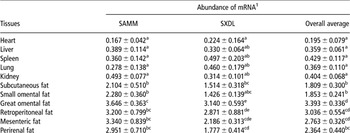
The relative abundance of ADRB3 mRNA in various ovine tissues from two different flocks is listed in Table 1. The expression of ADRB3 mRNA was not significantly different among non-adipose tissues in SAMM and in mixed flocks as indicated by the overall averages for each tissue. The least expression level for both flocks was found in the heart. There was no regular pattern for the difference in the specific tissue expression between the two flocks. For adipose tissues, the least expression was found in subcutaneous fat for SAMM (2.104) and in small omental fat for SXDL (1.426), with the second least in small omental fat for SAMM (2.280) and in subcutaneous fat (1.514) for SXDL, just the opposite order, while a significant difference exists between these two at deposits in SXDL. The greatest expression was found in great omental fat for both flocks (3.646 and 3.140 for SAMM and SXDL, respectively), and subsequently in mesenteric and retroperitoneal fat for SAMM and in reverse order for SXDL. There was higher expression in each adipose tissue of SAMM compared to the corresponding tissues of SXDL. ADRB3 mRNA was much more expressed in all adipose tissues than in non-adipose tissues from both flocks.
Abundance of ADRB3 mRNA (mean ± s.e.) in different ovine tissues

SAMM = South African Mutton Merino; SXDL = Shanxi Dam Line.
1Values represent the expression of ADRB3 relative to the house keeping gene (see Methods). Numericals with different superscripts at the same column differ significantly (P < 0.05).
Though not shown in Table 1, there was no significant difference for each tissue between the two flocks. The average abundance of ADRB3 mRNA for all tissues in SAMM (1.746 ± 0.233) was higher than for those in SXDL (1.340 ± 0.185), and the pattern of differences between tissues in SXDL was very different from that found in SAMM.
Expression of ADRB3 protein
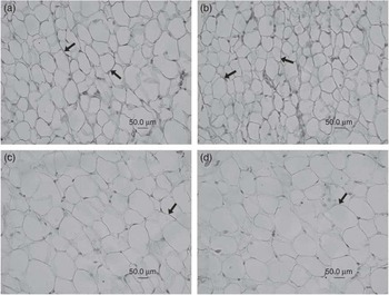
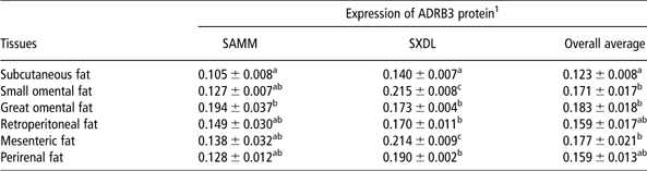
The presence of ADRB3, as defined by positive staining, was observed on the membrane of adipocyte (Figure 1). Figure 1a showed strong staining in great omental fat. In contrast, weak immunoreactivity was found in subcutaneous fat (Figure 1d). The gray level values for the immunopositive expression of ADRB3 in adipose tissues are presented in Table 2. The smallest gray level existed in subcutaneous fat for both flocks, and was significantly lower than that existing in any deep fat deposit.
The expression of ADRB3 in various ovine adipose tissues by immunohistochemistry technique. All images were taken at the same magnification (×400) and using the same light filter and luminous intensity. Expressions in (a) great omental fat, (b) retroperitoneal fat, (c) perirenal fat and (d) subcutaneous fat. The arrowheads indicate the positive ADRB3 expression (light brown).

Expression of ADRB3 (mean ± s.e.) in different ovine adipose tissues tested by immunohistochemistry
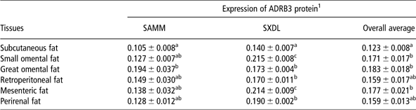
SAMM = South African Mutton Merino; SXDL = Shanxi Dam Line.
1Values represent ‘gray scale levels’ (pixel intensity of digital images of the tissue stain: see Methods). Numericals with different superscripts at the same column differ significantly (P < 0.05).
This result showed that ADRB3 was expressed less in subcutaneous fat than in other fats. For deep fat deposits, ADRB3 expressed much more in omental fats as seen in great and small omental fat for SAMM (0.194) and SXDL (0.215), respectively. There was no significant difference for the gray value detected in other fat deposits of SAMM, though the pattern was different in SXDL. The gray value for all fats in SAMM (0.140 ± 0.010) was lower than that in SXDL (0.184 ± 0.006).
Sources of expression differences
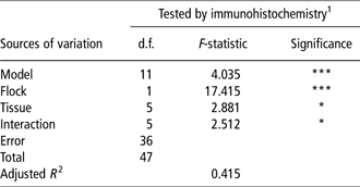
Table 3 lists the results of analysis of variance for the ADRB3 expression in adipose tissues detected by immunohistochemistry to explain the sources of expression differences. Highly significant expression difference (P < 0.001) existed between the flocks due to the flock differences in genetic basis and hereditary stability. Significant tissue effect (P < 0.05) was also found, which mainly resulted from the difference between subcutaneous and deep fats.
Analysis of variance for the ADRB3 expression tested by immunohistochemistry
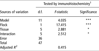
1Immunohistochemistry approach was used only in identification of ADRB3 in adipose tissues.
*P < 0.05; ***P < 0.001.
Discussion
Most of the evidence for expression of ADRB3 in various tissues has come from studies of response to receptor agonists. The ADRB3 agonist might lead to an increase in energy expenditure and decrease in deposit of fat in adipose tissues of humans (Larsen et al., Reference Larsen, Toubro, van Baak, Gottesdiener, Larson, Saris and Astrup2002), dogs (Sasaki et al., Reference Sasaki, Uchida, Niiyama, Yoshida and Saito1998b) and rodents (Weyer et al., Reference Weyer, Gautier and Danforth1999). The effects of ADRB3 agonists held interest in human medicine for the treatment of obesity and type 2 diabetes, but it was disappointing in clinical studies (van Baak et al., Reference Van Baak, Hul, Toubro, Astrup, Gottesdiener, DeSmet and Saris2002; Larsen et al., Reference Larsen, Toubro, van Baak, Gottesdiener, Larson, Saris and Astrup2002). Nevertheless, the agonists could offer useful insights into the pharmacology of ADRB3 (Arch, Reference Arch2008).
The relative abundance of ADRB3 mRNA in adipose tissues was much higher than in non-adipose tissues in this study. This result may explain why ADRB3 was identified mainly on the surface of white and brown adipocytes in early studies (Strosberg, Reference Strosberg1997). On the other hand, its higher expression in adipose tissue may be because this is necessary for it to mediate lipolysis and thermogenesis (Cannon and Nedergaard, Reference Cannon and Nedergaard2004).
The distribution in adipose tissues by the RT-PCR technique was reported also in other species, for example, in dogs (Sasaki et al., Reference Sasaki, Uchida, Niiyama, Yoshida and Saito1998a), in cattle (Sumner and McNamara, Reference Sumner and McNamara2007) and in humans. Chamberlain et al. (Reference Chamberlain, Jennings, Paul, Cordell, Berry, Holmes, Park, Chambers, Sennitt, Stock, Cawthorne, Young and Murphy1999) and de Matteis et al. (Reference De Matteis, Arch, Petroni, Ferrari, Cinti and Stock2002) demonstrated in humans (by immunohistochemistry) the existence of the ADRB3 protein in white adipocytes that came from subcutaneous fat and omental deposits. Likewise, as detected by both the RT-PCR of mRNA and immunohistochemistry techniques in this study, ADRB3 receptor was distributed in ovine subcutaneous, small and great omental, retroperitoneal, mesenteric and perirenal fat. Despite the differences due to the two experimental techniques, the distribution pattern of ADRB3 receptor in different adipose tissues had similarities. Generally, higher ADRB3 expression was found in deep deposits than in subcutaneous ones. This phenomenon is consistent with the studies in adult humans (Krief et al., Reference Krief, Lönnqvist, Raimbault, Baude, Van Spronsen, Arner, Strosberg, Ricquier and Emorine1993; Chamberlain et al., Reference Chamberlain, Jennings, Paul, Cordell, Berry, Holmes, Park, Chambers, Sennitt, Stock, Cawthorne, Young and Murphy1999).
A lower level of mRNA expression of the ADRB3 gene was identified in ovine liver in this study. However, earlier studies have suggested that there was an absence of ADRB3 in the liver of humans (Berkowitz et al., Reference Berkowitz, Nardone, Smiley, Price, Kreutter, Fremeau and Schwinn1995), rats (Evans et al., Reference Evans, Papaioannou, Bonazzi and Summers1996), dogs (Sasaki et al., Reference Sasaki, Uchida, Niiyama, Yoshida and Saito1998a) and pigs (McNeel and Mersmann, Reference McNeel and Mersmann1999). The slight difference may be attributed either to experimental techniques and/or species differences. Recently, Trebicka et al. (Reference Trebicka, Hennenberg, Schulze Pröbsting, Laleman, Klein, Granzow, Nevens, Zaagsma, Heller and Sauerbruch2009) detected the presence of ADRB3 in the liver of humans and rats, and found that ADRB3 was involved in portal hypertension in liver cirrhosis.
Though being the lowest in abundance, the expression of ADRB3 mRNA in the heart of sheep was comparable with the studies reported by Chamberlain et al. (Reference Chamberlain, Jennings, Paul, Cordell, Berry, Holmes, Park, Chambers, Sennitt, Stock, Cawthorne, Young and Murphy1999) and Skeberdis et al. (Reference Skeberdis, Gendviliene, Zablockaite, Treinys, Macianskiene, Bogdelis, Jurevicius and Fischmeister2008) in humans and by Evans et al. (Reference Evans, Papaioannou, Bonazzi and Summers1996) in rats. The function of ADRB3 in the heart includes increasing the contractility of myocardial tissue as indicated in studies of patients with heart failure (Moniotte et al., Reference Moniotte, Kobzik, Feron, Trochu, Gauthier and Balligand2001; Morimoto et al., Reference Morimoto, Hasegawa, Cheng, Little and Cheng2004) and dogs with pacing-induced heart failure (Cheng et al., Reference Cheng, Zhang, Onishi, Ukai, Sane and Cheng2001).
Expression of ADRB3 mRNA was low in the lung, spleen and kidney in this study. A similar result was found in the lungs of pigs (McNeel and Mersmann, Reference McNeel and Mersmann1999). However, earlier studies have testified to the absence of mRNA expression in the lung and kidney of humans (Krief et al., Reference Krief, Lönnqvist, Raimbault, Baude, Van Spronsen, Arner, Strosberg, Ricquier and Emorine1993), rats (Evans et al., Reference Evans, Papaioannou, Bonazzi and Summers1996) and dogs (Sasaki et al., Reference Sasaki, Uchida, Niiyama, Yoshida and Saito1998a). No study has reported its distribution and expression in the spleen to our knowledge. Much work for the expression and function of ADRB3 in these tissues is needed.
The newly developed Shanxi sheep dam line was a composite flock with complicated genetic background and lower hereditary stability. These may be a reflection of the significance of expression differences among tissues in SXDL. Significantly higher ADRB3 mRNA expression in adipose tissues of SAMM suggested that a potentially stronger lipolytic function existed in the flock. SAMM sheep have a larger size and stronger constitution than do SXDL sheep, needing more energy and hence ADRB3 expression may be needed to maintain the basic metabolism (Sheridan et al., Reference Sheridan, Ferreira and Hoffman2003). Theoretically, higher mRNA abundance corresponds to higher protein expression. However, the average expression of ADRB3 mRNA and protein in adipose tissues differed between SAMM and SXDL, suggesting that this relationship differed for at least one flock. This discrepancy may have been a consequence of the immuno-histochemistry technique. Though it is widely used in basic research for understanding the distribution and localization of biomarkers and differentially expressed proteins in biological tissues, its quantification by a digital computer-assisted method was regarded as semi-quantitative (Matos et al., Reference Matos, Stabenow, Tavares, Ferraz, Capelozzi and Pinhal2006) and thus not accurate enough. If a post-transcriptional modification does affect the protein expression deserves further study.
In conclusion, the mRNA of the ADRB3 gene was expressed in larger amounts in adipose tissues, and with lesser but easily detectable amounts in non-adipose tissues. Greater amounts were found in deep deposits than in subcutaneous fat. In addition to its established roles in lipolysis and thermogenesis in adipose tissues, and relaxation of the vascular smooth muscle in the cardiovascular system, detection of ADRB3 mRNA in the lung, spleen and kidney of sheep suggests that the ADRB3 receptor may conduct other functions in these tissues.
Acknowledgments
The authors thank the staff of the Laboratory of Animal Genetics, Breeding and Reproduction, Shanxi Agricultural University. This study was supported by the project of the Chinese National Natural Science Foundation (no. 30972084), the Program for New Century Excellent Talents in University, Ministry of Education of China (no. NCET-05-0269) and the Excellent Innovation Project for Graduate in Shanxi Province (no. 20093011).