Introduction
The current treatment of Trypanosoma cruzi infection is based on the nitro-heterocyclic compounds benznidazole or nifurtimox (Sosa-Estani et al. Reference Sosa-Estani, Viotti and Segura2009). However, the need of safer and more effective drugs against this parasite has stimulated the search for alternative Chagas disease chemotherapies. In this regard, several studies have reported that either natural, modified or synthetic naphthoquinones (NQs) are active against T. cruzi by producing free radicals and affecting mitochondrial function (de Castro et al. Reference de Castro, Batista, Batista, Batista, Daliry, de Souza, Menna-Barreto, Oliveira, Salomão, Silva, Silva and Soeiro2011; Salas et al. Reference Salas, Faúndez, Morello, Maya and Tapia2011 for Review). NQs are compounds widely distributed in plants with known anticancer, antibacterial, antimalarial and fungicide activities exerted by reactive oxygen species (ROS) generation after their intra-cellular reduction (Koyama, Reference Koyama2006). The β-lapachone (β-Lap) is an ortho-NQ (o-NQ) isolated from the heartwood of trees of the Bignoniaceae family found to be trypanocidal and cytotoxic to a variety of human cancers. The trypanocidal action of β-Lap is mediated by generation of ROS through formation of the semiquinone radical, leading to lipid peroxidation and inhibition of nucleic acid and protein synthesis (Docampo et al. Reference Docampo, Lopes, Cruz and Souza1977; Goijman and Stoppani, Reference Goijman and Stoppani1985; Molina Portela et al. Reference Molina Portela, Fernandez Villamil, Perissinotti and Stoppani1996). On the other hand, it has been demonstrated that the effect of β-Lap in cancer cells depends on the expression and activity of the NAD(P)H:quinone oxidoreductase-1 (NQO1), an enzyme with elevated levels of expression in a variety of tumours. The NQO1 reduces β-Lap which undergoes a futile redox cycle resulting in cell death by programmed necrosis (Ahn et al. Reference Ahn, Lee, Bai and Song2013; Park et al. Reference Park, Min, Lee, Yoo, Kim and Kwon2014).
We have previously described a novel NADPH-dependent aldo-keto reductase from T. cruzi (TcAKR) which has quinone oxido-reductase (QOR) activity specific to o-NQs, among them β-Lap, with concomitant generation of free radicals (Garavaglia et al. Reference Garavaglia, Cannata, Ruiz, Maugeri, Duran, Galleano and Garcia2010). Here, we characterized the trypanocidal effect of two o-NQs which act as TcAKR substrates, β-Lap and 9,10-phenanthrenequinone (9,10-PQ), and studied the participation of this enzyme in their mechanism of action. In order to achieve these aims, we evaluated trypanocidal activity and death phenotypes induced by these compounds using two approaches: (i) two T. cruzi strains with differential TcAKR expression, CL Brener and Nicaragua, and (ii) TcAKR-overexpressing epimastigotes. In order to learn about cell death mechanisms induced by these drugs in T. cruzi, we evaluated the following death phenotypes in CL Brener and Nicaragua epimastigotes: ROS production, mitochondrial membrane potential, phosphatidylserine exposition and the presence of autophagosomes. Our results showed that monodansylcadaverine (MDC)-positive cytoplasmic vacuoles, which is indicative of autophagy, were the common phenotype induced by these o-NQs. Regarding TcAKR, we concluded that although TcAKR is unlikely to be a major influence on susceptibility to o-NQs, it seemed to be involved in the mechanism of action of β-Lap, which may lead to ROS production and mitochondrial membrane depolarization. By contrast, TcAKR did not seem to participate in 9,10-PQ metabolism and the trypanocidal effect of this drug was not necessarily associated with ROS.
Materials and methods
Trypanosoma cruzi strains and culture conditions
Trypanosoma cruzi epimastigote forms from CL Brener (DTU VI), Nicaragua (DTU I) (Grosso et al. Reference Grosso, Bua, Perrone, González, Bustos, Postan and Fichera2010) and Adriana (DTU I) transfected with plasmids pLEW13 (Taylor and Kelly, Reference Taylor and Kelly2006) and pTcIndex-TcAKR (Garavaglia et al. Reference Garavaglia, Laverrière, Cannata and García2016) were cultured at 28 °C in BHT medium (33 g L−1 Brain Heart Infusion medium, 3 g L−1 bacto tryptose, 0.3 g L−1 glucose, 5.4 mm KCl, 28.2 mm Na2HPO4, 0.002% (w/v) hemin) supplemented with 10% (v/v) of fetal bovine serum (FBS). Additionally, transfected Adriana stock was cultured with 200 µg mL−1 G418 and hygromycin. TcAKR overexpression was induced by the addition of 5 µg mL−1 tetracycline (Garavaglia et al. Reference Garavaglia, Laverrière, Cannata and García2016).
Trypomastigote forms of the Nicaragua isolate were maintained in Vero cells monolayers cultured in RPMI medium containing 10% (v/v) FBS.
o-NQ toxicity assays on T. cruzi epimastigotes
Assays to determine the sensitivity to o-NQs of epimastigote forms were performed according to Garavaglia et al. Reference Garavaglia, Laverrière, Cannata and García2016. Briefly, epimastigotes (5 × 10⁶ cells mL−1) were seeded into BHT-10% FBS in 96-well culture flasks (200 µL well−1) in the presence of increasing amounts of drugs (0–12 µ m for β-lap and 0–6 µ m for 9,10-PQ) and cultured for 72 h. To induce TcAKR overexpression in transfected parasites, 5 µg mL−1 of tetracycline were added one day before the start of the assay. Parasite number was determined by optical microscopy counting in a Neubauer chamber. Parasite growth in the absence of o-NQs was considered as 100% and the concentration of drug that produces death to 50% of parasites (IC50) was calculated by a dose–response curve using non-linear regression analysis carried out with Prism 5.0 Software (GraphPad, San Diego, CA, USA). The final IC50 value for each drug comes from at least three independent survival curves performed in duplicate.
Effect of o-NQs in T. cruzi infection of Vero cells
To evaluate the o-NQ effect on trypomastigote and amastigote forms, Vero cells were seeded on 12 mm cover slips in 24-well tissue plates (5000 cells/well) in RPMI-10% FBS 3 h before the infection. Two protocols of treatment were performed: (i) trypomastigotes were treated with 9,10-PQ or β-Lap (1 µ m) 3 h prior to and during the overnight (ON) infection step and (ii) 9,10-PQ or β-Lap (1 µ m) was added after the infection step and maintained until the end of the experiment. In all cases, infection was carried out ON with 5 × 10⁴ trypomastigotes of Nicaragua strain per well. Unattached trypomastigotes were then removed by washing with phosphate-buffered saline (PBS) and cells were kept in RPMI-10% FBS for additional 72 h. Afterwards, glass cover slips were removed, cells were fixed in methanol and stained with Giemsa. The number of infected cells and the number of amastigotes per cell were quantified using optical microscopy. An average of 300 cells, from at least six randomly selected microscopic fields, was counted per cover slip. Each sample was tested in two to four replicates, in at least two independent experiments.
Intracellular ROS and mitochondrial membrane potential (Ψm) detection
After 72 h of treatment with o-NQs, epimastigotes were washed and pellets were resuspended in PBS with 3% glucose. To evaluate intracellular ROS, cells were incubated with 10 mm dichlorodihydrofluorescein diacetate (H2DCF-DA) and to evaluate the ΔΨm, cells were incubated with 10 mg mL−1 rhodamine 123 (Rho 123) for 30 min at 28 °C. Hydrogen peroxide (0.5 mm) and 20 µ m carbonyl cyanide m-chlorophenyl hydrazone (CCCP) were used as positive controls for ROS production and Ψm detection, respectively. Fluorescence was detected in a flow cytometer (FACSCalibur, Becton Dickinson & Co., NJ, USA) and Cyflogic software Version 1.2.1 was used for the data analyses. A total of 10 000 events were acquired in the region previously established as the one that corresponded to the parasites. Results come from at least three independent experiments performed in duplicate (for H2DCF-DA) or triplicate (for Rho 123).
Annexin V and propidium iodide labelling
After 24 h of treatment with o-NQs, epimastigotes were washed, resuspended in BHT medium and were double-stained for annexin V (AV)-fluorescein isothiocyanate and propidium iodide (PI) using the ‘Annexin-V Apoptosis Detection Kit I’ (BD Pharmingen™) according to manufacturer's instructions. Treatment of epimastigotes with 5 mm hydrogen peroxide for 1 h at room temperature was used as positive control. Representative quadrant dot plot analysis of positive control samples yielded the following percentage of cells average: AV+/PI−: 1.2%, AV−/PI+: 2% and AV+/PI+: 0.65%. Fluorescence was detected in a flow cytometer (FACSCalibur, Becton Dickinson & Co., NJ, USA) and Cyflogic software Version 1.2.1 was used for the data analyses. A total of 10 000 events were acquired in the region previously established as the one that corresponded to the parasites. Results come from at least three independent experiments performed in duplicate.
MDC labelling
After 24 h of treatment with o-NQs, epimastigotes were washed in PBS and incubated with 150 µ m MDC for 15 min, at room temperature before examination. Stained parasites were visualized using an Olympus BX51 fluorescent microscope at a magnification of 1000×. Images were taken with a digital camera and analysed with NIH-ImageJ software. MDC+ parasites were quantified in an average of 150 cells from 10 randomly selected microscopic fields per sample from at least three independent experiments.
TcAKR expression in pTcIndex-TcAKR transfected parasites
TcAKR overexpression in pTcIndex-TcAKR transfected epimastigotes was induced with 5 µg mL−1 tetracycline for 72 h. As previously described, TcAKR expression in non-induced and tetracycline-induced epimastigotes was evaluated by Western blot using a mouse anti-recombinant TcAKR serum. Equivalence in protein loading was controlled by immunodetection of the 19 kDa cyclophilin (TcCyp19). The relative intensity was calculated as follows: intensity of the signal obtained with anti-TcAKR serum/intensity of the signal obtained with anti-TcCyp19 serum (Garavaglia et al. Reference Garavaglia, Laverrière, Cannata and García2016).
AKR enzymatic activity
AKR activity was determined in reaction mixtures of 0.25 mL containing 5–150 µg of the soluble fraction from epimastigote lysates in 100 mm Tris–HCl buffer, pH 6.5, with 1 mm 4-nitrobenzaldehyde (4-NBA) and 0.2 mm NADPH. NADPH oxidation was measured at 340 nm (ε = 6270 M−1 cm−1) at 30 °C in a Beckman Coulter DU-640UV instrument.
Statistical analysis
Statistical analysis was conducted with GraphPad Prism 5 software (GraphPad Software, Inc., San Diego, CA, USA). Normally distribution of the data and homogeneity of variances were analysed by Kolmogorov–Smirnov and Bartlett tests, respectively. Once the parametric assumption was satisfied, the comparison between untreated, β-Lap- and 9,10-PQ-treated groups was performed by one-way analysis of variance (ANOVA). Survival curves of non-induced and tetracycline-induced transfected parasites were analysed by two-way ANOVA. Differences between groups were assessed with Tukey's post-test. Quantitation of MDC-labelled parasites was analysed by the non-parametric test Kruskal–Wallis and Dunn's as post-test. Comparisons between the data obtained in CL Brener and Nicaragua strains and the IC50 values from non-induced and tetracycline-induced parasites were performed by Mann–Whitney test. Differences with P < 0.05 were considered as statistically significant.
Results
Trypanocidal activity and mechanism of action of o-NQ substrates of TcAKR
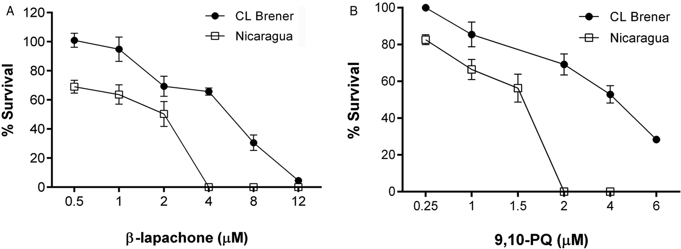
We have previously shown that TcAKR reduces o-NQs such as β-Lap and 9,10-PQ with concomitant ROS generation. With the aim to evaluate whether there is a direct relationship between the trypanocidal effect of these o-NQs and TcAKR activity, we assessed the effect of these compounds in two T. cruzi strains with differential TcAKR expression, CL Brener and Nicaragua, of which CL Brener has 2.2-fold higher TcAKR expression than Nicaragua (Garavaglia et al. Reference Garavaglia, Laverrière, Cannata and García2016). Epimastigote survival curves after 72 h of treatment with different concentrations of this TcAKR o-NQ substrates showed that CL Brener had higher IC50 values to β-Lap and 9,10-PQ than Nicaragua (Fig. 1 and Table 1). The fact that CL Brener which has higher TcAKR levels than Nicaragua is more resistant to these compounds indicates that greater TcAKR activity is unlikely to be a major influence on o-NQ toxicity. This suggests either this enzyme may not activate o-NQs in intact parasites or its activity may not be relevant in o-NQs metabolism.
Susceptibility of CL Brener and Nicaragua epimastigotes to TcAKR o-NQ substrates. Representative survival curves of epimastigotes of CL Brener and Nicaragua strains of T rypanosoma cruzi cultured for 72 h in the presence or absence of different concentrations of β-Lap (A) or 9,10-PQ (B). A survival rate of 100% corresponds to the mean number of duplicate samples of untreated epimastigotes.

Susceptibility to o-NQ substrates of TcAKR of two T rypanosoma cruzi strains with differential expression of TcAKR

a NADPH-dependent AKR enzymatic activity using 4-NBA as a substrate (data from Garavaglia et al. Reference Garavaglia, Laverrière, Cannata and García2016).
b IC50s calculated by linear regression analysis of the plot of the growth constant vs drug concentration.
c Detection of intracellular ROS production by flow cytometry using the H2DCF-DA probe. Values correspond to the relative fluorescence units (RFU) calculated as the median fluorescence intensity (MFI) of o-NQ-treated parasites/MFI of untreated parasites.
d Drug concentration used in the assay.
e Measurement of change in mitochondrial membrane potential (ΔΨm) by flow cytometry using rhodamine 123. Values correspond to RFU calculated as MFI of NQ-treated parasites/MFI of untreated parasites.
f Phosphatidylserine exposure measured by double staining with annexin-V-fluorescein (AV) and propidium iodide (PI) after 24 h of treatment. Values correspond to the ratio between % AV+/PI− in o-NQ-treated and untreated epimastigotes.
g Autophagosomes evaluated by MDC staining. Values correspond to the ratio between % of MDC+ in NQ-treated and untreated epimastigotes.
*P < 0.05, for the difference between results with CL Brener and Nicaragua.
**P < 0.05 with respect to untreated controls.
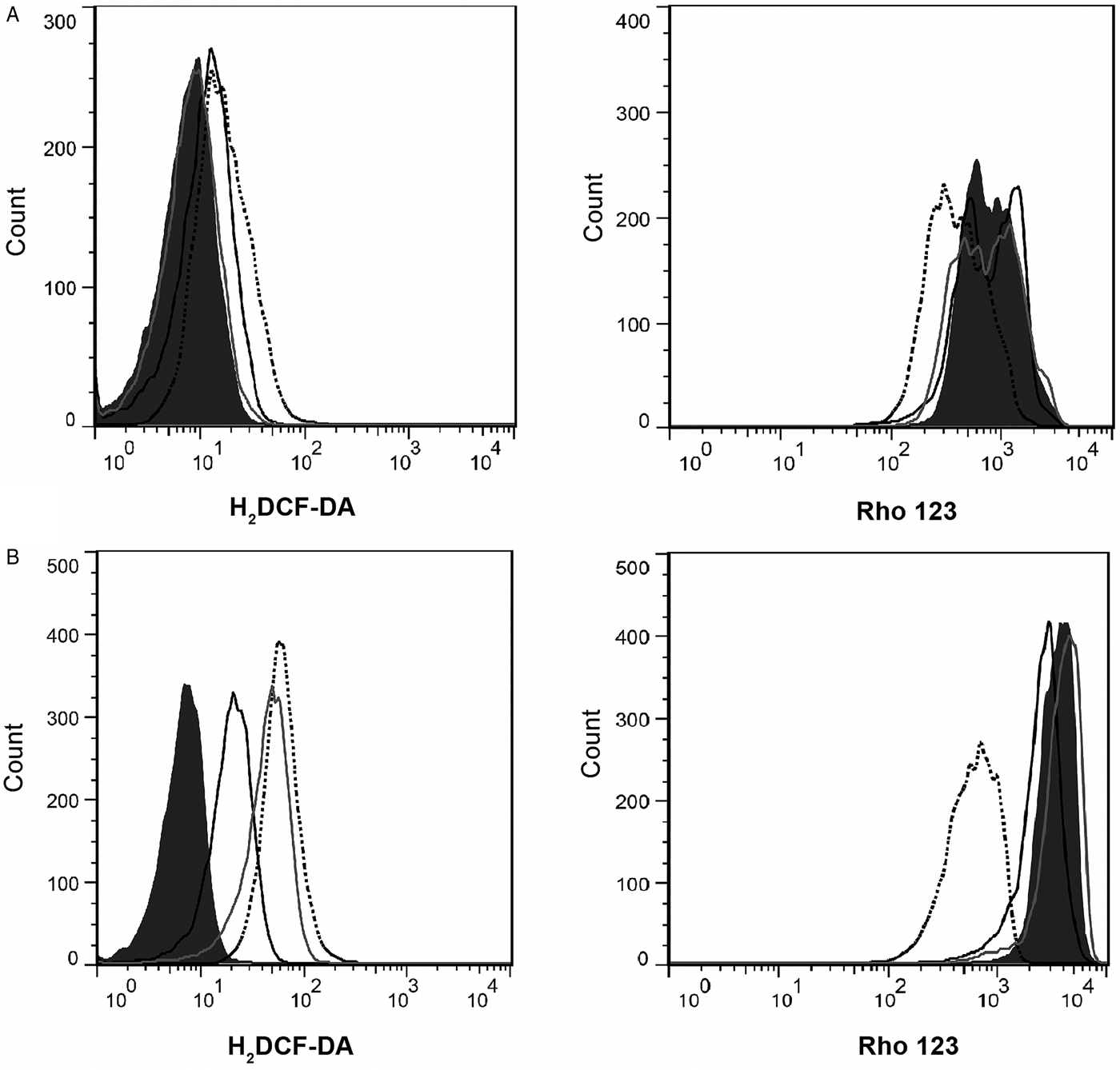
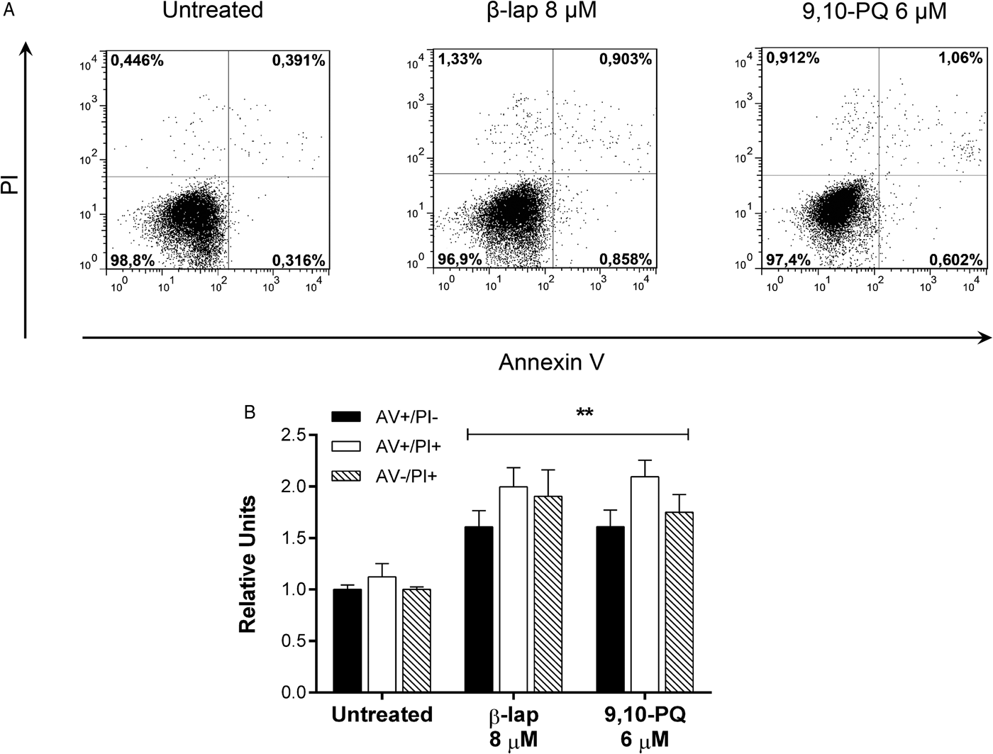
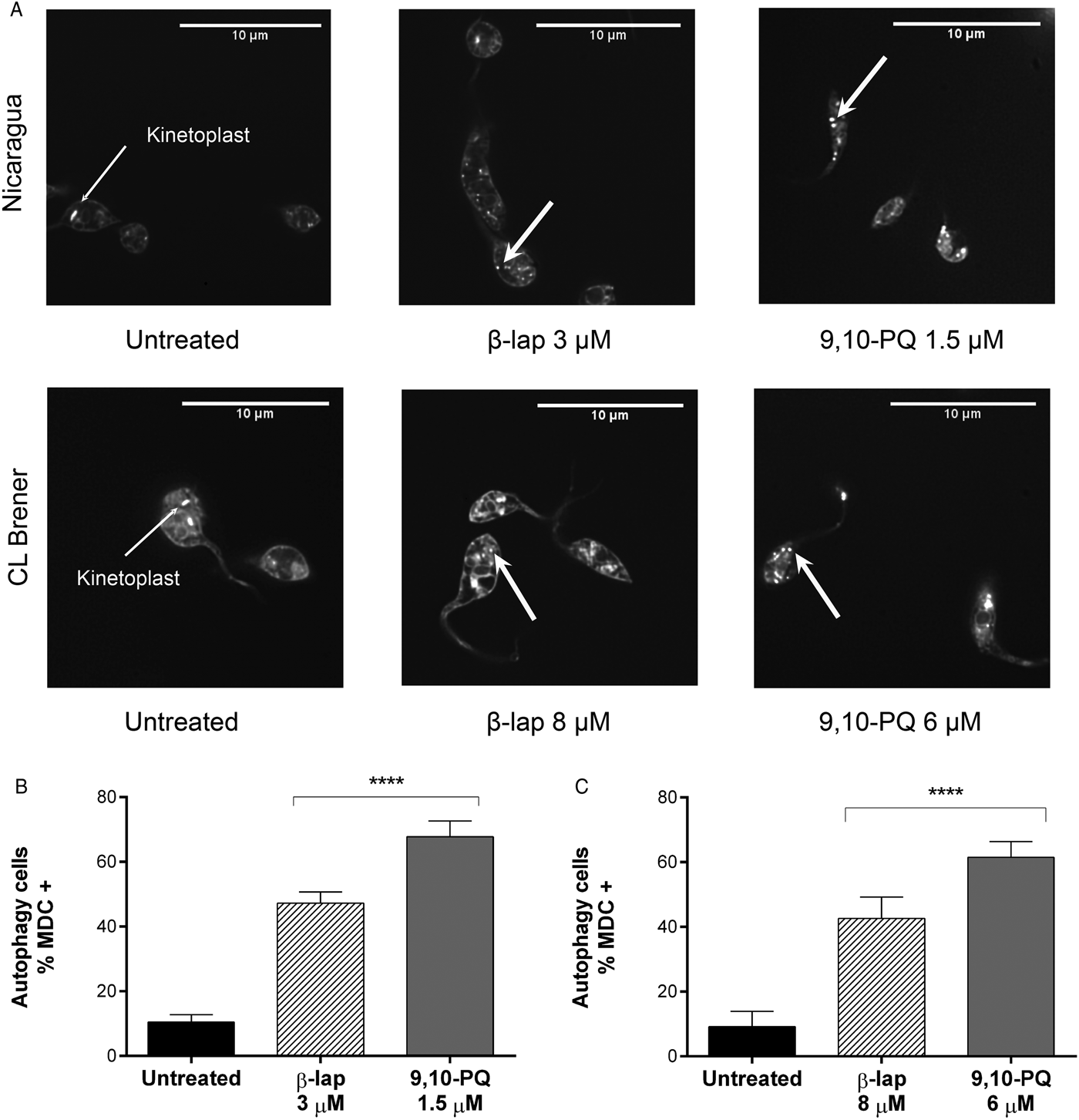
With the aim to gain knowledge about the mechanism of action of these TcAKR o-NQ substrates, different parameters associated with cell death were evaluated in epimastigotes of CL Brener and Nicaragua strains after treatment with these drugs: (i) ROS production and decrease of mitochondrial membrane potential (ΔΨm), (ii) exposition of phosphatidylserine residues on plasma membrane surface and (iii) presence of autophagosomes. (i) ROS production and ΔΨm measured by flow cytometry using specific probes showed that β-Lap induced ROS production in both strains at the rather same extent but a decrease of Ψm potential only in CL Brener strain. On the other hand, 9,10-PQ induced ROS in CL Brener but not in Nicaragua and no Ψm change was detected in either of the strains (Fig. 2 and Table 1). (ii) Phosphatidylserine exposition was evaluated by double staining with AV and PI and analysed by flow cytometry. Treatment with β-Lap and 9,10-PQ induced an increase in the percentage of epimastigotes AV+/PI− (representative of early apoptosis), AV+/IP+ (representative of late apoptosis or necrosis) and AV−/IP+ (characteristic of a type of necrosis with exaggerated membrane damage) only in the CL Brener strain (Fig. 3 and Table 1). (iii) Autophagosomes were detected with MDC labelling, a fluorescent probe that accumulates in autophagic vacuoles (Biederbick et al. Reference Biederbick, Kern and Elsasser1995), and analysed by fluorescence microscopy. An increase in MDC-positive cytoplasmic vacuoles was induced by both o-NQs in CL Brener as well as Nicaragua strain (Fig. 4 and Table 1). Altogether, these results indicate that TcAKR o-NQ substrates induce several death phenotypes which may vary depending on the combination of drug and T. cruzi strain used. Among these death phenotypes, cytoplasmic vacuoles were common to all combinations.
ROS production and loss of mitochondrial membrane potential (ΔΨm) of Nicaragua and CL Brener epimastigotes treated with TcAKR o-NQ substrates. Measurement of ROS generation and ΔΨm after o-NQs treatment for 24 h using H2DCF-DA and Rho123, respectively, in epimastigotes of Nicaragua (A) and CL Brener (B) strains. Shown are representative histograms corresponding to fluorescence analysis. The grey histograms represent untreated parasites; the dotted line corresponds to a positive control performed with either H2O2 (0.5 mm) for ROS production or CCCP (20 µ m) for ΔΨm detection. The black line corresponds to parasites treated with β-lap (2 µ m for Nicaragua and 8 µ m for CL Brener) and the grey line corresponds to parasites treated with 9,10-PQ (1.5 µ m for Nicaragua and 6 µ m for CL Brener).

Annexin V (AV) and propidium iodide (PI) labelling of CL Brener epimastigotes after treatment with TcAKR o-NQ substrates. Phosphatidylserine exposition was measured by double staining with AV and PI in CL Brener epimastigotes treated with β-Lap or 9,10-PQ for 24 h. (A) Flow cytometry dot plots of a representative experiment. (B) Relative quantification of epimastigote phenotypes. The bars represent the mean and s.e.m. of three independent experiments. Relative units were calculated for each quadrant as follows: % of epimastigotes in o-NQ-treated samples/% of epimastigotes in untreated samples. **P < 0.001 with respect to untreated controls.

Detection of autophagosomes by the probe monodansylcadaverine (MDC). (A) Fluorescence microscopy of MDC-labelled Nicaragua and CL Brener epimastigotes. Controls (untreated) present poorly and diffusely labelling showing only bright stain of kinetoplast (thin arrows). β-Lap- and 9,10-PQ-treated parasites display numerous and often closely apposed strongly labelled compartments (thick arrows). (B and C) Quantitation of parasites displaying MDC-labelled compartments by fluorescence microscopy. Data represent the mean and s.e.m. of a representative experiment using Nicaragua (B) and CL Brener (C) epimastigotes. (****P < 0.0001 with respect to untreated controls).

Effect of TcAKR o-NQ substrates in T. cruzi infection of Vero cells
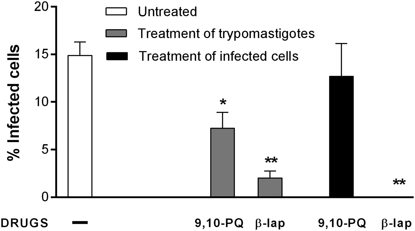
The lack of enough information in the literature related to trypanocidal activity of 9,10-PQ prompted us to evaluate its effect against the mammalian stages of the parasite. Treatment of trypomastigotes with 9,10-PQ, 3 h prior to and during the infection of Vero cells, caused a decrease in the number of infected cells indicating this drug may affect trypomastigote viability and/or infectivity. On the other hand, when trypomastigotes were treated with β-Lap, a greater reduction of infected cells was observed (Fig. 5), endorsing the inhibitory effect of β-Lap on trypomastigote viability described by others (Lopes et al. Reference Lopes, Cruz, Docampo, Vasconcellos, Sampaio, Pinto and Gilbert1978; Salas et al. Reference Salas, Tapia, Ciudad, Armstrong, Orellana, Kemmerling, Ferreira, Maya and Morello2008). It is important to note that an average of 25 amastigotes per cell were observed after 72 h of infection with untreated trypomastigotes as well as with 9,10-PQ- and β-Lap-treated trypomastigotes.
Effect of TcAKR o-NQ substrates on T. cruzi infection of Vero cells. Monolayers of Vero cells were infected with trypomastigotes of Nicaragua strain. Treatment with o-NQs was performed according to two protocols: (1) trypomastigotes were incubated 3 h prior to and during the infection process with 9,10-PQ or β-Lap (1 µ m), then drugs were washed away and infection was allowed to continue for 72 h (grey bars) and (2) 9,10-PQ or β-Lap (1 µ m) was added to Vero cells after the infection and maintained for 72 h (black bars). Infection of Vero cells in the absence of drugs was used as control (white bar). Three days post-infection, cells were fixed, stained with Giemsa and the number of infected cells was counted by optical microscopy. Results are expressed as the mean and s.e.m. of a representative experiment. *P < 0.05 and **P < 0.01 compared to the untreated control.

To further evaluate whether these o-NQs are able to affect the intracellular form of T. cruzi, Vero cells were treated after the infection process for 72 h. Treatment of infected cells with 9,10-PQ did not modify either the percentage of infected cells or the number of amastigotes per cell with respect to untreated infected controls (Fig. 5). As it has been demonstrated that 9,10-PQ can enter the cell (Taguchi et al. Reference Taguchi, Fujii, Yamano, Cho, Kamisuki, Nakai, Sugawara, Froines and Kumagai2007; Matsunaga et al. Reference Matsunaga, Arakaki, Kamiya, Endo, El-Kabbani and Hara2009), this result suggests that 9,10-PQ cannot affect amastigote replication, at least in the concentration used in this assay. Conversely, when infected Vero cells were treated with β-Lap, no infected cells were found after 72 h of treatment (Fig. 5), even using doses as low as 0.5 µ m. This result suggests that β-Lap can inhibit trypomastigote intracellular differentiation and/or amastigote replication. It is worth mentioning that doses of both drugs used in these experiments did not affect viability of Vero cells, evaluated by thiazolyl blue tetrazolium bromide (MTT) assay and microscopic observation (data not shown).
Trypanocidal effect of the o-NQ substrates of TcAKR on TcAKR-overexpressing epimastigotes
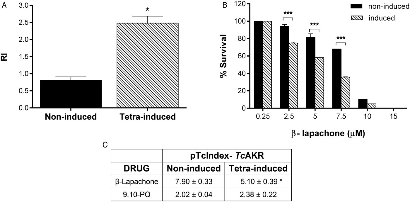
In order to evaluate if TcAKR participates in o-NQs activation in vivo, the effect on survival of β-Lap and 9,10-PQ was tested in epimastigotes transfected with the recombinant plasmid pTcIndex-TcAKR (Garavaglia et al. Reference Garavaglia, Laverrière, Cannata and García2016). These transfected parasites overexpressed this enzyme about 2.5-fold after 72 h of tetracycline induction (Fig. 6A). TcAKR-overexpressing parasites were more susceptible to β-Lap, showing a significant decrease in the IC50 value with respect to non-induced controls (Fig. 6B and C). However, no changes were observed in survival between non-induced and tetracycline-induced epimastigotes after treatment with 9,10-PQ (Fig. 6C).
Effect of o-NQ substrates of TcAKR on the survival of TcAKR-overexpressing epimastigotes. Non-induced and tetracycline-induced pTcIndex-TcAKR-transfected parasites were cultured for 72 h in the presence or absence of different concentrations of β-Lap or 9,10-PQ. (A) Semiquantitative analysis of TcAKR expression in untreated parasites evaluated by Western blot with a mouse anti-recombinant TcAKR serum. RI, relative intensity. (B) Survival curve of β-Lap-treated epimastigotes. Parasite growth in the absence of drug was considered a survival rate of 100%. (C) IC50 values (μ m) calculated by the dose–response curves using non-linear regression analysis. *P < 0.05 and ***P < 0.001, for results in non-induced vs tetracycline-induced epimastigotes.

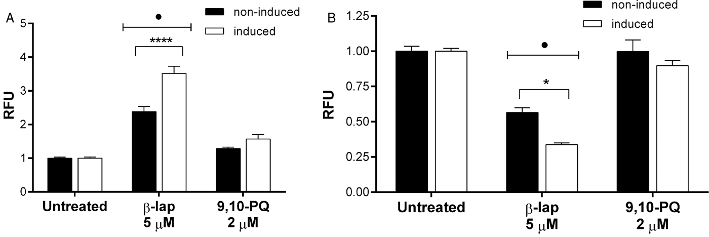
As these results suggest that TcAKR participates, at least, in β-Lap activation, ROS production and ΔΨm were evaluated in non-induced and tetracycline-induced TcAKR-transfected epimastigotes after treatment with the o-NQ substrates of TcAKR. Treatment with β-Lap induced a greater ROS production and Ψm decrease in TcAKR-overexpressing parasites than in non-induced controls (Fig. 7A and B), endorsing the putative participation of this enzyme in the metabolism of β-Lap. On the other hand, treatment with 9,10-PQ did not induce changes in neither ROS nor ΔΨm in transfected parasites, either non-induced or tetracycline-induced ones (Fig. 7A and B). In addition to survival curves results, the lack of ROS induction in TcAKR-overexpressing parasites indicates that TcAKR does not participate in 9,10-PQ activation in vivo. Otherwise, these results strengthen the fact that 9,10-PQ may not necessarily use a mechanism of action involving ROS. It is worth to note that transfected epimastigotes belong to the same lineage as Nicaragua strain, DTU I, in which no ROS and ΔΨm induction by 9,10-PQ was observed (Table 1).
Induction of ROS and ΔΨm in transfected epimastigotes overexpressing TcAKR. Non-induced and tetracycline-induced transfected epimastigotes were treated for 72 h with β-lap or 9,10-PQ. Detection of intracellular ROS production (A) and ΔΨm (B) by flow cytometry using H2DCF-DA and rhodamine 123 probes, respectively. Values correspond to the relative fluorescence units (RFU) calculated as the median fluorescence intensity (MFI) of o-NQs-treated parasites/MFI of untreated parasites. The asterisks indicate significant differences (****P < 0.0001 and *P < 0.05) with respect to the corresponding non-induced control. The dots indicate significant differences with respect to the corresponding untreated group (•P < 0.0001).

Discussion
We have previously identified TcAKR from T. cruzi, a NADPH-dependent reductase specific for o-NQs (Garavaglia et al. Reference Garavaglia, Cannata, Ruiz, Maugeri, Duran, Galleano and Garcia2010) which has been recently described to be involved in benznidazole resistance (Garavaglia et al. Reference Garavaglia, Laverrière, Cannata and García2016; González et al. Reference González, García-Huertas, Triana-Chávez, García, Murta and Mejía-Jaramillo2017). β-Lap and 9,10-PQ are o-NQs that can be reduced by TcAKR with concomitant production of free radicals (Garavaglia et al. Reference Garavaglia, Cannata, Ruiz, Maugeri, Duran, Galleano and Garcia2010) and whose trypanocidal activity has been previously documented by others. While the effect of β-Lap against T. cruzi has been extensively studied (Docampo et al. Reference Docampo, Lopes, Cruz and Souza1977; Goijman and Stoppani, Reference Goijman and Stoppani1985; Molina Portela et al. Reference Molina Portela, Fernandez Villamil, Perissinotti and Stoppani1996; Pieretti et al. Reference Pieretti, Haanstra, Mazet, Perozzo, Bergamini, Prati, Fato, Lenaz, Capranico, Brun, Bakker, Michels, Scapozza, Bolognesi and Cavalli2013; Ogindo et al. Reference Ogindo, Khraiwesh, George, Brandy, Brandy, Gugssa, Ashraf, Abbas, Southerland, Lee, Bakare and Fang2016), as far as we know, only one publication has reported the trypanocidal effect of 9,10-PQ (Silva et al. Reference Silva, Ferrioli-Filho, Kanesiro, Ferreira, Santos, Pinto, Fonseca, Mizrahy, Gilbert, Pinto, Ribeiro and Pinto1992). Based on this knowledge, the main goal of this work was to elucidate whether TcAKR participates in o-NQ mechanism of action. In order to achieve this aim, we firstly characterized the trypanocidal effect of the o-NQ substrates of the TcAKR β-Lap and 9,10-PQ in two T. cruzi strains with differential expression of this enzyme: CL Brener (DTU VI) and Nicaragua (DTU I). We have previously established, by Western blotting and by measuring AKR and QOR NADPH-dependent enzymatic activities, that CL Brener has higher TcAKR levels than Nicaragua strain (Garavaglia et al. Reference Garavaglia, Laverrière, Cannata and García2016). Here, we showed that CL Brener was more resistant than Nicaragua to both o-NQs. Therefore, we may infer that TcAKR is unlikely to be a major influence on o-NQ susceptibility and hypothesize that other reductases may overcome its QOR activity. In this regard, it has been documented that NQs act as subversive substrates of several T. cruzi redox enzymes such as old yellow enzyme, trypanothione reductase and lipoamide dehydrogenase (Salmon-Chemin et al. Reference Salmon-Chemin, Buisine, Yardley, Kohler, Debreu, Landry, Sergheraert, Croft, Krauth-Siegel and Davioud-Charvet2001; Kubata et al. Reference Kubata, Kabututu, Nozaki, Munday, Fukuzumi, Ohkubo, Lazarus, Maruyama, Martin, Duszenko and Urade2002; Ramos et al. Reference Ramos, Garza, Krauth-Siegel, Bader, Martinez and Maldonado2009).
The characterization of cell death phenotypes induced by β-Lap and 9,10-PQ in CL Brener and Nicaragua strains of T. cruzi showed that these o-NQs promoted different phenotypes depending on the combination of drug and strain analysed. Some of the phenotypes we studied here, such as ROS production, changes in mitochondrial membrane potential and exposition of phosphatidylserine in the outer leaflet of an intact plasma membrane (cells stained A+/IP−), are considered features of apoptosis (Smirlis et al. Reference Smirlis, Duszenko, Ruiz, Scoulica, Bastien, Fasel and Soteriadou2010). o-NQ treatment of parasites for evaluation of these parameters was performed at drug concentrations corresponding to an average of 1.6 of the IC50 value at 72 h. In these conditions, β-Lap and 9,10-PQ were able to induce these apoptotic characteristics mainly in CL Brener strain. By AV and PI staining, an elevated percentage of cells AV+/IP−, characteristic of early apoptosis, as well as AV+/IP+ and AV−/IP+ were detected only in CL Brener strain. It is important to note that IP-positive staining implies loss of plasma membrane integrity, a characteristic which is associated with necrotic mechanisms (Jamison et al. Reference Jamison, Gilloteaux, Taper, Calderon and Summers2002; Zong and Thompson, Reference Zong and Thompson2006; Silva, Reference Silva2010). Placing the focus on ROS production, β-Lap induced ROS in all of the evaluated T. cruzi strains while 9,10-PQ only did it in CL Brener strain. This finding indicates that, unlike β-Lap, 9,10-PQ trypanocidal effect was not necessarily associated with ROS production and suggests that the high toxicity of 9,10-PQ in Nicaragua and Adriana strains may be mediated by other mechanisms. The decrease in the mitochondrial potential was another parameter which varied according to the drug and T. cruzi strain analysed. In this case, β-Lap induced ΔΨm in CL Brener and Adriana and not in Nicaragua, whereas 9,10-PQ in neither of the evaluated strains. Studies with different drugs suggested that the increase in ROS is both a cause and a consequence of mitochondrial dysfunction (Lazarin-Bidóia et al. Reference Lazarin-Bidóia, Desoti, Ueda-Nakamura, Dias Filho, Nakamura and Silva2013). In this regard, it has been documented that triazolic NQ derivatives are able to induce an increase in ROS levels without Ψm alterations (Fernandes et al. Reference Fernandes, Da Silva, Pinto, De Castro and Menna-Barreto2012), which is coincident with the effects produced by β-Lap in Nicaragua strain. Altogether these results suggest that the T. cruzi strains evaluated here may have different targets for β-Lap and 9,10-PQ and/or different detoxification enzymes for each of semiquinone-free radicals. On the other hand, conversely to the variability observed in the induction of apoptotic and necrotic features, the strong increase in MDC labelling after treatment with both o-NQs suggests that autophagy may be a common death mechanism used by β-Lap and 9,10-PQ. Although various trypanocidal drugs, including NQs, have shown to trigger autophagy-like processes (Menna-Barreto et al. Reference Menna-Barreto, Salomão, Dantas, Santa-Rita, Soares, Barbosa and de Castro2009; Sengupta et al. Reference Sengupta, Chowdhury, Bosedasgupta, Wright and Majumder2011; Veiga-Santos et al. Reference Veiga-Santos, Barrias, Santos, de Barros Moreira, de Carvalho, Urbina and de Souza2012; Salomão et al. Reference Salomao, De Santana, Molina, De Castro and Menna-Barreto2013; Dos Anjos et al. Reference Dos Anjos, Sobral Alves, Gonçalves, Fontes, Nogueira, Suarez-Fontes, Neves da Costa, Rios-Santos and Vannier-Santos2016), ultrastructural and functional studies using specific inhibitors of the process have to be further investigated to confirm that autophagy is responsible of parasite death induced by these TcAKR o-NQ substrates.
In the second part of the work, we studied whether TcAKR participates in o-NQ activation in vivo by evaluating the effect of these drugs in TcAKR-overexpressing epimastigotes. We have previously shown that a 2.5-fold increase in TcAKR expression was enough to shift the IC50 value for benznidazole from 9.90 to 17.45 µ m suggesting this enzyme is involved in Bz detoxification (Garavaglia et al. Reference Garavaglia, Laverrière, Cannata and García2016). Recently, it has been described that TcAKR is upregulated in clones of T. cruzi naturally resistant to benznidazole and its expression is increased 1.7-fold in an induced resistant clone (González et al. Reference González, García-Huertas, Triana-Chávez, García, Murta and Mejía-Jaramillo2017), thus validating our previously obtained results with TcAKR-transfected epimastigotes. TcAKR-overexpressing parasites treated with β-Lap evidenced the following changes with respect to non-induced controls: (i) enhanced susceptibility (decrease in the IC50 value), (ii) increased intracellular ROS production and (iii) decreased Ψm. None of these parameters varied when TcAKR-overexpressing parasites were treated with 9,10-PQ. Therefore, these results suggest that TcAKR is involved in β-Lap but not 9,10-PQ activation. These findings let us hypothesize that TcAKR participation in β-Lap activation may be comparable to NQO1 role in cancer cells (Pink et al. Reference Pink, Planchon, Tagliarino, Varnes, Siegel and Boothman2000). However, unlike NQO1 whose expression was found to be a principal determinant of therapy response, our studies in two T. cruzi strains with differential TcAKR expression suggest that this enzyme is unlikely to be a major influence on susceptibility to β-Lap. In this sense, it is important to note that previous studies regarding resistance to benznidazole using the same strains of T. cruzi, CL Brener and Nicaragua, also concluded that the action of TcAKR was not a determinant of resistance to this drug in wild-type strains (Garavaglia et al. Reference Garavaglia, Laverrière, Cannata and García2016). By contrast, TcAKR showed to be a relevant enzyme in benznidazole resistance when studies were performed on clones derived from a natural T. cruzi population resistant to benznidazole (González et al. Reference González, García-Huertas, Triana-Chávez, García, Murta and Mejía-Jaramillo2017). A possible explanation for these contradictory results may arise from the nature of these strains, since whereas CL Brener is a clone coming from the CL strain (Zingales et al. Reference Zingales, Pereira, Oliveira, Almeida, Umezawa, Souto, Vargas, Cano, da Silveira, Nehme, Morel, Brener and Macedo1997), Nicaragua is an isolate recently obtained from a Triatomine bug in which may co-exist parasites with different genetic background (Grosso et al. Reference Grosso, Bua, Perrone, González, Bustos, Postan and Fichera2010).
Given the emergence of parasite isolates resistant to currently used drugs to treat T. cruzi infection, benznidazole and nifurtimox, and the controversy about their efficacy during the chronic phase of the infection, it is important to characterize new trypanocidal compounds in order to find alternative chemotherapeutic drugs. In this context, the following conclusions achieved in this work indicate that β-Lap could be a good candidate: (i) Nicaragua strain that is moderately resistant to benznidazole (Grosso et al. Reference Grosso, Bua, Perrone, González, Bustos, Postan and Fichera2010; Garavaglia et al. Reference Garavaglia, Laverrière, Cannata and García2016) was highly sensitive to β-Lap and 9,10-PQ, (ii) β-Lap affected trypomastigote infectivity and amastigote replication and (iii) TcAKR, an enzyme with no counterpart in normal mammalian cells, may be involved in β-Lap activation. Furthermore, experience taken from phase I/II clinical trials of β-Lap in cancer field (Gerber et al. Reference Gerber, Arriaga, Beg, Dowell, Schiller, Frankel, Leff, Meek, Bolluyt, Fatunde, Martinez, Vo, Fattah, Sarode, Zhou, Xie, McLeod, Schwartz and Boothman2014; Beg et al. Reference Beg, Huang, Silvers, Gerber, Bolluyt, Sarode, Fattah, Deberardinis, Merritt, Xie, Leff, Laheru and Boothman2017) may facilitate the implementation of β-Lap chemotherapy for other pathologies.
Acknowledgements
M.F.R., L.E.F and G.A.G. are members of the Research Career of the National Research Council of Argentina (CONICET). M.L. had fellowships from AMSUD-PASTEUR, ANPCyT and CONICET, and his work was done in the laboratory of J. J. Cazzulo (Instituto de Investigaciones Biotecnologicas IIB-INTECH). The authors thank John Kelly and Martin Taylor (London School of Tropical Medicine, London, United Kingdom) for kindly providing the pTcINDEX vector and Patricia Bustos (INP-ANLIS ‘Dr. Carlos G. Malbrán’) for helpful discussion of the manuscript.
Financial support
This work was supported by FOCANLIS 2011 and 2013 from the Instituto Nacional de Parasitología ‘Dr. Mario Fatala Chaben’ – ANLIS ‘Dr. Carlos G. Malbrán’.