Atresia of the common pulmonary vein is an exceedingly rare form of totally obstructed anomalous pulmonary venous drainage. This definition, first used by Lucas et al.1 in 1962, describes a condition in which atresia of the initially common pulmonary venous channel occurs before its absorption into the left atrium, and following obliteration of any potential collateral venous channels.2
In our case, the diagnosis was made at necropsy in a one-day-old infant. During life, the diagnosis of totally obstructed anomalous pulmonary venous connection had been made at cross-sectional echocardiography, and subsequently by cardiac catheterization.
Case report
A full term newborn girl, weighing 3 kilograms, was born at a secondary level referral hospital. The infant was the product of an uneventful delivery, with unremarkable obstetric history. The Apgar score was 9 at 1 minute, albeit that shortly thereafter the baby developed respiratory distress and cyanosis refractory to administration of 100 percent oxygen. Worsening of cyanosis, with ensuing metabolic acidosis and a state of low cardiac output, required intubation and high frequency oscillating ventilation was used. The cyanosis proved refractory to the use of surfactant and inhalation of nitric oxide. The infant was transferred to our department for further evaluation. On admission, fifteen hours after delivery, the infant was profoundly cyanosed. The precordium was quiet, and the peripheral pulses were not palpable. A grade 2 out of 6 systolic murmur was heard at the left sternal border. Sampling arterial blood gas showed marked metabolic acidosis, with pH at 6.53, and severe hypoxemia, with arterial partial tension of oxygen at 18 millimetres of mercury despite the use of an inspired oxygen fraction at 100 percent. A chest radiograph showed ground glass opacities over both lung fields, but the heart was of normal size. Cross-sectional echocardiography showed normal cardiac structures, with gross dilation of both the right atrium and ventricle. A right-to-left shunt was present across a patent oval foramen, and there was a large persistently patent arterial duct, with signs of pulmonary hypertension. The pulmonary veins could not be demonstrated on either side. Colour-flow mapping failed to visualise any pulmonary venous return to the heart. We made the diagnosis of obstructed anomalous pulmonary venous drainage, and catheterization was carried out as an emergency to detail the site of the drainage. A right ventriculogram (Fig. 1) showed normally branching, albeit small, pulmonary arteries, and a large patent arterial duct, which fed both the descending aorta and the ascending aorta in a retrograde fashion. The pulmonary veins, the left atrium, and the left ventricle were never opacified. The infant died of intractable metabolic acidosis before any surgical treatment could be carried out.

Figure 1. Right ventricular injection shows opacification of both the ascending and descending aorta through a patent arterial duct. RV: right ventricle; AAo: ascending aorta; PAD: patent arterial duct.
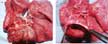
At necropsy, the right and left pulmonary veins were found to join an atretic common pulmonary venous channel, which pointed at the posterior wall of the left atrium. There was no communication of the pulmonary veins either with the heart, or with the systemic venous circulation (Figs. 2a and b).

Figure 2. Posterior heart and lung view. (a) The right and left pulmonary veins (*) are seen to join into a fibrous strand (⇒) extending to the posterior wall of the left atrium. (b) A probe is advanced into the left atrium to show the absence of any connection with the atretic common pulmonary vein. LA: left atrium; LPVs: left pulmonary veins; RPVs: right pulmonary veins.
Discussion
The common pulmonary vein is an embryonic structure that connects with the venous elements of the early pulmonary vasculature, which ultimately become incorporated into the left atrium.2 When atresia of the common vein occurs late in the embryogenesis, the collateral venous channels are already obliterated, and the individual pulmonary veins empty into a blind confluence, with no direct connection with either the left heart or the systemic veins.3 Atresia of the common pulmonary vein is probably less rare than expected, because in the neonatal period4 its clinical and radiologic features mimic strictly those of severe pulmonary parenchymal disease. The difficulty in establishing a diagnosis during life is supported by the fact that almost half of reported cases were diagnosed only at autopsy.5 Echocardiographic diagnosis of this anomaly is more difficult to establish than that of obstructed totally anomalous pulmonary venous drainage, especially when the pulmonary confluence is hypoplastic or atretic,4, 6 and in some cases echocardiography has failed to distinguish atresia of the common pulmonary vein from noncardiac conditions.4 Cardiac catheterization may become necessary if the diagnosis cannot be established,4 but angiography is often poorly tolerated by these very fragile neonates.
As suggested in the literature, once the diagnosis is made, the majority of newborns with this malformation are amenable to surgical repair.4, 7, 8 Haemodynamic stabilization prior to surgery4 is, therefore, of crucial importance, but cardiovascular collapse and death often occur before any procedure can be attempted. Prompt diagnosis of this rapidly life-threatening cardiac anomaly, therefore, is mandatory if any attempt is to be made to provide a life-saving surgical repair.