Introduction
Protective envelopes of metacestodes, corresponding to the outer and inner envelopes (the exo- and endocyst), together with the tail appendage, constitute the cercomer (Janicki, Reference Janicki1920; Jarecka, Reference Jarecka1975; Krasnoshchekov, Reference Krasnoshchekov1980; Jarecka et al., Reference Jarecka, Michajlow and Burt1981). There is some controversy about the meaning of the term ‘cercomer’ in recent literature. Other authors (for example Freeman, Reference Freeman1973; Gulyaev, Reference Gulyaev1996; Chervy, Reference Chervy2002) consider the cercomer to be the ‘tail appendage’. We use the term ‘cercomer’ in this paper for the tail appendage and protective envelopes to emphasize their common attributes: (1) they have microvilli, not microtriches; and (2) they are lost after the parasite is ingested by the definitive host.
During the course of evolution of cyclophyllid metacestodes the tail appendage in a number of cases has been transformed into a cellular exocyst having a complex structure, for example, in the diplocyst of Aploparaksidae (Bondarenko & Kontrimavichus, Reference Bondarenko and Kontrimavichus2006). The presence of a non-cellular exocyst has long been considered to be a characteristic feature of cysticercoids in the family Dilepididae, namely a monocercus (Gabrion, Reference Gabrion1975; Gabrion & Gabrion, Reference Gabrion and Gabrion1976; Krasnoshchekov & Tomilovskaya, Reference Krasnoshchekov and Tomilovskaya1978; Gabrion & Jourdan, Reference Gabrion and Jourdan1979; Gabrion & Helluy, Reference Gabrion and Helluy1982; Jarecka et al., Reference Jarecka, Bance and Burt1984; Gulyaev & Kornienko, Reference Gulyaev and Kornienko1998; Gulyaev & Ishigenova, Reference Gulyaev and Ishigenova2003). However, those authors who studied the post-embryonic development of hymenolepidids also noted the presence of non-cellular outer envelopes, so that the cysticercoids of Hymenolepididae with a non-cellular outer envelope were distinguished as an independent modification of the cysticercoid, known as a cyclocercus (Kotel'nikov, Reference Kotel'nikov and Ershov1965, Reference Kotel'nikov and Ershov1971; Krasnoshchekov & Pluzhnikov, Reference Krasnoshchekov and Pluzhnikov1984; Regel, Reference Regel1986; Grytner-Ziecina, Reference Grytner-Ziecina1994; Nikishin & Lebedev, Reference Nikishin and Lebedev2009, Reference Nikishin and Lebedev2011).
In general, cestodes (Hymenolepididae s.l.) of waterfowl from north-eastern Asia use crustaceans or oligochaetes as intermediate hosts (Regel, Reference Regel1986, Reference Regel2000; Bondarenko & Kontrimavichus, Reference Bondarenko and Kontrimavichus2006). Hence, the identification of a hymenolepidid metacestode in caddisworms (Insecta: Trichoptera) attracted special attention, and this metacestode was described as a new morphological modification of cysticercoids – a tectacyst (Regel & Kashin, Reference Regel and Kashin1995). The most characteristic feature of a tectacyst is the presence of a non-cellular outer envelope (exocyst), which starts its formation at the earliest stages of post-embryonic development (before the primary lacuna). Differences between two hymenolepidid cysticercoids having a non-cellular exocyst (cyclocercus and tectacyst) are as follows: in a cyclocercus the non-cellular exocyst completely covers the cysticercoid (including the tail appendage), whereas in a tectacyst the tail appendage perforates the exocyst and develops outside, commencing during early scolexogenesis (Kotel'nikov, Reference Kotel'nikov and Ershov1971; Regel, Reference Regel1986; Regel & Kashin, Reference Regel and Kashin1995). The functional significance of this difference is not clear.
The origin and structure of the outer non-cellular envelope in metacestodes remain controversial. It has been interpreted as a capsule (Gabrion, Reference Gabrion1975; Jarecka et al., Reference Jarecka, Bance and Burt1984), as a larval envelope, being derived from the oncospheral membrane (Gabrion & Jourdan, Reference Gabrion and Jourdan1979), as an exfoliated hyaline layer of the cyst wall (Maksimova, Reference Maksimova1990) and as an envelope of unknown nature (Bondarenko & Kontrimavitchus, Reference Bondarenko and Kontrimavichus2004). Such disagreements are not surprising, since ultrastructure of the outer non-cellular envelope has been studied in only a small number of metacestodes (Gabrion et al., Reference Gabrion, Plateaux and Quentin1976; Gabrion & Jourdan, Reference Gabrion and Jourdan1979; Jarecka et al., Reference Jarecka, Bance and Burt1984; Nikishin & Lebedev, Reference Nikishin and Lebedev2009). Light-microscopic observations of the metacestodes of Fimbriaria fasciolaris did not reveal the outer non-cellular envelope (Valkounova, Reference Valkounova1985), although it has been described by other authors (Krasnoshchekov & Pluzhnikov, Reference Krasnoshchekov and Pluzhnikov1984) using electron microscopy. Probably, the use of only light-microscopic methods of investigation is not always sufficient for identification of the non-cellular outer envelope in cysticercoids.
Protective envelopes are regarded as critical adaptive structures of metacestodes, for providing their normal development in the intermediate host (exocyst) and preserving the parasite's prospective parts (the scolex and neck) during transition through the initial sites of the alimentary tract in the definitive host (endocyst). Envelopes are more variable parts of cysticercoids, so additional information about them can give more data relevant to the discussion of phylogenetic interrelations and systematics among the cestodes.
This study aims to clarify the morphology of protective envelopes and the tail appendage of the tectacyst of Microsomacanthus paraparvula Regel, 1994 from the haemocoel of the caddisworm Grensia praeterita Walker, 1852.
Materials and methods
Collection and examination of metacestodes
Initially, metacestodes of M. paraparvula were found in naturally infected caddisworms G. praeterita from the Chaun lowland (north-western Chukotka, Russia). Experimental infections of the definitive and intermediate hosts allowed determination of the species of metacestodes, and exploration of their life circle – from metacestode to metacestode (Regel & Kashin, Reference Regel and Kashin1995). Images of living metacestodes in physiological saline (0.65% NaCl) from experimentally infected caddisworms were obtained using the light microscope ‘Amplival’ with ‘MF-matic’ (Carl Zeiss, Jena, Germany) photomicrographic eguipment.
The mature proglottids of M. paraparvula separate as a single unit (block, according to Denny, Reference Denny1969) and in this form are ingested by the caddisworm; thus the latter may contain from 10 to 100 metacestodes. When retrieved from the caddisworm, long tail appendages of cysticercoids form a mycelium-like tangle that binds the cysticercoids together.
Four groups of developed cysticercoids of M. paraparvula (about 30 samples) were isolated from four naturally infected caddisworms G. praeterita and fixed in 4 % glutaraldehyde in 0.1 M phosphate buffer (pH 7.2) from 2 h to several days at approximately 4°C. The material was postfixed in a 2 % solution of osmium tetroxide (OsO4) for 12 h, stained with 1 % uranyl acetate in 70 % ethanol (for 10 h) during dehydration, and embedded in Epon and Araldite. The obtained blocks were sectioned with a glass knife on LKB Bromma 2088 and LKB Nova ultratomes (Uppsala, Sweden). Ultra-thin sections (90 nm) were placed on pioloform-coated copper, single-slot grids and examined using JEM-100C and JEM-1011 (Jeol, Japan) transmission electron microscopes (TEM) operating at 80 kV and a Libra-120 (Carl Zeiss) TEM operating at 120 kV.
Semi-thin sections (1–2 μm) were cut using a LKB Bromma 2088 microtome, stained with methylene blue using the method of Morgenstern (Reference Morgenstern1969) and viewed through an Olympus CX41 light microscope (Olympus Corporation, Japan) with an Olympus E-420 digital camera.
Results
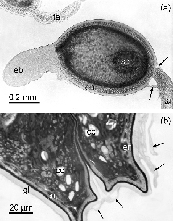
The outer envelope (exocyst) of the developed cysticercoids was found to be closely adjacent to the endocyst's glycocalyx, except for the areas around the poles. A clearly visible exocyst bubble remains after the invagination of the prospective part of the metacestode at its anterior pole (fig. 1a). On the posterior pole fragments of the exocyst after perforation by the tail appendage can be seen (fig. 1a and b).
The tectacyst of Microsomacanthus paraparvula. (a) Live tectacyst in physiological saline from the experimentally infected caddisworm Grensia praeterita showing the scolex (sc), endocyst (en), exocyst bubble (eb) and fragments of broken exocyst (arrows) after release of the tail appendage (ta). (b) Semi-thin section of the posterior pole showing the endocyst (en) with glycocalyx (gl), calcareous corpuscles (cc) and peeled exocyst (arrows).

Endocyst
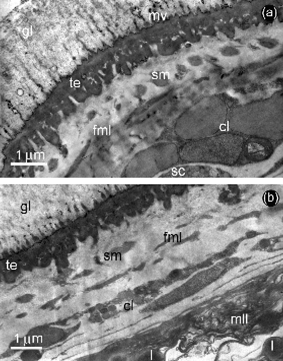
The endocyst of fully developed metacestodes of M. paraparvula (fig. 2a and b) consists of the tegument, which is covered with microvilli and a layer of glycocalyx, and is underlain by the basal matrix, a fibrous–muscular layer, cellular layer and a myelin-like layer. The glycocalyx, 3–6 μm thick, is formed by a fibrillar meshwork material with inclusions of vesicles of different sizes. The distal cytoplasm of the endocyst tegument, approximately 1 μm thick, is filled with electron-dense material. A thin basal matrix is not always apparent. A fibrous layer (consisting of two mutually perpendicular layers of fibres) with inclusions of muscular processes is transpierced by cytoplasmic bridges connecting the distal cytoplasm with more deeply located tegumental cytons. In the structure of the cellular layer the tegumental cytons and elements of the osmoregulatory system (terminal cells and collecting channels) were found, along with muscular and secretory cells producing round, dense granules, up to 350 nm in diameter (fig. 2a). A loose myelin-like layer with lipid droplets (fig. 2b) is located on the border of the neck, which contains large, calcareous corpuscles.
The endocyst of Microsomacanthus paraparvula. (a) Anterior pole with glycocalyx (gl), tegument (te), microvilli (mv), fibrous–muscular (fml) layer, containing superficial muscles (sm) and cellular (cl) layer, containing secretory cells (sc). (b) Posterior pole showing all the listed layers, and myelin-like layer (mll) with lipid droplets (l).
Exocyst
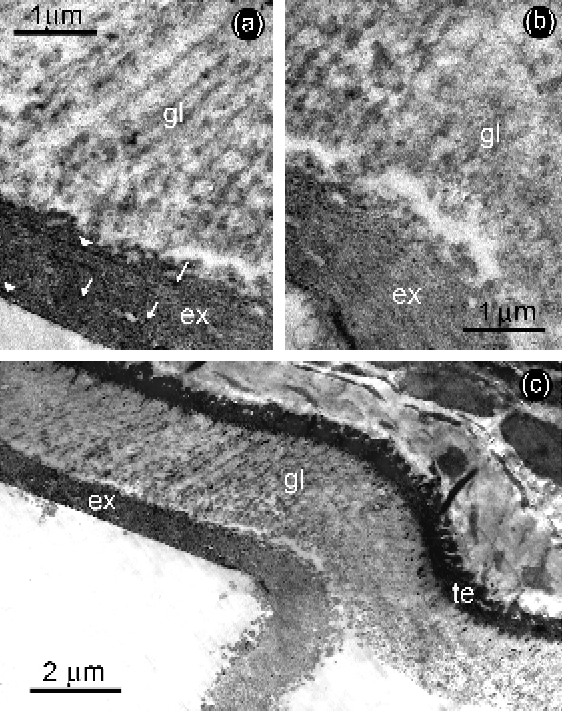
The exocyst, 0.5–3 μm thick, is formed of granular material with the inclusion of small vesicles (fig. 3). The outer surface of the envelope is represented by a thin membrane-like layer of electron-dense material. The attached and disconnected exocyst has differences in its thickness and density, although we do not exclude that these differences may be due to the preparation of material peculiar to the present investigation. The exocyst adjacent to the glycocalyx surface of the endocyst (fig. 3a) is thinner (0.5–1.5 μm), being composed of denser granular material. Granules and parallel layers of electron-dense material (fig. 3a) occur inside the adjacent exocyst. When the exocyst detaches from the glycocalyx surface (fig. 3b and c), its structure becomes disordered, the granular material loosens and the exocyst disperses into separate fragments. The thickness of the loosened, detached exocyst reaches 2.5–3 μm.
The exocyst of Microsomacanthus paraparvula. (a) Exocyst (ex) with dense granules (arrows) and layers of electron-dense material (arrowheads) attached to the glycocalyx (gl). (b) Initial stage of separation of the exocyst (ex) from the endocyst glycocalyx (gl). (c) General view of the site with the attached and detached exocyst (ex) showing the tegument of the endocyst (te) and the glycocalyx (gl).
Tail appendage
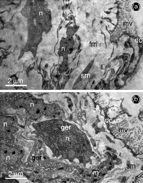
The tail appendage is restricted by a thin, dense layer of distal cytoplasm and covered with sparse microvilli. The loose fibrous matrix of the tail appendage is situated immediately below the distal cytoplasm. This matrix includes small, disconnected clusters of superficial musculature (fig. 4a) and cellular elements: cytons of tegument, muscle and small, poorly differentiated cells. A major proportion of tegumental cytons and the muscle cells in the terminal part of the tail appendage have signs of high synthetic activity, such as multiple Golgi zones and developed granular endoplasmic reticulum (GER). Another characteristic feature of cells in this area is the presence of numerous condensed mitochondria. Muscle cells are characterized by widened cisternae of GER with a small-granule content of moderate density (fig. 4b). On occasion these cisternae were seen to occupy the major part of the cytoplasm, and were often seen to fuse together. In this case, the nucleus was seen located in the central patch of cytoplasm, surrounded by the solid mass of synthesized material.
The tail appendage of Microsomacanthus paraparvula. (a) Proximal part where the fibrous matrix (fm) is limited by the distal cytoplasm (dc) with microvilli (mv). The matrix contains bundles of superficial muscles (sm) and nucleus (n) of muscle cells and cytons. (b) Distal part where muscle cells and cytons with nucleus (n), widened cisternae of granular endoplasmic reticulum (ger) and condensed mitochondria (m), superficial muscles (sm), distal cytoplasm (dc) and microvilli (mv) are visible.
Tegumental cytons appear to produce a secretion of low electron density, which enters the distal cytoplasm. On the surface of the tegument clear vesicles are released. The space between adjacent caudal appendages is filled with loose granular material containing vesicles.
Discussion
The morphological features of protective envelopes of the tectacyst of M. paraparvula are similar to those of other hymenolepidid cysticercoids with a non-cellular outer envelope, which are generally referred to as of the cyclocercus type (Krasnoshchekov & Pluzhnikov, Reference Krasnoshchekov and Pluzhnikov1984). However, a cyclocercus differs from a tectacyst due to the non-cellular exocyst of the former that covers the whole cysticercoid together with the tail appendage, while the tail appendage of the latter perforates the exocyst and develops outside. Probably, the increased synthetic activity of cells in the distal part of the tail appendage is related to its intense rate of growth (Regel & Kashin, Reference Regel and Kashin1995).
The exocyst of M. paraparvula consists mainly of granular material, whereas the exocyst of F. fasciolaris cyclocercus consists of several layers of fibrillar material (Krasnoshchekov & Pluzhnikov, Reference Krasnoshchekov and Pluzhnikov1984). These authors note that the morphology of the cyclocercus is typical for hymenolepidid metacestodes, but the presence of the outer non-cellular envelope brings it closer to the monocercus of Dilepididae.
The source of the non-cellular exocyst's formation remains unclear. Given the early appearance of the exocyst in ontogeny (before the primary lacuna), we consider that the hypothesis suggesting that the exocyst is formed by the means of glandular secretions from the oncosphere is plausible (Krasnoshchekov & Tomilovskaya, Reference Krasnoshchekov and Tomilovskaya1978; Kashin, Reference Kashin1986).
Presently available published data suggest that non-cellular envelopes are widely distributed at least within the families Hymenolepididae and Dilepididae (Kotel'nikov, Reference Kotel'nikov and Ershov1965, Reference Kotel'nikov and Ershov1971; Gabrion & Gabrion, Reference Gabrion and Gabrion1976; Krasnoshchekov & Tomilovskaya, Reference Krasnoshchekov and Tomilovskaya1978; Gabrion & Jourdan, Reference Gabrion and Jourdan1979; Jarecka et al., Reference Jarecka, Bance and Burt1984; Krasnoshchekov & Pluzhnikov, Reference Krasnoshchekov and Pluzhnikov1984; Regel, Reference Regel1986; Maksimova, Reference Maksimova1990; Grytner-Ziecina, Reference Grytner-Ziecina1994; Regel & Kashin, Reference Regel and Kashin1995; Gulyaev & Kornienko, Reference Gulyaev and Kornienko1998; Gulyaev & Ishigenova, Reference Gulyaev and Ishigenova2003; Nikishin & Lebedev, Reference Nikishin and Lebedev2009). Moreover, a transparent outer envelope, reminiscent of a non-cellular exocyst, was found in cercoids of the tetraphyllids Pseudanthobothrium hanseni and Pseudanthobothrium sp. (Jarecka & Burt, Reference Jarecka and Burt1984). The apparent absence of the exocyst in some metacestodes (Valkounova, Reference Valkounova1985) can be explained by its loss during the preparation of the specimens, or by structural and mechanical differences between exocysts of various species that influence the lifetime of this envelope.
A review of the nomenclature of metacestodes by Chervy (Reference Chervy2002) does not mention the non-cellular outer envelope in cysticercoids, although some publications concerning the description of this envelope are cited (Gabrion & Gabrion, Reference Gabrion and Gabrion1976; Gabrion et al., Reference Gabrion, Plateaux and Quentin1976; Jarecka et al., Reference Jarecka, Bance and Burt1984; Krasnoshchekov & Pluzhnikov, Reference Krasnoshchekov and Pluzhnikov1984). Probably, the presence of a non-cellular exocyst cannot be considered as a ‘key character differentiating the types of larval cestodes’ (Chervy, Reference Chervy2002), but its presence or absence provided evidence of heterogeneity in the genus Microsomacanthus (Regel, Reference Regel1986), which was subsequently confirmed (Galkin & Regel, Reference Galkin and Regel2010). In order to understand whether it is possible to use the non-cellular exocyst as a systematic character, further ultrastructural studies of uninvestigated cyclophyllid metacestodes are needed. In addition, new data on the ultrastructure of metacestodes can be utilized for the analysis of phylogenetic relationships among cestodes (Beveridge, Reference Beveridge, Littlewood and Bray2001; Swiderski, Reference Swiderski2007).
Acknowledgements
The authors extend their sincere thanks to Marshall Lightowlers (University of Melbourne) for his kind assistance in revising the manuscript. The authors would also like to thank the staff of the Centers of Electron Microscopy (Institute of Marine Biology, Vladivostok and Institute of Biology of Inland Waters, Borok, Russia) for technical assistance. We also thank the reviewers for their constructive and benevolent comments and suggestions. The present study was supported by the Russian Foundation for Basic Research (N 11-04-00870).