Introduction
The first documented partial thyroidectomy was carried out by Pierre Joseph Desault in 1791.Reference De Santo, Bisaccia, De Santo, Cirillo and Richet1 Thyroid surgery has evolved since then, and techniques now include endoscopic and robotic-assisted, minimally invasive surgery.
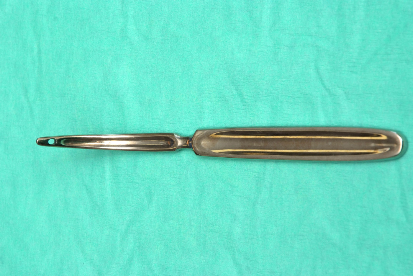
More than a century has passed since Emil Theodor Kocher (1841–1917), a Nobel laureate for his work on thyroid surgery, first described the use of ‘Kocher's thyroid dissector’ (Figure 1) to secure the superior thyroid pedicle.Reference Ellis2 Despite the technological advances in thyroid surgery, we still find the dissector to be an extremely useful instrument. We have adapted its originally described application. The current paper describes how this modified use of the dissector facilitates safer and easier surgery.
‘Kocher's thyroid dissector’.

Technique
Because of the extremely rich blood supply of the thyroid gland, thyroid surgery involves meticulous devascularisation of the gland. Planned ligation of the superior and inferior pedicles is required prior to excision of the thyroid lobe.
When dealing with the superior pedicle, the plane between the superior pole laterally and the underlying cricothyroid muscle medially is developed. The external branch of the superior laryngeal nerve is related to the structures of the upper pole of the thyroid gland. It passes through the potential avascular cricothyroid space (space of Reeve), and over the inferior constrictor muscle to reach and supply the cricothyroid muscle. Dissection in the right plane, followed by ligation of the individual branches of the artery on the superior pole of the thyroid gland, minimise the risk of damage to the nerve and of resultant voice change.
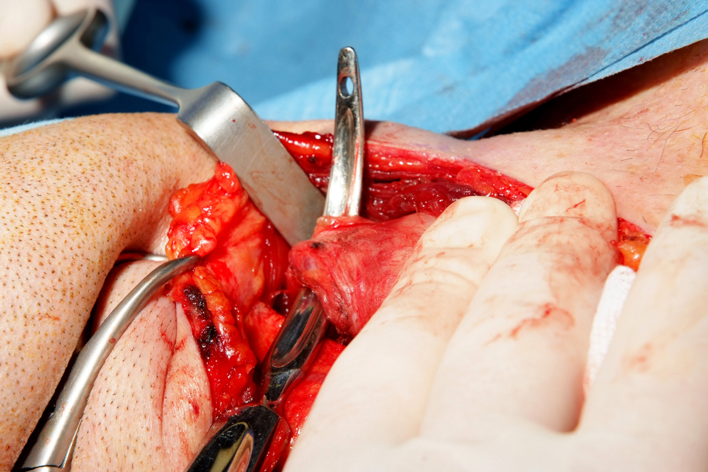
Once the plane has been identified, the tip of the Kocher's dissector is passed carefully under the superior pedicle, with the convex surface facing upwards, and used to elevate the superior pedicle (Figure 2).
Kocher's dissector elevating the superior pedicle.

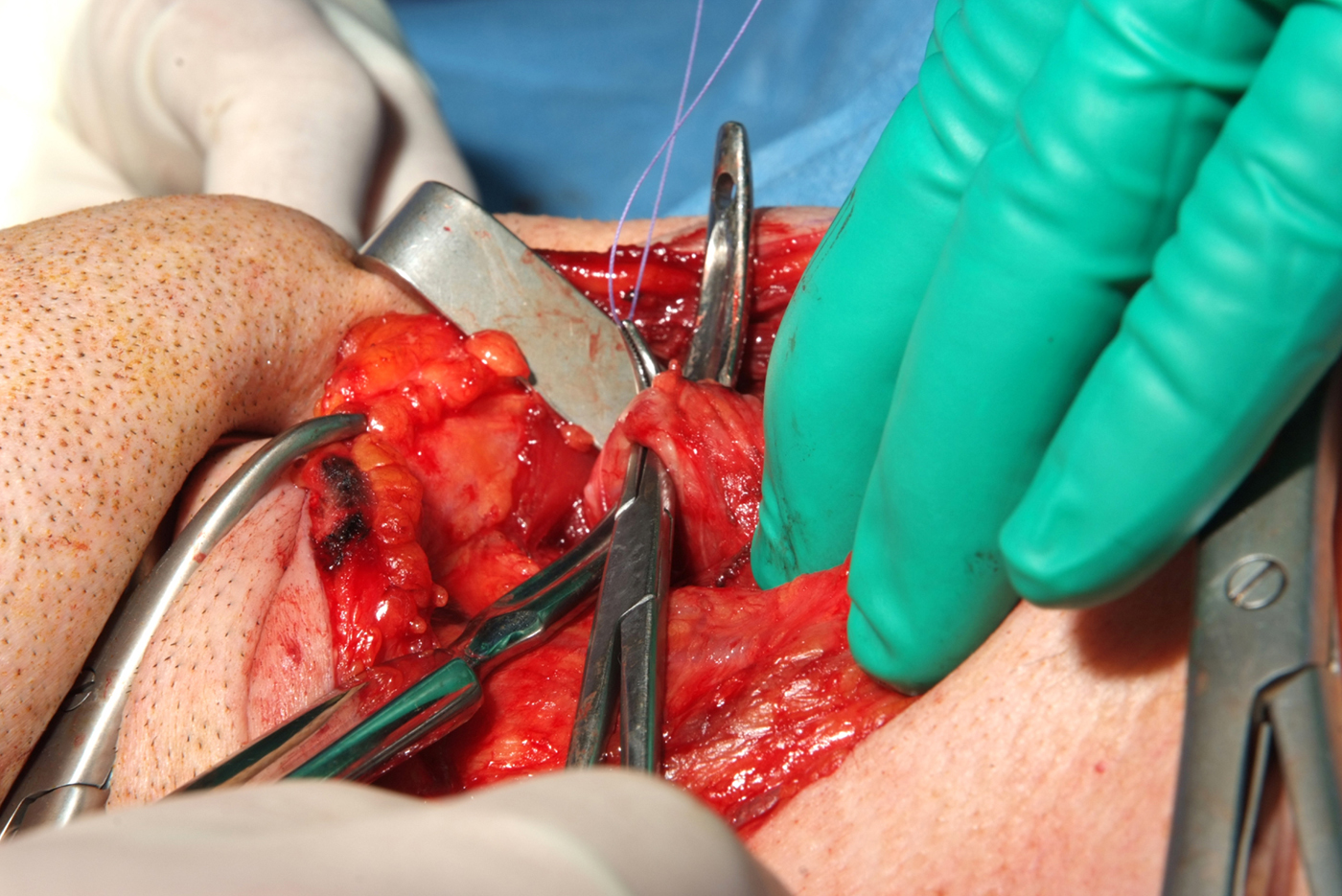
Individual branches of the superior thyroid vessels can then be opened to expose adequate length for ligation in continuity (Figure 3). Kocher originally described passing a ligature through the eye in the blade of the dissector and withdrawing the dissector, ensuring the suture is passed though the thyroid pedicle.
Baby Adson's forceps passing through the grooves on the dissector handle, allowing the application and tying of the ligature under direct vision.

We find that by keeping the dissector stationary, with the superior pedicle elevated, and passing Baby Adson's forceps through the grooves on the dissector handle, we can apply the ligature and tie it under direct vision. This is safer and easier than the conventional method of tying the thyroid pedicle. Three sutures are passed around the superior thyroid pedicle and tied. The superior pedicle is divided, keeping two ligatures towards the upper pole. This technique prevents repeatedly passing and withdrawing the dissector from under the superior thyroid pedicle for the three ligatures.
Conclusion
We feel that the application of this simple technique can improve the safety and efficiency of thyroid surgery, with negligible financial outlay.