Introduction
Aspergillosis involving the paranasal sinuses is a common and well-known occurrence. The maxillary sinus is the most commonly involved sinus, followed by the ethmoid and sphenoid sinuses. Frontal sinus involvement often occurs with the progression of aspergillosis in association with other sinuses. Primary frontal sinus aspergillosis is rare. There are only a few reports in the English literature, with most authors advocating an external approach for its management.Reference Stevens1–Reference Panda and Reddy8 The clinical presentation of such a lesion is usually non-specific and misleading.
We report our experience of patients who were successfully managed using endoscopic surgery. We believe that this is the first retrospective study of isolated frontal sinus aspergillosis cases in which a transnasal endoscopic surgical approach was used.
Materials and methods
This study comprised 16 cases of primary frontal sinus aspergillosis. The patients had presented to the out-patient services of the Department of Otolaryngology and Head and Neck Surgery, Post Graduate Institute of Medical Education and Research, Chandigarh, India, between January 1999 and July 2011. They had complaints of heaviness and intermittent pain in the frontal head region. All patients underwent a detailed general physical examination, a local examination and computed tomography (CT; coronal, axial and sagittal cuts). Other investigations included a fungal smear, fungal serology, and culture and histopathological examination.
The diagnosis of allergic fungal rhinosinusitis was made on the basis of criteria described by Bent and Kuhn.Reference Bent and Kuhn9 This included type 1 hypersensitivity to fungi, nasal polyposis, a characteristic radiographic finding of serpiginous areas of high attenuation, eosinophilic mucin and positive fungal stain. The histopathological criteria included the presence of allergic mucin, Charcot—Leyden crystals and no fungal hyphae within the soft tissues. In addition, radiology in the form of contrast enhanced CT and magnetic resonance imaging (MRI) were performed on patients with intracranial or intraorbital extension of disease. A fungal smear was taken for all patients, for which tissue was subjected to 10 per cent potassium hydroxide solution. This was followed by the microscopic examination of tissue for fungal hyphae. The fungal smear was positive in 12 patients (75 per cent).
All patients underwent transnasal endoscopic clearance of the disease under general anaesthesia. They were given antibiotics for one week and oral steroids, which were tapered post-operatively over the following three to six weeks. Systemic steroids were administered post-operatively on the assumption that pre-operative steroids may confuse the diagnosis of allergic fungal rhinosinusitis and lead to resolution of the typical allergic mucin required for a histopathologic diagnosis.
All patients were subjected to nasal endoscopic examination and saline nasal douching. Nasal suction was carried out after one week for four successive weeks, followed by weekly suction for four weeks, and one a month thereafter for six months. Topical nasal steroids were started in all patients one week after surgery and were continued for three months. Computed tomography was repeated after one month of surgery to inspect for residual disease. All patients received regular follow up.
The success of disease management was determined during the follow-up period on the basis of symptomatic improvement, and on post-operative radiological findings and nasal endoscopic scores. Clinical symptoms were graded according to four categories: 0 = no symptoms, 1–4 = few symptoms, 5–6 = moderate symptoms, and more than 7 = severe symptoms. Radiological outcomes were assessed on the basis of CT and MRI, and were scored using the Lund–Mackay staging system (0 point = no abnormality; 1 point = partial opacification; 2 points = total opacification).Reference Lund and Mackay10 Nasal endoscopic scores were as follows: 0 = no evidence of disease; 1 = mucosal oedema with or without allergic mucin; 2 = polypoidal mucosa with or without allergic mucin; and 3 = polyposis with allergic mucin.
Results
Ten (62.5 per cent) of the 16 patients were males and 6 (37.5 per cent) were females. The mean age of patients was 43.3 years, ranging from 7 years to 62 years.
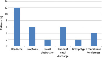
Symptomatically, 12 (75 per cent) patients suffered headache, out of which 4 (33.3 per cent) had left-sided frontal headache, 2 (16.6 per cent) had right-sided frontal headache and 6 (50 per cent) had vague symptoms of deep-seated headache (Figure 1). Six (37.5 per cent) of the patients had proptosis on the left side and two patients had presented with left nasal obstruction.
Clinical features of patients (n = 16) with isolated primary frontal sinus aspergillosis.

On clinical examination, six patients had purulent nasal discharge in the left nasal cavity and middle meatus, two had a small greyish polyp in the left middle meatus, four had unilateral frontal sinus tenderness, and two had bilateral frontal sinus tenderness.
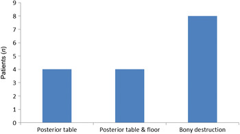
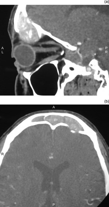
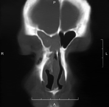
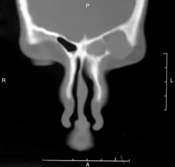
Computed tomography of the nose and paranasal sinuses showed bony destruction of the floor of the frontal sinus in eight cases (50 per cent). There was destruction of the posterior table of the frontal sinus in four (25 per cent) of the patients, and four (25 per cent) of the patients had posterior table and frontal sinus floor destruction (Figures 2–4). The remaining eight patients showed no destruction of the frontal sinus (Figure 5). Magnetic resonance imaging was conducted in eight patients with intracranial, intraorbital, or both intracranial and intraorbital extension of disease. The diagnosis of aspergillosis was based upon hypo-intense signals on T2-weighted MRI images.
Frontal sinus destruction in patients (n = 16) with isolated primary frontal sinus aspergillosis (determined radiologically).

Sagittal (a) and axial (b) computed tomography images of the nose and paranasal sinuses showing a heterogeneous soft tissue density in the left side of the frontal sinus, and erosion of the floor and posterior table. A = anterior, L = left, P = posterior, R = right

Coronal computed tomography image showing a mass with homogeneous density in the right frontal sinus. P = posterior, R = right, L = left, A = anterior

Coronal computed tomography image showing a mass with homogeneous density destroying the floor of the left frontal sinus. P = posterior, R = right, L = left, A = anterior

The intact bulla technique was used in 4 of the 16 cases, and endoscopic uncapping of the bulla was performed in 12 cases. One of the patients, who was seven years old, had intracranial extension of the disease through the posterior table of the frontal sinus. Following debridement of the affected area, the patient suffered cerebrospinal fluid rhinorrhoea which was repaired endonasally in the same sitting.
The fungal smear was positive for septate hyphae in 12 (75 per cent) of the patients. Fungal culture was positive for Aspergillus flavus in 14 (87.5 per cent) of the patients. Histopathology revealed allergic fungal rhinosinusitis in 12 (75 per cent) of the patients, and a ‘fungal ball’ was observed in 4 (25 per cent) patients.
The period of follow up ranged from 6 to 48 months (median follow up was 25.4 months). The disease recurred in 3 (18.75 per cent) of the patients following surgery; this was managed using the modified Lothrop procedure, and the patients had regular follow up for 28 months with no evidence of recurrent disease. The overall success rate of disease management was 82.25 per cent, which was determined on the basis of symptomatic improvement, radiological outcome and nasal endoscopic scores.
Discussion
Paranasal sinus aspergillosis is a potentially aggressive diseaseReference Rowe-Jones11 that can be invasive or non-invasive. Non-invasive infections include allergic sinusitis and sinonasal fungal balls. Bony erosion may occur due to decalcification, but with no histological evidence of invasion of tissue or bone.
Patients with the allergic variant of fungal rhinosinusitis usually display the following features: excessive sneezing, rhinorrhoea, raised immunoglobulin E levels, and Charcot—Leyden crystals and allergic mucin on histopathology. The diagnosis is based on histopathology, which in the allergic variant reveals a lack of fungal hyphae within the soft tissues, as well as the presence of allergic mucin and Charcot—Leyden crystals. In contrast, a fungal ball is usually found in immunocompetent patients. The diagnosis is based chiefly on histological examination as fungal cultures are frequently negative, which shows numerous entangled hyphae with 45-degree branching.
The maxillary sinus is the most commonly involved sinus in aspergillosis, followed by the anterior ethmoid, posterior ethmoid and sphenoid sinuses; however, isolated involvement of the frontal sinus is rare. Fungal sinusitis is endemic in Sudan, south-western states of the USA and northern parts of India.Reference Chakrabarti, Sharma and Chander12–Reference Washburn, Kennedy, Begley, Henderson and Bennett14 Prolonged exposure to contaminated vegetable detritus is regarded as a secondary risk factor and poses a high risk to populations living in southern parts of the world which have hot and humid climates.Reference Warder, Chikes and Hudson4–Reference Gupta, Udhayakumar, Rao, Madhaven and Das Gupta7 In its localised form, the disease originates in one of the sinuses and spreads to other sinuses through focal bony erosion. The frontal sinus ostium is located in the anterosuperior part of the nasal cavity and as such it is the least accessible site for the inhaled spores to enter and cause fungal sinusitis.Reference Swoboda and Ullrich3
Gupta et al. (1973) were the first to report primary frontal sinus aspergillosis,Reference Gupta, Udhayakumar, Rao, Madhaven and Das Gupta7 followed by others in the early 1980s.Reference Bhalla, Kumar, Pal, Malhotra and Dhingra5, Reference Sekula, Popielski and Betkowski15 In 2003, Gupta et al. investigated sinonasal aspergillosis in a series of children and reported only one case of isolated frontal sinus disease.Reference Gupta, Ghosh and Gupta16 In most of the published reports, the first clinical sign presents following involvement of the orbit or intracranial extension of the disease.Reference Bhalla, Kumar, Pal, Malhotra and Dhingra5, Reference Chakrabarti, Sharma and Chander12 The first case of primary frontal sinus aspergillosis was an incidental finding during roentgenography for epistaxis.Reference Stevens1
Varied sinus complaints and asymptomatic sinus opacity should raise the suspicion of paranasal sinus aspergillosis. Fungus involving the frontal sinus usually presents with vague symptoms such as unexplained epistaxis, a pressure sensation over the sinus,Reference Stevens1 frontal or periorbital pain,Reference Chen and Chen2 heaviness over the frontal and temporal region, and intermittent obstruction of the nasal cavity, as seen in our patients. In the present series, four patients had proptosis, three had a small greyish polyp in the middle meatus and the other nine patients had vague complaints of headache. Aspergillosis was diagnosed in the patients on the basis of computerised tomography of the nose and paranasal sinuses.
Computed tomography can raise a suspicion of paranasal sinus aspergillosis due to presence of heterogeneous expansile lesions with central hyperattenuation.Reference Manning, Merkel, Kriesel, Vuitch and Marple17 Bony destruction of the frontal sinus walls is commonly a result of pressure atrophy and dissolution by proteolytic enzymes and cytokines that are produced as part of the inflammatory process,Reference Corey, Delsuphe and Ferguson18 as seen in our cases. Computed tomography was an important tool in deciding the surgical approach for the frontal sinus. In our series, the CT findings were typical of fungal sinusitis. All patients except two had heterogeneous opacity with bony destruction. There was a statistically significant correlation between radiology and the surgical findings (r = 1).
There are characteristic MRI features of aspergillosis of sinonasal origin too. The aspergillus mass can have iso- to hypo-intense signals on T1-weighted images and extremely hypo-intense signals on T2-weighted images. There can be a bright homogeneous contrast enhancement of the mass, both in the sinus and its extension into the adjacent cranial and orbital cavities. The MRI hallmark of an aspergillosis diagnosis is the extremely hypo-intense signals on T2-weighted images.
The goal of treatment for primary frontal sinus aspergillosis is debridement and restoration of aeration.Reference Stevens1, Reference Warder, Chikes and Hudson4, Reference Stammberger, Jakse and Beaufort19 Almost all previous studies advocated the use of external approaches to remove the disease, including external fronto-ethmoidectomy, osteoplastic flapReference Panda and Reddy8 and trephination of the frontal sinus.Reference Swoboda and Ullrich3 In this study, all patients underwent endonasal frontal sinusotomy. Only one previous study (by Kodama et al.) advocated the role of the modified Lothrop procedure for frontal sinus mucoele with contralateral frontal sinus aspergillosis.Reference Kodama, Moriyama, Okamoto, Hirano and Suzuki20 We utilised the modified Lothrop procedure in three patients who presented with recurrence of symptoms. The involvement of the most lateral parts of the frontal sinus, or destruction of the posterior table of the frontal sinus (from where the disease can spread intracranially), were thought to be limitations for an endoscopic surgical approach. The surgical approach to treat disease in the frontal sinus with intracranial extension is difficult as it necessitates surgical clearance of the disease from the skull base and frontal sinus. With the availability of angled endoscopes and expertise in endoscopic surgery it has also become possible to approach these areas endonasally.
Aspergillus flavus was the causative organism in most of our cases, as described in earlier series.Reference Panda and Reddy8, Reference Chakrabarti, Sharma and Chander12, Reference Gupta, Ghosh and Gupta16 Rowe-Jones advocated the role of itraconazole post-operatively.Reference Rowe-Jones11 We did not give any antifungal drugs to our patients as histopathology revealed no tissue invasion.
We opted for short-term systemic steroids, as post-operative nasal endoscopy showed normal sinus mucosa in most of the cases. Systemic steroids are known to reduce the number of circulating eosinophils, inhibit the migration of eosinophils to tissue sites of inflammation, decrease eosinophil survival time and suppress the production of eosinophil-specific cytokines. In addition, steroids inhibit the production of arachidonic acid metabolites by interfering with phospholipase A2 enzymatic activity. The steroids thereby reduce chemotaxis, mucous secretion, vascular permeability and cell proliferation.
The success rate for patients with isolated frontal sinus aspergillosis, with follow up ranging from 6 to 48 months, was 82.25 per cent. This was determined on the basis of symptomatic improvement, radiological outcome and nasal endoscopic scores. There were no major complications during or after surgery. Three patients (18.75 per cent) had a recurrence of the disease and underwent revision surgery using the modified Lothrop procedure for clearance of the disease; these patients received subsequent follow up for a minimum of 26 months. Because the symptoms of isolated frontal sinus aspergillosis are non-specific, clinicians may occasionally miss the diagnosis, which is likely to result in complications.
We had difficulties in removing the disease from the most lateral region of the frontal sinus; angled endoscopes and other instruments have made it possible to reach these areas. The chance of residual disease is greater in cases where there is extensive intracranial disease to be removed through a small defect in the posterior table of the frontal sinus.
Patients who experience recurrence of disease in the frontal sinus should be considered for the endoscopic modified Lothrop procedure to optimise disease clearance. In our study, the disease recurred in 18.75 per cent of patients; all other patients were successfully managed with endoscopic frontal sinusotomy.
A pre-operative radiological evaluation helps to reduce the chance of residual disease. Frontal Kuhn's cells, pneumatisation of the most lateral part of the frontal sinus, and posterior table defects with extensive intracranial extension predispose the patient to residual disease. We found the indication for the endoscopic modified Lothrop procedure was limited to patients with recurrence of the disease. These observations are contradictory to published reports advocating the modified Lothrop procedure for primary frontal sinus diseases.
• This retrospective study examined 16 patients (10 males) with rare primary frontal sinus aspergillosis
• Diagnosis was based on clinical presentation, radiology and histopathology
• Complaints were vague, including frontal headache in 12 patients
• Disease was removed by endoscopic frontal sinusotomy, with a success rate of 82.25 per cent
• Disease recurred in three patients, which was dealt with using a modified Lothrop procedure
In summary, isolated frontal sinus aspergillosis should be considered in patients who present with non-specific symptoms. Minimally invasive procedures such as endoscopic frontal sinusotomy can provide an excellent cure rate and obviate the need for extensive external approaches, as is evident from the current series success rate of 82.25 per cent. This is the largest retrospective study on isolated primary frontal sinus aspergillosis managed with frontal sinusotomy using an endonasal endoscopic approach. This approach provided comparable post-operative results to the external approaches advocated in previous literature.
Conclusion
Minimally invasive endonasal frontal sinusotomy was found to be a safe and effective approach for managing primary frontal sinus aspergilloma, even in cases with more lateral and larger bony defects involving the posterior table of the frontal sinus.