Introduction
Canfieldite is a mineral species first discovered at Potosí, Bolivia in 1893 by S. L. Penfield and named in honour of F. A. Canfield, a mining engineer and mineral collector from Dover, New Jersey, USA (Penfield, Reference Penfield1893). Since its discovery and description, canfieldite has been widely reported as an important resource of silver in various metallic ore deposits (e.g. Harris and Owens, Reference Harris and Owens1971; Nekrasova and Borodaev, Reference Nekrasova and Borodaev1972; Kvaček et al., Reference Kvaček, Novák and Drábek1975; Soeda et al., Reference Soeda, Watanabe, Hoshino and Nakashima1984; Lu et al., Reference Lu, Mao, Gao, Su and Zheng2012). It also attracted attention from chemical and material scientists as a potential superionic and semiconducting material (e.g. Evain et al., Reference Evain, Gaudin, Boucher, Petříček and Taulelle1998; Slade et al., Reference Slade, Gvozdetskyi, Wilde, Kreyssig, Gati, Wang, Mudryk, Ribeiro, Pecharsky, Zaikina, Bud’ko and Canfield2021; Pogodin et al., Reference Pogodin, Pop, Shender, Studenyak, Filep, Malakhovska, Kokhan, Babuka, Stercho, Rubish and Kopčanský2022). Despite the long history and industrial importance, the crystal structure of natural canfieldite remains undetermined to date. Based on powder X-ray diffraction data of synthetic Ag8SnS6, Wang (Reference Wang1978) provided a structure model according to that of synthetic Ag8GeS6 by Eulenberger (Reference Eulenberger1977), indicating it to be orthorhombic with space group Pna21. Slade et al. (Reference Slade, Gvozdetskyi, Wilde, Kreyssig, Gati, Wang, Mudryk, Ribeiro, Pecharsky, Zaikina, Bud’ko and Canfield2021) confirmed the orthorhombic Pna21 structure of synthetic canfieldite below 460 K and found a phase transition to a Pmn21 structure at 120 K. On the other hand, Bindi et al. (Reference Bindi, Nestola, Guastoni, Zorzi, Peruzzo and Raber2012) and Zhai et al. (Reference Zhai, Bindi, Voudris, Liu, Tombros and Li2019) respectively reported the crystal structures of Te-rich canfieldite and Se-rich canfieldite, showing them to be cubic with a space group F  $\bar 4$3m. These works raise critical questions: Is the crystal structure of natural canfieldite (and by extension, argyrodite) orthorhombic or cubic? and how are they compared to their synthetic counterparts? This paper seeks to explore and clarify these questions.
Samples and experiments
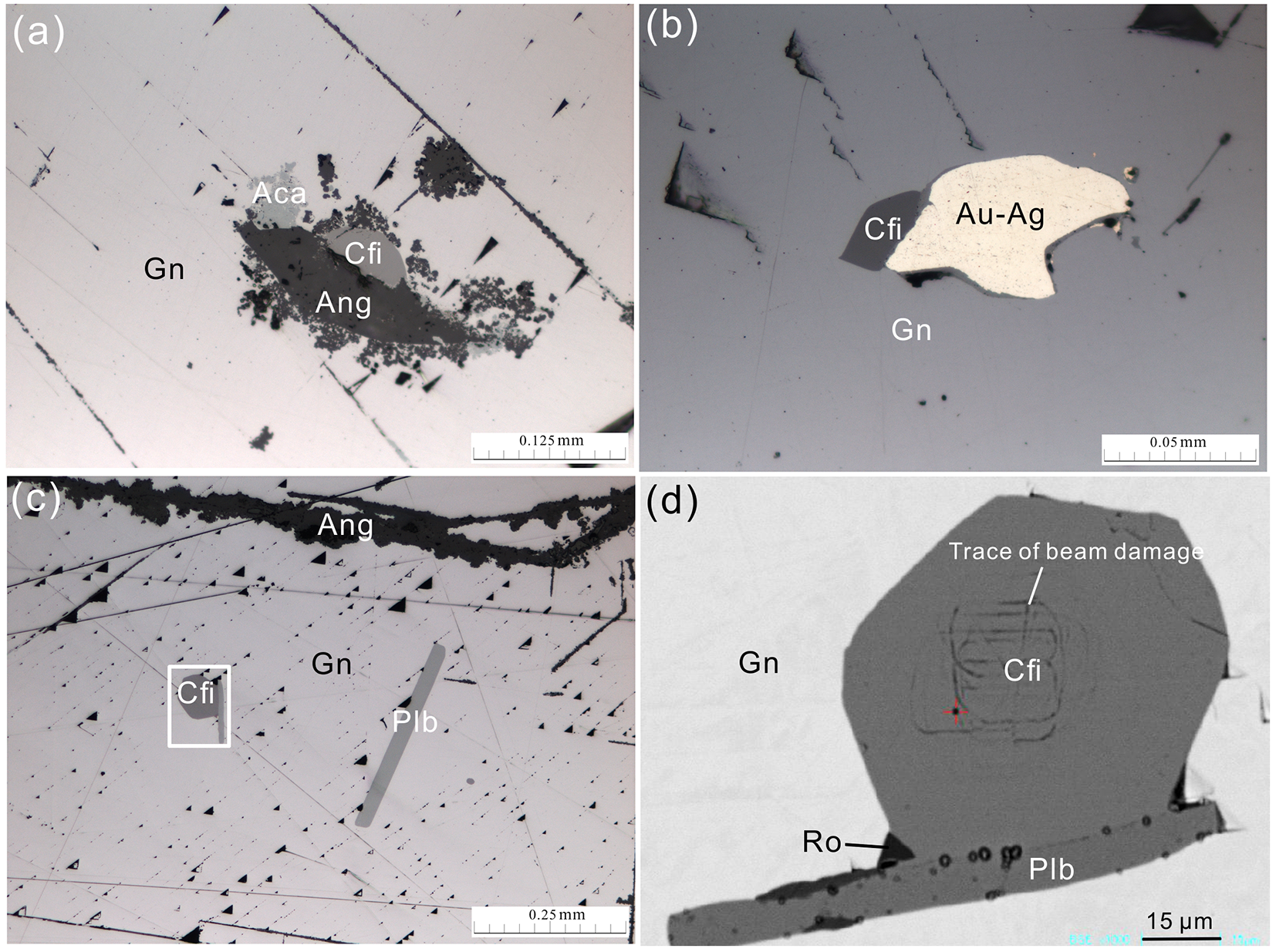
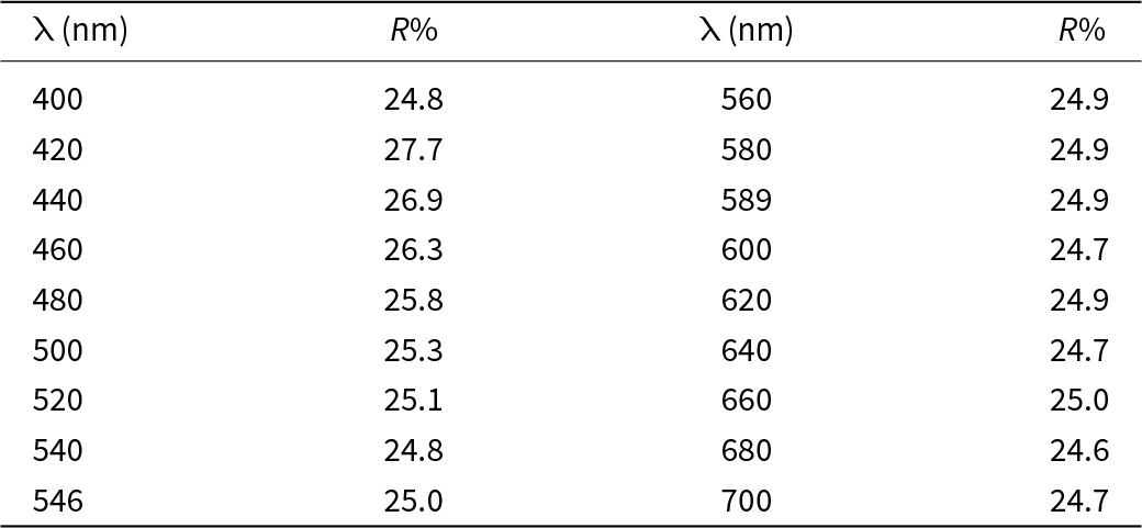
The samples for this study were obtained from the Youqialang lead-silver mine in Nagqu, Tibet, China, which is a magmatic-related mesothermal deposit in a NE-extending alteration rupture zone hosted by Jurassic slate and quartzite, with a Pb reserve of ∼90,000 tons (average Pb grade 18.47%) and Ag reserve of ∼300 tons (average Ag grade 434 g/t) (Feng et al., Reference Feng, Lü and Wang2011). The ores are classified into Pb–Ag, Zn–Pb and CuBi types. Canfieldite occurs with brecciated Pb–Ag ores in which the crystals of galena are strongly deformed in preferred orientation. In reflected light microscopy, canfieldite occurs as subhedral to euhedral grains, 30 to 60 μm in size, embedded in galena. It is associated with a suite of minerals including polybasite, acanthite, electrum and roquesite, with occasional replacement by anglesite (Fig. 1). It displays an isotropic nature with a pale grey colour. The reflectance data, measured in air with a Tidas MSP400 microphotometer using SiC as a standard, are shown in Table 1.
Optical microscope images (a–c) and back-scattered electron image (d) of canfieldite and associated minerals. Key: Aca = acanthite, Au-Ag = electrum, Gn = galena, Plb = polybasite, Ro = roquesite. In image (d), the dark lines within canfieldite are due to electron beam damage during analysis.

Reflectance data for canfieldite*

* The colour index in ICE C illumination is: Y = 25.1%; λd = 466 nm; Pe = 2.88%.
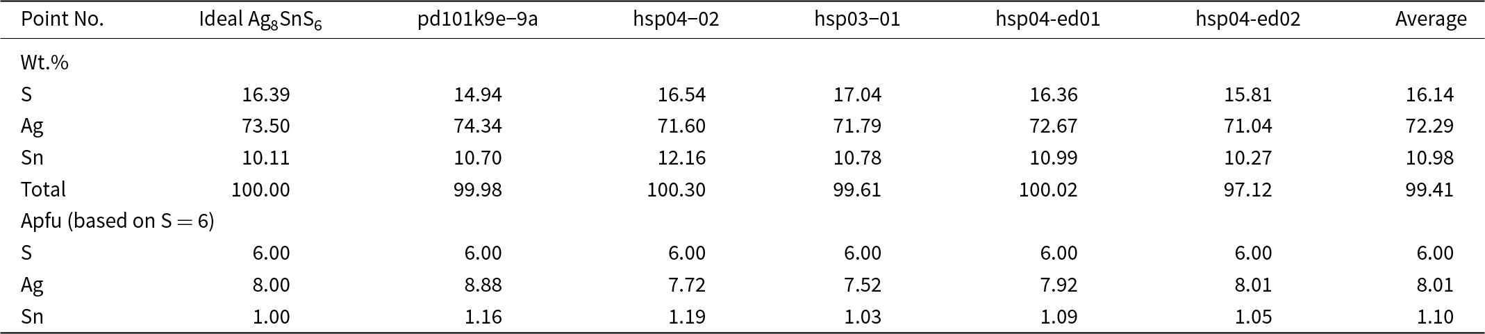
The Shimadzu EPMA-1720 electron probe microanalyser (15 kV, 10 nA and beam size 1 μm) was used for qualitative and quantitative analysis of canfieldite. In qualitative analysis, only S, Ag and Sn were detected and Te, Se, Cu, Pb, Bi and As were below the detection limits (0.01%). Pure metal Ag, SnS and FeS2 were used as standards for WDX quantitative analysis using the ZAF4 correction program provided with the machine. However, the quantitative results were strongly affected by substantial electron beam damage (as shown in Fig. 1d), leading to a considerable variation of the composition, with the average empirical formula being Ag8.01Sn1.10S6 from 5 analyses based on S = 6 as shown in Table 2.
Microprobe analyses (wt.%) for canfieldite

Notes: Apfu – atoms per formula unit
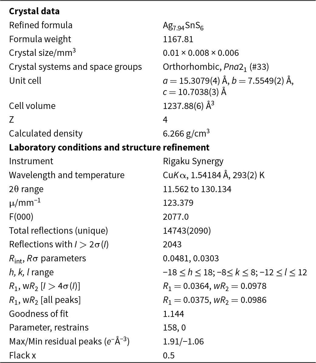
The crystal of canfieldite (0.01 × 0.008 × 0.006 mm) in Fig. 1d was extracted for single-crystal X-ray diffraction study using a Rigaku Synergy-DS diffractometer (CuKα, 50 kV, 1 mA, 0.2 mm focal spot, 0.5º slit width, frame exposure time 12–39 s and total 38 h). Diffraction data were collected in a hemisphere range, data reduction and absorption correction were performed using the instrument’s proprietary software CrysAlisPro. The diffraction data indicated the sample to be orthorhombic and suggested possible space groups Pna21 or Pnma. The structure was solved by ShelXT with the space group Pna21 as the only solution and refined using the ShelXL module embedded in Olex2 software (Dolomanov et al., Reference Dolomanov, Bourhis, Gildea, Howard and Puschmann2009; Sheldrick, Reference Sheldrick2015a, Reference Sheldrick2015b).
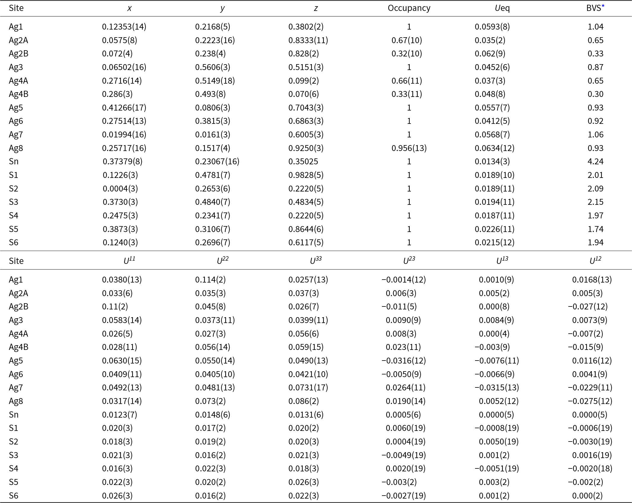
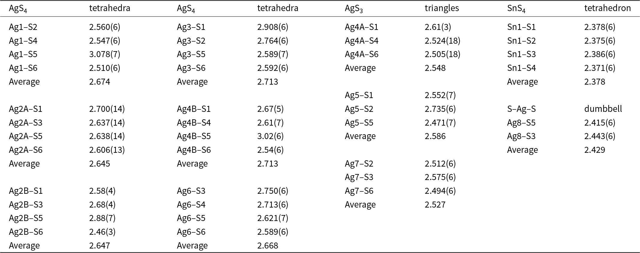
The initial structure conforms to that of Eulenberger (Reference Eulenberger1977) for synthetic Ag8GeS6 and Slade et al. (Reference Slade, Gvozdetskyi, Wilde, Kreyssig, Gati, Wang, Mudryk, Ribeiro, Pecharsky, Zaikina, Bud’ko and Canfield2021) for synthetic Ag8SnS6 in space group Pna21, which has one site for Sn atoms, six sites for S atoms and eight sites for Ag atoms, corresponding to the ideal formula Ag8SnS6. The initial structure was refined to R 1 = 0.0418 for all the 2090 unique reflections with significant residual electron densities near some Ag sites (up to 2.15 e –/Å3). The subsequent free refinement for the occupancies of Ag atoms indicated that Ag occupancies of the eight sites vary from 0.89 (Ag8) to 0.99 (Ag1), yielding an empirical formula Ag7.53SnS6. Consequently, Ag atoms were assigned to the two main residual peaks near Ag2 and Ag4, and free refinement was made for the sites with low occupancies (Ag2, Ag4 and Ag8). The structure was then refined as a 2-component perfect inversion twin with a twin law of (–1 0 0, 0 –1 0, 0 0 –1). This led to decreased R 1 = 0.0375 and a refined empirical formula Ag7.94SnS6 and site splitting of Ag2 and Ag4 sites (labelled as Ag2A and Ag2B, Ag4A and Ag4B) with splitting distances ranging from 0.26 to 0.42 Å. The final information on the crystal and structural refinement is summarised in Table 3, and the atomic coordinates and displacement parameters are given in Table 4. Selected bond distances are presented in Table 5. The structure file (canf.cif) and the checkcif file from International Union of Crystallography (IUCr) are available as supplementary material (see below).
Summary of crystal data and structure refinement for canfieldite

R int = Σ|F o2–F o2(mean)|/Σ[F o2], GooF = S = {Σ[w(F o2–F c2)2]/(n–p)}1/2. R 1 = Σ||F o|–|F c||/Σ|F o|, wR 2 = {Σ[w(F o2–F c2)2]/Σ[w(F o2)2]}1/2; w = 1/[σ2(F o2) + (aP)2 + bP] where a is 0.049 and b is 25.3, and P is [2F c2 + Max(F o2,0)]/3.
Fractional atomic coordinates, site occupancies, anisotropic displacement parameters (in Å2) and bond valence sums (BVS, in valence units) for canfieldite

* Bond valence sums are calculated using the ECoN21 program (Ilinca, Reference Ilinca2022).
Selected bond distances (d, Å) for canfieldite

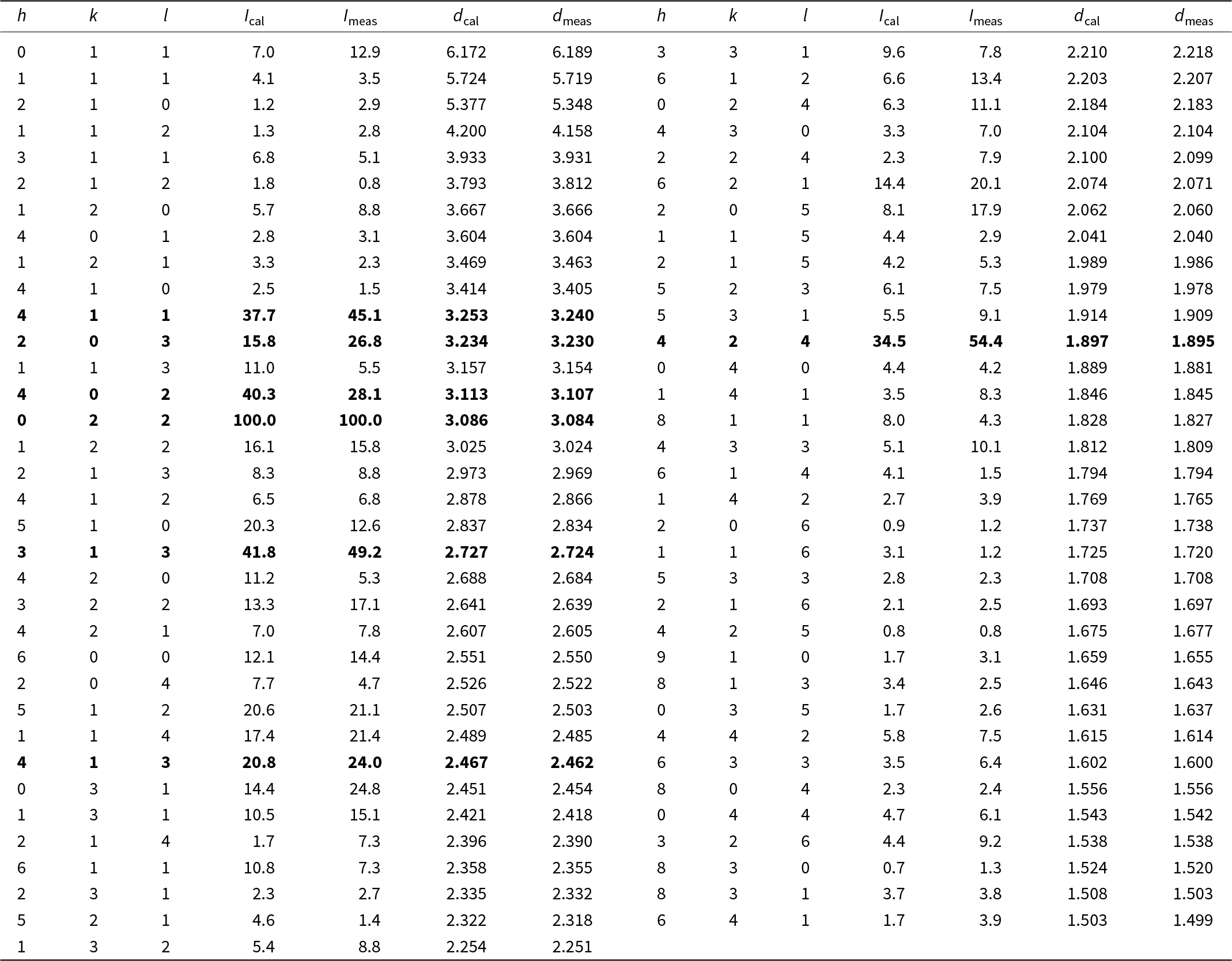
Powder X-ray diffraction for polycrystal grains of canfieldite was carried out in situ on the polished section using a Rigaku D/max Rapid IIR microdiffractometer (CuKα; 40 kV, 250 mA, 0.03 mm collimator and exposure time 6 h). The peak data are listed in Table 6, and the unit-cell parameters were refined according to the orthorhombic Pna21 structure using the program of Holland and Redfern (Reference Holland and Redfern1997) as: a = 15.2808(5) Å, b = 7.5464(2) Å, c = 10.7045(3) Å, V = 1234.40(5) Å3 and Z = 4.
Powder X-ray diffraction data (d in Å, I in %) for canfieldite

Note: Bold data are the 7 strongest lines.
Results
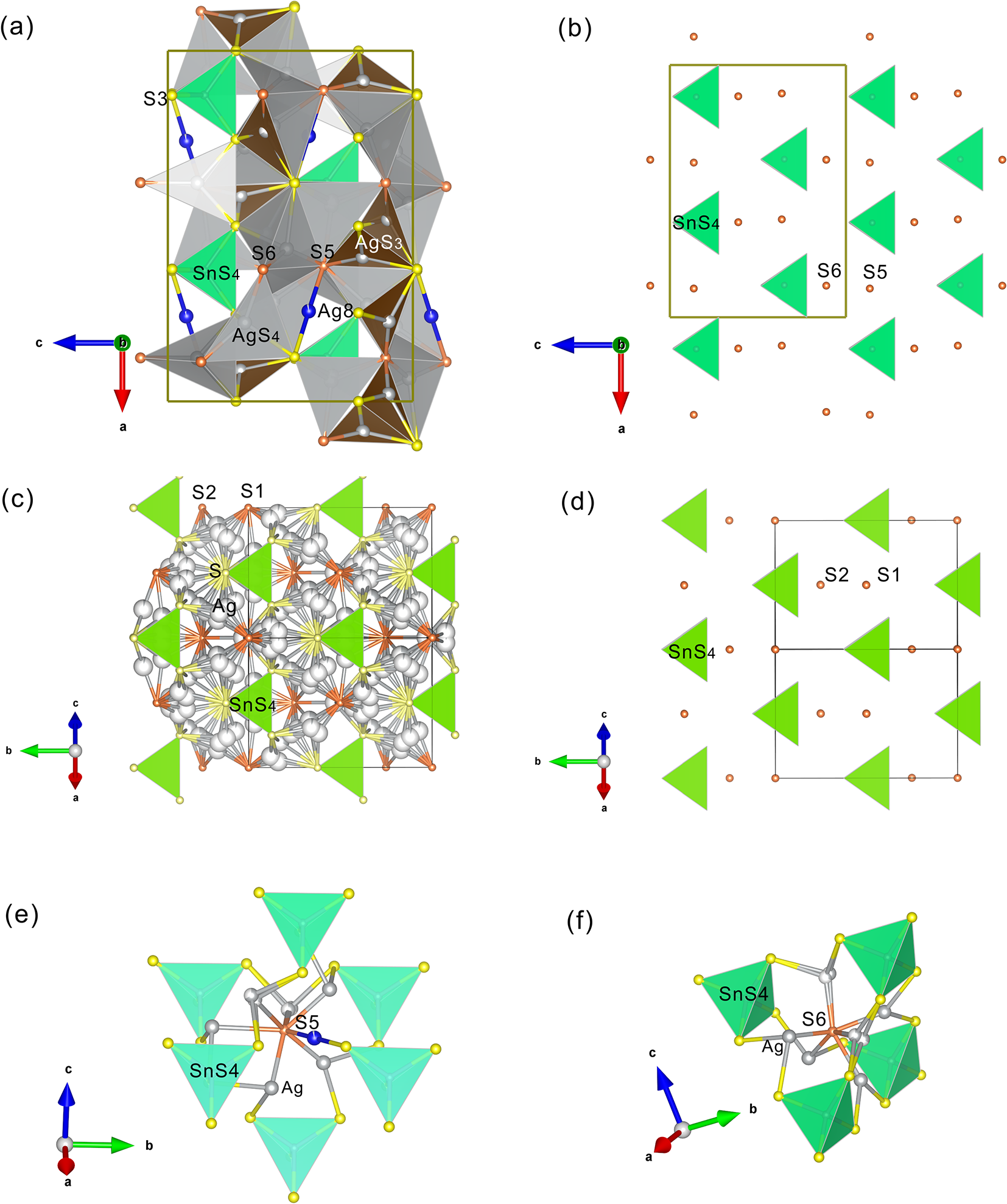
This study shows that natural canfieldite has an orthorhombic Pna21 structure, comparable to synthetic Ag8GeS6 (Eulenberger, Reference Eulenberger1977) and synthetic Ag8SnS6 (Slade et al., Reference Slade, Gvozdetskyi, Wilde, Kreyssig, Gati, Wang, Mudryk, Ribeiro, Pecharsky, Zaikina, Bud’ko and Canfield2021). It is composed of isolated SnS4 distorted tetrahedra (average Sn–S bond length 2.378 Å) arranged in three-dimensional space following the orthorhombic symmetry, in which the distances between SnS4 tetrahedra (represented by Sn–Sn length) range from 7.464 Å to 7.750 Å (the distance along the b-axis is equal to b = 7.5549 Å). The two sulfur sites S5 and S6 outside the SnS4 tetrahedron are located respectively in the centre of 6 SnS4 tetrahedra and 4 SnS4 tetrahedra (Fig. 2e, f). The S5 atom is bonded to 7 Ag atoms with an average bonding length 2.714 Å, while the S6 atom is bonded to 6 Ag atoms with an average bonding length 2.537 Å. The Ag atoms are located in the space between SnS4 tetrahedra and S5, S6 in tetrahedral coordination (Ag1, Ag2A, Ag2B, Ag3, Ag4B and Ag6), triangular coordination (Ag4A, Ag5 and Ag7) and linear coordination (Ag8), with the average Ag–S distance being 2.682 Å for tetrahedral coordination, 2.554 Å for triangular coordination and 2.429 Å for linear coordination (Table 4). The AgS4 tetrahedra, AgS3 triangles and linear AgS2 dumbbells share corners and/or edges to form a framework which are connected to SnS4 tetrahedra through sharing S corners (Fig. 2a).
The crystal structure of canfieldite from Youqialang viewed along the b-axis (a, b), compared with the crystal structure of synthetic cubic Ag8SnS6 by Gorochov (Reference Gorochov1968) viewed along [1 0 1] (c, d), plotted with Vesta 3 (Momma and Izumi, Reference Momma and Izumi2011). (a) The connection among the AgS4 tetrahedra (grey), AgS3 triangles (orange), SnS4 tetrahedra (green) and linear Ag8. The S atoms of SnS4 tetrahedra are yellow-coloured and the S atoms (S5, S6) outside SnS4 tetrahedra are pink-coloured. (b) The ordered arrangement of SnS4 tetrahedra and S5 and S6. (c) Disordered Ag atoms (grey-coloured) connected to S atoms. The S atoms outside SnS4 tetrahedra are pink-coloured and labelled as S1 and S2. (d) Similar arrangement of SnS4 tetrahedra and S1 and S2 to (b). (e) External S (S5) in the centre of 6 SnS4 tetrahedra in canfieldite. (f) External S (S6) in the centre of 4 SnS4 tetrahedra in canfieldite.

In comparison with the structures of the synthetic orthorhombic Pna21 Ag8SnS6 and Ag8GeS6 (Eulenberger, Reference Eulenberger1977; Wang, Reference Wang1978; Slade et al., Reference Slade, Gvozdetskyi, Wilde, Kreyssig, Gati, Wang, Mudryk, Ribeiro, Pecharsky, Zaikina, Bud’ko and Canfield2021), site splitting of Ag atoms (Ag2, Ag4) in natural canfieldite is present with the split distances ranging from 0.26 Å (Ag2A–Ag2B) to 0.42 Å (Ag4A–Ag4B). It is also noted that the linear Ag8 site is partially occupied (0.95) in natural canfieldite, leading to the Ag atoms per formula units of less than 8 in the refined formula Ag7.94SnS6.
Discussion
There exists a unit-cell transformation between the orthorhombic Pna21 structure and the cubic F  $\bar 4$3m structure, i.e. a or =
 $\sqrt 2 $a c, b or = a c/
 $\sqrt 2 $ and c or = a c. The orthorhombic Pna21 structure of natural canfieldite has comparable topology in the arrangement of SnS4 tetrahedra and the external S atoms with the high temperature cubic F
 $\bar 4$3m structure (a = 10.85 Å) of synthetic Ag8SnS6 of Gorochov (Reference Gorochov1968) and Slade et al. (Reference Slade, Gvozdetskyi, Wilde, Kreyssig, Gati, Wang, Mudryk, Ribeiro, Pecharsky, Zaikina, Bud’ko and Canfield2021) (Fig. 2b, d), which transformed reversibly to orthorhombic Pna21 Ag8SnS6 at 445–460 K. However, they are significantly different in the locations of Ag atoms. All the Ag atoms are partially occupied and clustered around S atoms in the cubic F
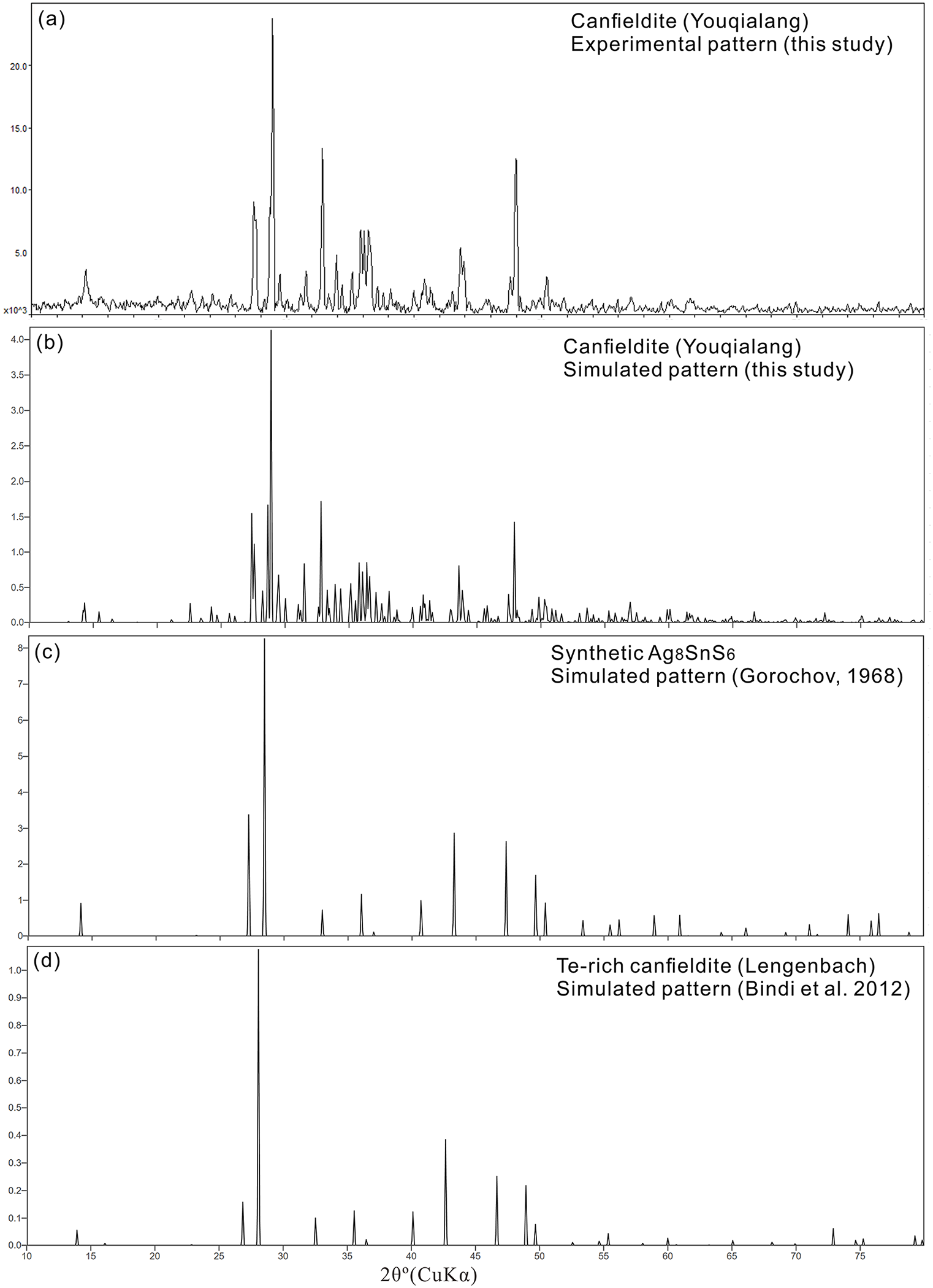
 $\bar 4$3m structure (Fig. 2c) in contrast to the ordered Ag atoms around S atoms in the orthorhombic Pna21 structure (Fig. 2a). Likewise, natural canfieldite is different from the cubic Te-rich canfieldite and Se-rich canfieldite by Bindi et al. (Reference Bindi, Nestola, Guastoni, Zorzi, Peruzzo and Raber2012) and Zhai et al. (Reference Zhai, Bindi, Voudris, Liu, Tombros and Li2019). They can be also easily distinguished by powder X-ray diffraction data (Table 6, Fig. 3). It is likely that the difference of space group between the orthorhombic canfieldite and the cubic Te-rich and Se-rich canfieldite is related to the difference in the formation temperature.
Comparisons of powder X-ray diffraction patterns of canfieldite: (a) and (b) from this study; (c) from Gorochov (Reference Gorochov1968); and (d) from Bindi et al. (Reference Bindi, Nestola, Guastoni, Zorzi, Peruzzo and Raber2012).

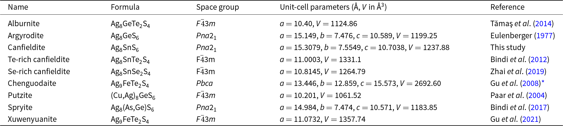
The argyrodite–canfieldite group includes argyrodite (Ag8GeS6), canfieldite (Ag8SnS6), putzite (Cu, Ag)8GeS6), alburnite (Ag8GeTe2S4), spryite [Ag8(As,Ge)S6], chenguodaite (Ag9FeTe2S4), and xuwenyuanite (Ag9FeTe2S4), as listed in Table 7. Similar compositions and structures of synthetic phases include Ag8GeTe6 (Rysanek et al., Reference Rysanek, Laruelle and Katty1976), Ag7PSe6 (Evain et al., Reference Evain, Gaudin, Boucher, Petříček and Taulelle1998), and Cu8SiS6 (Ishii et al., Reference Ishii, Onoda and Shibata1999). Within this group of minerals, isomorphic substitutions at the cationic positions Ge–Sn–As–Fe, Ag–Cu, and anionic positions S–Se–Te form independently named end-members. Apart from the orthorhombic Pna21 and cubic F  $\bar 4$3m structures, synthetic phases also possess structures with space groups P213 (Ag8GeS6, Ag8SnS6) (Gorochov, Reference Gorochov1968), R3m (Ag8GeTe6) (Von Unterrichter and Range, Reference Von Unterrichter and Range1978), and Pmn2₁ (Ag₈SnSe₆, Cu8GeS6) (Semkiv et al., Reference Semkiv, Ilchuk, Pawlowski and Kusnezh2017; Abbasova et al., Reference Abbasova, Alverdiyev, Rahimoglu, Mirzoyeva and Babanly2017; Slade et al., Reference Slade, Gvozdetskyi, Wilde, Kreyssig, Gati, Wang, Mudryk, Ribeiro, Pecharsky, Zaikina, Bud’ko and Canfield2021). Notably, the powder X-ray diffraction data of chenguodaite from Gu et al. (Reference Gu, Watanabe, Hoshino and Shibata2003, Reference Gu, Watanabe, Xie, Peng, Nakamuta, Ohkawa, Hoshino, Ohsumi and Shibata2008) could not be explained by any of the above structures and may possess a new structure, and the structural relationship between chenguodaite and xuwenyuanite is not analogous to that between orthorhombic Pna21 and cubic F
 $\bar 4$3m structures, for which the investigation is ongoing. On the other hand, significant substitutions of Te and Se for S seems to be related to high temperature as all of the minerals with (Te,Se)/S = 1/2 in this group have the cubic F
 $\bar 4$3m structure except for chenguodaite.
Crystal chemical data for the minerals in the argyrodite–canfieldite group

* New result from uncompleted study of the author (X. Gu).
Conclusions
This paper presents the first precise determination of the crystal structure of natural canfieldite sampled from the Youqialang lead-silver mine in Nagqu, Tibet, China. It has the orthorhombic Pna21 structure, composed of isolated SnS4 tetrahedra and ordered external S atoms arranged in three-dimensions, with the Ag atoms located in the space between SnS4 tetrahedra and external S atoms in tetrahedral, triangular and linear coordinations. The AgS4 tetrahedra, AgS3 triangles and linear AgS2 share corners and/or edges to form a framework which is corner-connected to SnS4 tetrahedra. Site splitting is present for some Ag atoms with the split distances from 0.26 Å to 0.42 Å. The orthorhombic Pna21 structure of natural canfieldite has similar arrangement of SnS4 tetrahedra and the external S atoms to the high temperature cubic F  $\bar 4$3m structure, but they are significantly different in the locations and Ag–S bonding styles of Ag atoms with all the Ag atoms being disordered in the latter.
Supplementary material
The supplementary material for this article can be found at https://doi.org/10.1180/mgm.2025.17.
Acknowledgements
The manuscript was improved from the comments of Prof. Peter Leverett and an anonymous reviewer.
Funding statement
This work was supported by the National Natural Science Foundation of China (Grant No. 42072054, 41172042 to X.G.).
Competing interests
The authors declare none.