Implications
Several external markers that can be recovered in faeces are used in ruminant studies to estimate dry matter intake (DMI). However, analytical procedures are tedious and time-consuming. Polyethylene glycol (PEG) has been successfully tested as a faecal marker determined with the near-infrared reflectance spectroscopy (NIRS) technique. The present study has confirmed that PEG is a valuable faecal marker to estimate DMI in sheep with good accuracy when measured with the NIRS method. Because it is easily administered and quickly measured with NIRS, PEG can be used to satisfactorily estimate DMI and allows to work on a greater number of animals than other markers.
Introduction
In ruminants studies based on individual dry matter intake (DMI) assessment, DMI is estimated from total faeces output and DM digestibility (DMD) assessments. The DMD can be estimated using in vitro techniques and total faecal output by determining the dilution rate of external markers (Prigge et al., Reference Prigge, Varga, Vicini and Reid1981).
Polyethylene glycol (PEG) with high molecular weight (>3000) is generally used as a soluble marker to measure rumen fluid volume and output flow rate from the rumen (Bauman et al., Reference Bauman, Davis, Frobish and Sachan1971). It has also been used as an external faecal marker to estimate total faecal output (Corbett et al., Reference Corbett, Greenhalgh, Gwynn and Walker1958; Hopson and McCroskey, Reference Hopson and McCroskey1972). However, when PEG is measured with colorimetric or turbidimetric methods, its recovery in faeces has been found to be variable and low (Corbett et al., Reference Corbett, Greenhalgh, Gwynn and Walker1958). These results have been attributed to diet composition (Clark et al., Reference Clark, Hembry, Thompson and Preston1972), feed digestibility or methodological difficulties (Alexander et al., Reference Alexander, Meyer and Bartley1969).
More recently, Landau et al. (Reference Landau, Friedman, Devash and Mabjeesh2002) demonstrated that PEG could be determined in goat faeces with the near-infrared reflectance spectroscopy (NIRS) method with good accuracy. Furthermore, Jones and Palmer (Reference Jones and Palmer2000) showed that PEG addition does not modify in vitro DMD. However, they pointed out that PEG binds with condensed tannins (CTs) and may indirectly increase CP digestibility. Compared with other analytical methods, NIRS method only requires the samples to be dried and ground, before scanned, and consequently a high number of samples (a hundred) can be determined daily. The use of PEG in combination with NIRS, allow the use of 10 animals in trials estimating DMI for periods of adequate length, which is difficult (costly and time-consuming analytical methods) to apply with PEG when measured by colorimetric or turbidimetric methods or by other external markers (Corbett et al., Reference Corbett, Greenhalgh, Gwynn and Walker1958; Hopson and McCroskey, Reference Hopson and McCroskey1972; Prigge et al., Reference Prigge, Varga, Vicini and Reid1981).
Considering the potential interest of PEG as a faecal marker and its rapid determination by NIRS, Hassoun et al. (Reference Hassoun, Fabre, Bastianelli, Bonnal and Bocquier2007a) set up a 1000-sample database with wide ranges of PEG content in faeces. In an in vivo experiment with sheep (Hassoun et al., Reference Hassoun, Bastianelli and Bonnal2007b), various PEG doses were tested. Regardless of PEG level of administration, the recovery rate was higher than 98%.
With external markers used to estimate total faeces excretion, two sources of errors have to be considered. The first one deals with total recovery of the marker in faeces, meaning that few or no administered marker disappeared until reaching rectal excretion. The second one deals with the daily excretion pattern of the marker that is linked to the grab sampling approach (i.e. faecal samples taken directly from rectum) to estimate total faeces output. Consequently, the marker concentration measured in grab samples must be as close as possible to the marker mean concentration in total faeces. These two potential sources of bias have been studied with chromium sesquioxide (Langlands et al., Reference Langlands, Corbett, McDonald and Reid1963) and PEG (Hopson and McCroskey, Reference Hopson and McCroskey1972; Landau et al., Reference Landau, Friedman, Devash and Mabjeesh2002). Langlands et al. (Reference Langlands, Corbett, McDonald and Reid1963) and Hopson and McCroskey (Reference Hopson and McCroskey1972) found that total faeces output was either under- or overestimated due to these two sources of bias according to the administration form of the marker (on paper or into a capsule) and daily administration frequency of the marker (once or twice daily). On the contrary, Landau et al. (Reference Landau, Friedman, Devash and Mabjeesh2002) stated that grab sampling would not bias significantly PEG recovery when goats were given 20g/day PEG.
The present experiment aimed to measure daily total PEG excretion in faeces, the diurnal excretion pattern in grab samples and to test three levels of PEG and two methods of administration in order to conclude on the usefulness of the PEG as external marker in forage intake studies.
Material and methods
Four trials were conducted. The first three trials were conducted at the Montpellier-Vauguières Experimental Unit (INRA–PHASE, UMR SELMET) in the south of France, and trial 4 at the La Fage experimental farm unit (INRA–GA–SAGA) in a semi-mountainous area. All trials were conducted indoors in January/February 2005 (trial 1), March 2006 (trial 2), October/November 2006 (trial 3) and April 2007 (trial 4). All experimental procedures were approved by the current French Animal Ethics Committee at the time that each trial was performed.
Trial design
For all trials, each adult dry and non-pregnant ewe was previously orally drenched against internal parasites with 14 ml of Supaverm® (Jansen-Cilag, Issy-les-Moulineaux, France) (trials 1 to 3) or 18 ml of Panacur® 2.5% (Intervet, Beaucouzé, France), and placed in individual metabolism crates. Within trials, groups were balanced on a BW basis.
Trial 1 was conducted on two groups of Merinos d'Arles (MA) (48 ± 3 kg). It consisted of a 14-day control period, followed by two measurement periods of 2 weeks. The control period consisted of 1 week of adaptation, followed by a 5-day DMD measurement period. The measurements periods consisted of 2 weeks during which 20 g/day (PEG20) and 40 g/day of PEG (PEG40) were administered as a solution in two equal parts (0800 and 1600 h) to five (PEG20) and four (PEG40) ewes. During the 1st week, the daily kinetics of the appearance of PEG in total faeces was recorded. In the 2nd week, PEG recovery was measured daily on total faeces output. The excretion pattern between 0800 and 1800 h was defined through grab samples collected every 2 h for 5 consecutive days. The diet DMD was measured over the same period. The PEG administration was then stopped and the kinetics of PEG disappearance in total faeces output was recorded for 1 week. Diet DMD was measured the 2nd week for 5 consecutive days
Trial 2 was conducted on two groups of four MA each (51 ± 5 kg). It was performed according to a cross-over design with two periods of 2 weeks, including 1 week of transition, followed by 1 week of measurement. Two groups of four ewes received 20 g/day of PEG in solution during the two periods. The first group (PEGsingle) received a single dose at 0800 h (period 1) and twice daily in split doses (period 2) at 0800 and 1600 h. The reverse procedure was applied for the second group (PEGtwice). The PEG recovery, diurnal kinetics of excretion and the DMD of the diet were measured during the 2nd week of each period for 5 consecutive days.
Trial 3 was conducted on two groups of five MA each (51 ± 2 kg) successively fed two Crau perennial meadow hays of different qualities. The control group (C3) was not dosed with PEG, and the experimental group (E3) received a dose of 20 g/day of PEG administered once a day (0800 h) on a daily basis. The trial consisted of a 2-week adaptation period, followed by 1 week for DMD determination with the medium quality hay (MH) and a 1-week adaptation period for PEG and one measurements week. A 1-week adaptation period was then used to adapt the animals to the low quality hay (LH), followed by one measurements week. The PEG recovery was measured on total faeces collected for 5 consecutive days, and grab samples were simultaneously collected at 0800 h.
Trial 4 was conducted on two groups of five dairy Lacaune ewes each (79 ± 17 kg). The control group (C4) received fresh oat-vetch forage without PEG and the experimental group (E4) received the same forage plus 10 g/day of PEG in a daily single dose (0800 h). The trial consisted of a 2-week adaptation period including PEG adaptation, followed by a 10-day measurement period (total and grab PEG recovery, DMD determination). Grab samples were collected at 0800 h.
Diets and feeding
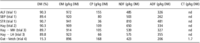
In trial 1, animals were fed 0.3 kg of dehydrated alfalfa pellets and 0.2 kg of dehydrated sugar beet pulp pellets, both provided in two equal parts at 0900 and 1600 h. In addition, they were fed 1.5 kg of molassed wheat straw pellets once a day at 0900 h. In trial 2, animals were fed a 5-year-old semi-mountainous meadow hay harvested in July 2004. The hay was chopped into 5-cm-long pieces and given for ad libitum intake (allowing 10% to 15% refusal on the DM basis) in two equal meals as in trial 1. In trial 3, animals were fed hay for ad libitum intake (allowing 10% to 15% refusal on the DM basis), chopped into 5-cm-long pieces. In trial 4, animals received fresh oat-vetch cut every day and given for ad libitum intake (allowing 20% refusal on the DM basis). Fresh water was freely available to all of the animals in all of the trials. The chemical composition of the feeds is presented in Table 1.
Chemical composition of the feeds provided in trials 1, 2, 3 and 4
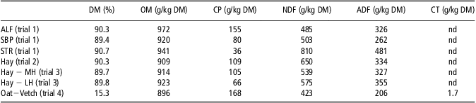
DM = dry matter; OM = organic matter; NDF = neutral detergent fibre expressed exclusive residual ash; ADF = acid detergent fibre expressed exclusive residual ash; CT = condensed tannins; ALF = dehydrated alfalfa; SBP = dehydrated sugar beet pulp; STR = molassed straw pellet; MH = medium-nutritive value hay; LH = low-nutritive value hay; nd = not determined.
Chemical analysis
All samples (feed and faeces) were ground through a 1-mm sieve. Total mineral content was determined by ashing in a muffle furnace for 5 h at 550°C. Total nitrogen was determined using the Kjeldahl procedure. Cell wall fractions (NDF, ADF) were determined sequentially according to the method of Goering and Van Soest (Reference Goering and Van Soest1970) with an amylolytic and protease pre-treatment. Cell wall fractions are expressed exclusive of residual ash. The CT (trial 4) were determined using the vanillin method (Burns, Reference Burns1971).
DMI and digestibility
Individual DMI was measured daily in the four trials. Refusals were recorded every morning before the first new meal. Offerings and refusals were sampled every day for DM content determination (48 h, 60°C). During the measurement periods, total faeces were collected, weighed and sampled for DM determination (48 h at 60°C) in order to calculate total faecal output. Apparent DMD, based on the individual average DMI and faecal output, was calculated on 5 to 7 consecutive days, depending on the trial.
PEG preparation, administration and NIRS measurement
The PEG used was PEG 6000 (molecular weight 6000 da, Panreac Qimica SA, Barcelona, Spain). It was administered in solution form with plastic syringes. The PEG solution, with a concentration of 333.33 g/l, was prepared a few days before in the laboratory with double distilled water. In order to precisely determine the quantity of PEG administered, each syringe was weighed to the nearest 0.1 g before and after administration. The concentration of PEG in faeces was estimated with the NIRS method. Calibration databases were built by adding known amounts of PEG in faeces samples collected before the first doses, in order to build PEG + faeces databases similar to collected samples. The PEG was added to faeces in solution within a range of 0 to 100 g of PEG/kg DM (by 5 or 10 g of PEG/kg DM steps). The PEG + faeces mixtures were dried (50°C until constant dry weight) and ground through a 1-mm sieve, similarly to the procedure applied for faeces preparation during trials.
The samples were scanned on a monochromator NIR spectrophotometer (NIRS 6500, Foss NIRSystems, Silver Spring, MD, USA). Measurement was done in reflectance mode in small circular cups (diameter: 50 mm) with quartz glass. Spectral data were collected every 2 nm from 400 to 2500 nm. Samples were scanned in duplicate (two different cup fillings) and spectra were averaged. Spectra were added to our existing database (Hassoun et al., Reference Hassoun, Fabre, Bastianelli, Bonnal and Bocquier2007a), leading to a global database of 527 samples of faeces samples collected from various origins (sheep breed and diets). The NIRS calibration was carried out using partial least squares regression (MPLS procedure, WinISI software, Infrasoft Int., Port Matilda, PA, USA). Only wavelengths in the 1100 to 2500 nm range were used, because of the unstability of models built with visible wavelengths. Mathematical pre-processing was applied to spectra with detrending and normalization (SNV) of data, and use of the second derivative calculated on five consecutive points with a smoothing also on a gap of five points. During the calibration process, prediction outliers (studentized residual T > 2.5) were discarded. Cross-validation of NIR equations was performed during the calibration process, dividing the database into four groups of samples of which three were used for calibration and the fourth for validation. Criteria for the evaluation of the prediction models are SEC (standard error of calibration) and SECV (standard error of cross-validation), which is an estimate of precision that can be expected in routine analysis. The RPD (=s.d./SECV) was calculated as an indicator of the quality of the models (Williams and Sobering, Reference Williams and Sobering1993). The Mahalanobis distance was used to compare spectra with the database and identify spectral outliers following Shenk and Westerhaus (Reference Shenk and Westerhaus1991).
PEG recovery and PEG content in grab samples
The PEG recovery (REC) was calculated with the formula: REC = PEGT (g/kg DM) × FT (g/kg DM)/PEGin (g/day), where PEGT is the PEG content estimated with NIRS in total faeces (FT) and PEGin the daily amount of PEG intake. The REC was calculated daily for each ewe. Faecal grab samples were collected at the rectal level the same day that total faeces were collected for REC measurement. The PEG content of grab samples (PEGg) collected at 0800 h (trials 1 to 4) and 1600 h (trials 1 and 2) was determined with NIRS. It was compared with PEGT as a percentage:
 $${\rm{(PE}}{{{\rm{G}}}_{\rm{g}}}\,{\rm{/}}\,{\rm{PE}}{{{\rm{G}}}_{\rm{T}}}{\rm{)}}\,\times \,{\rm{100}}{\rm{.}}$$
Estimated faecal output and DMI
Faecal output was calculated including the correction of the two sources of bias values (total and grab sample recovery) or without correction (i.e. assuming that PEG recovery was not different of 100%). Each bias was calculated within trial and factor of variation when applied or averaged for the four trials. Estimated faecal output, corrected (Fc) with the within trial biases or averaged trial biases or not corrected (Fnc), was calculated as follows:
 $$ { {{{\rm{F}}}_{\rm{c}}}\,{\rm{(kg}}\;{\rm{DM)}}\,{\rm{ = }}\,{\rm{[PE}}{{{\rm{G}}}_{{\rm{in}}}}\,\times \,{\rm{REC}}\,\times \,{\rm{(1000 - PE}}{{{\rm{G}}}_{\rm{g}}}{\rm{8}}\,{\rm{/}}\,{\rm{RE}}{{{\rm{C}}}_{\rm{g}}}{\rm{)}}\,{\rm{/}}\cr\qquad{\rm{(PE}}{{{\rm{G}}}_{\rm{g}}}{\rm{8}}\,{\rm{/}}\,{\rm{RE}}{{{\rm{C}}}_{\rm{g}}}{\rm{)]}}\,{\rm{/}}\,{\rm{1000}} \\ {\rm{FEC_{nc}}}\,{\rm{(kg}}\;{\rm{DM)}}\,{\rm{ = }}\,{\rm{[PEG_{in}}}\,\times \,{\rm{(1000 - PE}}{{{\rm{G}}}_{\rm{g}}}{\rm{8)}}\,{\rm{/}}\,{\rm{PE}}{{{\rm{G}}}_{\rm{g}}}{\rm{8]}}\,{\rm{/}}\,{\rm{1000}} $$
where PEGg8 (g/kg DM) is the PEG content measured in grab samples collected at 0800 h and RECg the PEG recovery amount measured in grab samples.
Estimated DMI using Fc calculated with each trial biases (DMIc), averaged trial biases (DMIavc) or using Fnc (DMInc) were calculated as follows:
 $${ {\rm{DM}}{{{\rm{I}}}_{\rm{c}}}\;{\rm{or}}\;{\rm{DM}}{{{\rm{I}}}_{{\rm{avc}}}}\,{\rm{(kg/d)}}\,{\rm{ = }}\,{{{\rm{F}}}_{\rm{c}}}{\rm{/(1 - DMD/100)}} \\ {\rm{DM}}{{{\rm{I}}}_{{\rm{nc}}}}\,{\rm{(kg/d)}}\,{\rm{ = }}\,{{{\rm{F}}}_{{\rm{nc}}}}{\rm{/}}{\rm{(1 - DMD}}\,{\rm{/}}\,{\rm{100))}}$$
Statistical treatment of results
In all trials, DMI, DMD and faecal DM content (FDMc) were compared between groups (PEG20 and PEG40, PEGsingle and PEGtwice, C3 and E3 and C4 and E4, for trials 1, 2, 3 and 4, respectively) at each period: before PEG (trials 1 to 3), with PEG (trials 1 to 4) and after PEG (trial 1). Data were averaged per period for each ewe. Consequently, because of the low number of ewes per treatment or period, the non-parametric Mann and Whitney U-test (Siegel and Castellan, Reference Siegel and Castellan1988; Sprent, Reference Sprent1992) was used to compare two independent samples. The REC was also compared at each measurement period between PEG20 and PEG40 (trial 1), PEGsingle and PEGtwice (trial 2) and E3 MH and E3 LH (trial 3), with the same non-parametric test. Because non-statistical intra-trial differences were found, the REC between trials was compared adding trial 4, with the non-parametric Kruskal–Wallis test for multiple independent samples (Siegel and Castellan, Reference Siegel and Castellan1988; Sprent, Reference Sprent1992). The PEG content measured on grab samples at 0800 h, 1600 h or averaged was compared with PEGT with the non-parametric Wilcoxon test for paired samples because sample numbers were small, from four to eight (Siegel and Castellan, Reference Siegel and Castellan1988; Sprent, Reference Sprent1992). For the same reason, actual DMI were compared with DMIc, DMIavc and DMInc, respectively, with the non-parametric Wilcoxon test for paired samples. All statistical analyses were performed using Statistica v9.1 for Windows (Statsoft 2010, www.statsoft.fr).
Results
NIRS calibration for PEG content
The calibration itself had an R 2 = 0.99 and a residual standard error (SEC) of 3.05 g/kg. No major bias was observed in the calibration process, and there were only 13 outlier samples, that is, 2.5% of the calibration database. The validation of the calibration model by cross-validation resulted in a standard error (SECV) of 3.47 g/kg, which is quite accurate for such a measurement. The RPD value was 9.1.
When applying the calibration to our experimental samples, it appeared that all samples to be predicted were inside the calibration database range. Only seven samples had an H (Mahalanobis) distance higher than 3, which was perfectly satisfactory.
Intake, digestibility and faecal DM content
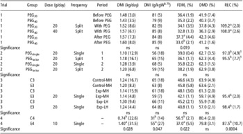
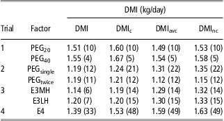
The average DMI, FDMc and DMD for trials 1 to 4 are presented in Table 2. In trial 1, DMI and DMD were not significantly different (P > 0.05) before PEG (control period), with 20 or 40 g of PEG, or after when PEG was stopped for 1 week (Table 2). The FDMc was not significantly different between PEG20 and PEG40 when PEG was administered. However, it decreased compared with the first period, from 36.4% to 34.1% for the PEG20 group, and from 35.3% to 32.8% for the PEG40 group. In contrast, FDMc was significantly different (P < 0.05) between PEG20 and PEG40 measured 1 week after PEG was stopped. During this period, the FDMc of PEG20 group increased and recovered its initial FDMc value (before the PEG period). On the other hand, the FDMc of PEG40 did not change and remained at the same value as during PEG administration.
Mean and relative standard deviation (values in parentheses) of DMI, FDMc, DMD and PEG recovery rate (REC) measured in the four trials

DMI = dry matter intake; FDMc = faecal dry matter content; DMD = dry matter digestibility; PEG = polyethylene glycol; MH = medium-nutritive value hay; LH = low-nutritive value hay.
Within trial and period for DMI, FDMc and DMD, values with different superscript letters in a column are significantly different (P < 0.05). For REC values with different superscript letters in the column are significantly different (P < 0.05).
1Average REC values per frequency.
In trial 2, there were no significant differences between single or split doses on any parameters (Table 2). The FDMc tended to be lower in both periods for ewes that first received PEG as a split dose and then as a single dose (36.1% and 35.8%) compared with the other group (39.0% and 38.2%), as presented in Table 2.
In trial 3, during the control period (Table 2), one ewe in the C3 group had a mouth problem and its results have been discarded for this period. After 3 days, the ewe recovered normal intake and had no more problems until the end of the trial. During the control period with MH hay, both DMI and DMD were not significantly different (P > 0.05) between the C3 and the E3 group (Table 2). Also, FDMc was not significantly different (P > 0.05) between C3 and E3. When PEG was administered to group E3 fed with MH or LH, DMI and DMD were not significantly different (P > 0.05) compared with the C3 group (Table 2). Only FDMc tended to be lower (P = 0.06) for E3 with MH but not with LH (P = 0.31).
In trial 4, during the 1st-week adaptation period, two ewes (one in each group) were replaced after 4 days because they did not adapt to the crates. This was the only problem that occurred. The DMI of E4 was significantly higher (P < 0.05) than the control group C4 (Table 2). In contrast, DMD was not significantly different (P = 0.84) between C4 and E4. The FDMc was significantly lower (P < 0.05) for the E4 compared with the C4 group: 37.0% v. 56.5%, respectively.
PEG recovery, daily and diurnal excretion pattern
The recovery (REC) measured in the total faeces collected over 7 (trial 1), 5 (trials 2 and 3) or 9 days (trial 4) are summarized in Table 2. The REC obtained in the first three trials ranged between 95% and 109%, with a similar variability of 6.2% to 8.9%. In trial 4, a lower mean value was obtained (88%) with a higher variability of 24%. No significant effect (P < 0.05) of the treatments was observed: dose, frequency and hay in trials 1, 2 and 3, respectively. However, when comparing trials, REC in trial 1 was significantly higher than in trial 2 (P < 0.05), trial 3 (P < 0.001) and trial 4 (P < 0.001). The three other trials were not significantly different (P > 0.05).
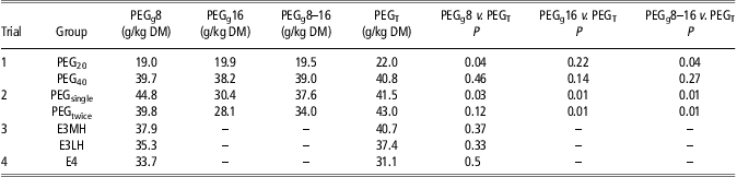
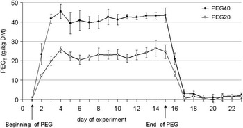
Faecal PEG content measured on grab samples (PEGg) collected at 0800 h (PEGg8) and 1600 h (PEGg16) or pooled (PEGg8-16) were compared with PEGT. Values and statistical results are summarized in Table 3. In all trials, PEGg8 values were not significantly different (P > 0.05) compared with PEGT, except in trial 1 when 20 g/day of PEG was administered (P = 0.04), and in trial 2 when 20 g/day of PEG was administered as a single dose (P = 0.03). The values of PEGg16 and PEGg8-16 were significantly different (P < 0.05) from those of PEGT in trial 2 but not in trial 1. The value of PEGg8 represented more than 90% of PEGT, except in trial 2 when PEG was administered in a split dose (79%). Average PEGT values measured in the four trials are summarized in Table 3. Values ranged from 22.0 to 43.0 g/kg DM with a similar standard deviation (s.d.) of 2.2 to 8.6, which gives a relative s.d. ranging from 5% to 28% (results not shown). Trial 4 gave the highest s.d. and relative s.d. The PEGT was followed from the 1st day until 1 week after the last PEG administration in trial 1. The excretion pattern is presented in Figure 1.
Average PEG content measured in grab samples collected at 0800 h (PEGg 8), 1600 h (PEGg16) or averaged (PEGg8–16), and in total faeces (PEGT), statistical result (P-value of the Wilkinson test) for the comparison of means
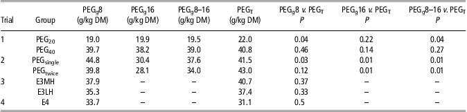
PEG = polyethylene glycol; MH = medium-nutritive value hay; LH = low-nutritive value hay.
Evolution of PEG content (PEGT) in total faeces during the experiment for groups dosed with 40 (PEG40) or 20 (PEG20) g of PEG per day (trial 1). PEG = polyethylene glycol.

Once PEG was administered, PEGT rapidly increased over the first 2 days and then remained quite stable although markedly different between doses, until the last day for both 20 and 40 g/day doses. When PEG administration was stopped, PEGT decreased as quickly as it had increased at the beginning. No PEG was recorded 2 to 3 days later. Although the values observed on subsequent days were not exactly null (1.1 and 1.3 g/kg DM for PEG20 and PEG40, respectively), they ranged within the NIRS standard error of prediction: SECV = 3.47 g/kg DM.
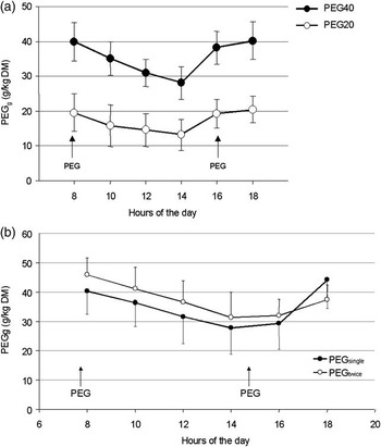
The diurnal excretion pattern was measured on grab samples in trial 1 (Figure 2a) and trial 2 (Figure 2b). When PEG was administered twice a day in trial 1, PEGg decreased from 0800 h to 1400 h, and then increased until 1800 h. The PEGg value at 0800 h and at 1800 h represented the highest daily values in trial 1. The PEGg value decreased and increased even more rapidly for PEG40 than for PEG20 (Figure 2a). In trial 2, regardless of the frequency of the dosed PEG, the highest PEGg was observed at 0800 h and then decreased until 1400 h, like in trial 1, and remained almost identical at 1600 h. The PEGg then increased in both groups but more quickly when administered as a single dose, almost reaching the initial value (i.e. at 0800 h), whereas with the split dose it remained lower (Figure 2b).
Kinetics of PEG content (PEGg) in grab faeces samples for groups dosed (a) with 40 (PEG40) or 20 (PEG20) g of PEG per day (trial 1) and (b) daily with 20 g of PEG administered once at 0800 h (PEGsingle) or split in two amounts at 0800 and 1600 h (PEGtwice; trial 2). PEG = polyethylene glycol.

Estimated DMI
Actual DMI compared with each estimated values of DMI (Table 4) are not statistically different (P > 0.05). The coefficient of variation (CV) of actual DMI in trials 1, 2 and 3 ranged from 4% to 12%. In trial 4, the CV reached 32%, respectively. The estimated values had a CV ranging from 5% to 22%, but was higher once again (48%) in trial 4.
Average and relative standard deviation (values in parentheses) of actual DMI and estimated values with each trial recovery bias (DMIc) or average recovery bias of the four trials (DMIavc) included or without (DMInc) recovery bias. Results were not significantly (P>0.05) different.
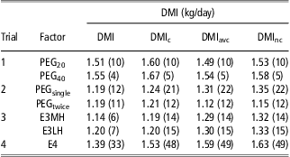
DMI = dry matter intake; PEG = polyethylene glycol; MH = medium-nutritive value hay; LH = low-nutritive value hay.
Discussion
The precision of NIRS calibration was high, with a low measurement error (3.47 g/kg) and high R 2 and RPD values. The analytical precision is of extreme importance in studies with markers because a small error in the quantification of markers in faeces has a strong effect on the calculation of feed intake. For this reason, it is essential to update the calibration database at each new experiment with some faecal samples obtained before PEG treatments and enriched in vitro with PEG in order to obtain concentrations within the range of those expected during the experiment.
The fact that recovery rates of PEG were close to 100% in the first three trials confirms that there was no major bias in PEG estimation. It validates the experimental calibration protocol, based on PEG addition in faeces, which was used to build and update the calibration database. This is also confirmed by the fact that the spectra of experimental samples were well fitted to the calibration database, that is, no major change was observed between PEG added in vitro and PEG having transited in vivo.
The estimation of PEG concentration in faeces by NIRS proved to be very efficient. As no alternative reference analysis method exists for quick PEG determination, it would have been impossible to measure PEG concentration in several hundred samples, as required in this type of studies. The ease of NIRS analysis makes it extremely convenient for intake studies, making it possible to analyse a very high number of samples at a low cost in terms of time and money.
Previously, PEG was used as faecal marker to estimate faecal output (Hopson and McCroskey, Reference Hopson and McCroskey1972; Landau et al., Reference Landau, Friedman, Devash and Mabjeesh2002). None of these studies reported any metabolism, intake or digestibility modifications due to PEG. In the present four trials, no effect of PEG was observed on DMI and DMD, except in trial 4 where E4 ewes ate twice as much as C4 ewes, 1.4 v. 0.74 kg DM, respectively. The PEG is known to alleviate the negative effect of CT on intake and digestibility (Silanikove et al., Reference Silanikove, Gilboa and Nitsan2001; Bhatta et al., Reference Bhatta, Shinde, Verma, Sankhyan and Vaithiyanathan2004). The CT content of oat-vetch in trial 4 was low (1.5 g/kg) compared with the values having a negative effect (i.e. >50 g/kg DM) on voluntary intake reported by Frutos et al. (Reference Frutos, Hervas, Giraldez and Mantecon2004). Therefore, it seems that PEG interaction with CT was not responsible for the higher DMI observed in trial 4. No valid explanation was found to interpret these results.
In all trials, FDMc significantly decreased for ewes dosed with PEG: −2.5 points (trial 1), −4 to −5.6 points (trials 2 and 3) and −19.5 points in trial 4. Landau et al. (Reference Landau, Friedman, Devash and Mabjeesh2002) and Teeter and Owens (Reference Teeter and Owens1983) observed with goats and steers, respectively, that FDMc tended to decrease with PEG. This FDMc decrease was also observed with other water soluble markers (Teeter and Owens, Reference Teeter and Owens1983). In sheep species, water is absorbed throughout the gut, from the small intestine to the rectum (Grovum and Hecker, Reference Grovum and Hecker1973; Grovum and Williams, Reference Grovum and Williams1973). Consequently, the digesta become drier from the reticulo-rumen to the rectum. The DM content of digesta in the caecum and proximal colon is low and progressively increases until the terminal colon and rectum (Hecker and Grovum, Reference Hecker and Grovum1971). Most of the water is absorbed at the caecum and proximal colon because digesta are retained for a relatively long time compared with the other sections of the colon (Hecker and Grovum, Reference Hecker and Grovum1971).
It is known that PEG has no effect on water movement at the rumen level and does not interfere with the physiological functions of ruminal microorganisms (Sperben et al., 1953, quoted by Sinha et al., Reference Sinha, Martz, Johnson and LeRoy Hahn1970). As the intestine cannot secrete free water (Lord, Reference Lord1999), the lower FDMc observed with PEG addition is probably due to the osmotic effect of PEG (Schiller et al., Reference Schiller, Emmett, Santa Ana and Fordtran1988), which sequesters water in the intestine, inhibiting water absorption (Davis et al., Reference Davis, Santa Ana, Morawski and Fordtran1980). The osmotic effect of the PEG is probably higher at the caecum and proximal colon level. Considering the great difference observed in DMI but not in DMD in trial 4, Blaxter et al. (Reference Blaxter, Graham and Wainman1956) observed that DMD (79.1% to 80.3%) was not modified when sheep were fed dry grass offered in a long form at 600 g or 1200 g/day. In the same time, they observed that DM content of the faeces tended to decrease (−3.7 points) from 42.3% to 38.6% when DMI increases. Grovum and Williams (Reference Grovum and Williams1973) observed that the quantity of water reaching the proximal colon is higher for sheep given 1200 g/day of lucerne chaff than those given 400 g/day but water absorption from the large intestine is also higher. They observed the same water content of digesta in the rectum and concluded that the level of DMI does not influence the water content in the rectum. These results can explain partly why in trial 4 the DM content of the faeces in E4 is lower as more water reaching the colon is not fully absorbed because of the osmotic effect of PEG.
These results could be elucidated in offering two levels the same forage (fresh and as hay) at two levels of offered with or without PEG.
The REC was high in the first three trials, ranging from 95% to 109%. The same PEG recovery ranges (96% to 113%) were measured indoors with cattle (Corbett et al., Reference Corbett, Greenhalgh, Gwynn and Walker1958; Teeter and Owens, Reference Teeter and Owens1981), sheep (Hassoun et al., Reference Hassoun, Bastianelli and Bonnal2007b; Caja et al., Reference Caja, Ralha and Albanell2009) and goats (Landau et al., Reference Landau, Friedman, Devash and Mabjeesh2002). In trial 4, REC was lower (87.5%), with an average range of 74.5% to 102.3%, as reported by Caja et al. (Reference Caja, Ralha and Albanell2009) when ewes were allowed to graze Italian ryegrass for 6 h (REC = 81.6%) in addition to being fed dry food indoors, compared with ewes exclusively fed indoors (REC = 101%). However, it was demonstrated that PEG with a high molecular weight (>3000) is not absorbable in the intestines (Schiller et al., Reference Schiller, Santa Ana, Porter and Fordtran1997) and not found in the urine (Teeter and Owens, Reference Teeter and Owens1981 and Reference Teeter and Owens1983). The REC results of trial 4 may be attributed to the low PEG level (10 g/day) or to the nature of the diet (fresh forage with low DM content), but this remains to be clarified.
Marker excretion must be as stable as possible from day to day. In trial 1, daily PEG excretion was rather constant over the 14-day trial. In trial 1, PEG reached its maximum level in faeces 2 days after the first administration with a rapid steady state. Moreover, PEG rapidly disappeared 3 to 4 days after the last PEG administration (Figure 1). These results are in agreement with Corbett et al. (Reference Corbett, Greenhalgh, Gwynn and Walker1958) and Hopson and McCroskey (Reference Hopson and McCroskey1972) who observed a total PEG recovery within 2 to 3 days with steers dosed with 50 to 400 g/day of PEG. The pattern of PEG excretion is similar to other water-soluble markers (Cr-EDTA, Co-EDTA) excreted within 48 h after the first administration (Aharoni et al., Reference Aharoni, Brosh and Holzer1999). The diurnal excretion pattern in trials 1 and 2 showed a curvilinear excretion with two high PEG content values at 0800 and 1800 h, and one lower value at 1400 h (i.e. 6 h after the morning dose). Corbett et al. (Reference Corbett, Greenhalgh and Florence1958 and Reference Corbett, Greenhalgh, Gwynn and Walker1959) found the same diurnal excretion pattern of PEG on cows dosed once or twice daily, with the lowest value observed 6 to 8 h after the morning dose and the highest at 0800 and 1800 h. Hopson and McCroskey (Reference Hopson and McCroskey1972), administrating various PEG levels in a single dose (0800 h) or twice daily (0800 and 1630 h) to steers, observed a similar diurnal excretion pattern but with a more pronounced difference between the highest and lowest PEG content with a single dose than with a split dose. Landau et al. (Reference Landau, Friedman, Devash and Mabjeesh2002), using 20 and 40 g PEG/day administered in a single dose at 0800 h to goats, obtained the same excretion pattern with 40 g/day that observed in the present experiment with 20g PEG/day administered in a single or split dose (trial 2), but a different excretion pattern with 20 g PEG/day with less hourly variability.
When a marker is used for assessing total faecal output through grab sampling, the marker content must be as close as possible to the mean content in total faeces. Otherwise, marker content of grab samples must be corrected for this systematic bias. The PEGg8 values, which represented 86% to 108% of the corresponding PEGT values, were not significantly different from actual PEGT in all trials, except in trial 1 (PEG20) and in trial 2 when PEG was administered in a single dose (Table 3). Averaging PEGg8 and PEGg16 did not reduce this bias. Hopson and McCroskey (Reference Hopson and McCroskey1972) obtained slightly higher values (92% to 116%) when PEG content in gathered grab samples (0800 and 1630 h ) were compared with PEG in total faeces collection. An average CV of 14% and 13% was found when measured at 0800 and 1600 h, respectively, that is similar to those observed by Landau et al. (Reference Landau, Friedman, Devash and Mabjeesh2002) with 16.4% and 15.3% with 20 and 40 g PEG/day, respectively. The CV was only higher in trial 4 with green forage, with 38%. Similarly, CV calculated on PEGT ranged from 3% to 19%, except in trial 4 (30%). Considering these results, it seems advisable to collect grab samples only in the morning, which is easier from the practical point of view (less work and animal handling). Our results also reveal that there is no major difference between administering PEG once or twice daily.
The actual DMI variations between ewes in trials 1, 2 and 3 are in good agreement with other values observed under various conditions (Andueza et al., Reference Andueza, Picard, Pradel, Egal, Hassoun, Peccatte and Baumont2011). However, the values were much higher (32%) with fresh green forage than those observed for other species of fresh green forage (Dulphy, Reference Dulphy1971). Consequently, if the diet does not contain tannins, the DMI can be estimated with good accuracy, corrected or not with the recovery biases. Landau et al. (Reference Landau, Friedman, Devash and Mabjeesh2002) obtained the same results without any correction.
Conclusions
These results confirm that faecal PEG content can be estimated by NIRS with good accuracy, provided that faeces obtained from sheep fed the diet on which measurements are to be made are included in the general NIRS database for calibration. Because PEG is rapidly excreted, it is recommended that faeces be collected within 1 week after the start of administration. The PEG recovery rate in total faeces is high and comparable with other results found in the literature concerning dry feed. Using PEG as a faecal marker at levels of 20 to 40 g/day does not modify DMD and DMI when sheep are fed dry diets. Surprisingly, PEG modifies the DMI but not the DMD of a fresh forage diet. Further experiments with fresh forage must be conducted because most of the results in the literature deal with dry diets. Consequently, PEG estimated by NIRS can be a good faecal external marker, allowing a rapid determination, no extraction, no use of health hazards for technicians (in contrast with other external marker like ytterbium or alkanes) and no problem of disposal. Briefly, the practical procedure proposed is to dose animals with PEG solution once a day (20 g of PEG/day) and to collect grab faeces samples in the morning for 5 days at the same time the animals are dosed. Collected faeces should be dried (at a temperature not exceeding 50°C) and gathered per animal and per period before being NIRS scanned.
Acknowledgements
The authors would like to thank J.M. Capron, S. Douls, V. Thiers and the technical staff of La Fage for their expertise in animal management and sample preparation. They are also grateful to L. Bonnal for his assistance in NIRS analysis.