Traumatic ventricular septal defects occur following penetrating and blunt trauma to the thorax, as well as iatrogenic procedures.Reference Dehghani, Ibrahim and Collins 1 Patients with penetrating cardiac traumas are usually referred for emergency operations owing to profound shock caused by cardiac rupture or tamponade. A few reports have described the transcatheter closure of traumatic ventricular septal defects.Reference Suh and Kern 2 – Reference Cowley and Shaddy 4 This report describes the closure technique and imaging of a traumatic ventricular septal defect with emphasis on the usefulness of transcatheter closure.
Case report
A 27-year-old man with a history of two stab wounds in the anterior chest wall by knife was referred to the emergency department. With the diagnosis of cardiac tamponade having been made, the patient was immediately transferred to the operating room for thoracotomy. Right ventricular rupture was discovered, which was repaired, as well as a laceration in the lingular lobe of the left lung. The post-operative course was unremarkable, and he was discharged after 15 days. After 40 days, he was referred to a cardiologist owing to atypical chest pain and cardiac murmur. Transthoracic echocardiography revealed a muscular ventricular septal defect with a left-to-right shunt. The defect was 5 millimetres in size in the off-axis four-chamber and parasternal long-axis views, and the shunt flow was estimated to be 2.5. Owing to high surgical risk, the consultant surgeon recommended device closure. Therefore, transcatheter closure was planned for the patient.
Procedure
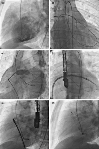
Traumatic ventricular septal defect was confirmed via right and left heart catheterisation and oximetry and was localised by left ventriculography in the upper anterior muscular septum in 115-degree left anterior oblique and 75-degree right anterior oblique projections (Fig 1a). Continuous transesophageal echocardiographic monitoring was conducted during the procedure. The procedure was performed under local anaesthesia using an arteriovenous loop between the right femoral artery and vein, and permitting a sheath passage across the interventricular septum. Unfractionated heparin was injected as a bolus dose of 5000 units and was continued in order to maintain the Activated Clotting Time between 250 and 300. With the use of Judkins right 5 × 4 catheter and 0.035 inch × 260 centimetres Angioflex® hydrophilic guide wire (Kimal, Arundel Road, England), the ventricular septal defect was negotiated. The Judkins catheter was then advanced over the Angioflex® wire to the pulmonary artery. The guide wire was snared using a 20-millimetre wire snare to the inferior vena cava and then externalised (Fig 1b). The defect size was 13 millimetres in balloon sizing (Fig 1c). Thereafter, the venous sheath was exchanged with a 10-French delivery sheath, and the sheath and dilator were advanced over the wire across the defect and towards the left ventricular apex (Fig 1d). A 14-millimetre Amplatzer muscular ventricular septal defect occluder was advanced. After the deployment of the left ventricular disk, the device was found in the right ventricle; perhaps it was too small for the defect. The device was then recaptured. We decided to use an atrial septal defect occluder device. The atrial septal defect occluder was deployed successfully (Fig 1e). The procedure was terminated despite the presence of a minimal jet of contrast from the left to the right ventricle (Fig 1f). The hospital stay was uneventful, and the patient was discharged home with aspirin and clopidogrel for the next 6 months. The only remaining physical finding at the time of discharge was a soft murmur at the lower left sternal border.
( a ) The traumatic ventricular septal defect is seen in the upper anterior part of the interventricular septum in 115-degree left anterior oblique projection; ( b ) snaring of the wire in the pulmonary artery to create arteriovenous loop; ( c ) balloon sizing of the ventricular septal defect; ( d ) the delivery sheath is passed through the ventricular septal defect from the right to the left ventricle. Also note the transesophageal echocardiographic probe; ( e ) the atrial septal defect occluder device before release, right anterior oblique projection; ( f ) 115-degree left anterior oblique view shows the atrial septal defect occluder device in position after release: there is a small left-to-right shunt.

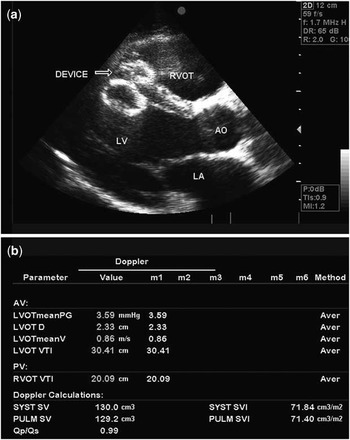
After 2 months, the patient demonstrated good functional capacity without any symptoms. No murmur was detected. Transthoracic echocardiography showed the device in its proper position without any shunt (Fig 2). All of the chambers and valves had normal size and function. Biochemical and haematologic tests were all normal and without any evidence of haemolysis.
( a ) Transthoracic echocardiography 2 months after device implantation shows that the device is in right position; ( b ) Qp/Qs study 2 months after traumatic ventricular septal defect closure shows no shunts.

Discussion
For several reasons, this case report is outstanding: First, the diagnosis of the traumatic ventricular septal defect was delayed perhaps owing to the emergency situation or later development of the defect following the first surgical operation. Second, the size of the traumatic ventricular septal defect was underestimated echocardiographically and by left ventriculography owing to its bizarre shape. Third, traumatic ventricular septal defect following penetrating cardiac trauma is very rare – overall incidence = 4.5% of all traumatic ventricular septal defects.Reference Pesenti-Rossi, Godart and Dubar 5 Fourth, the use of an atrial septal defect occluder device for percutaneous closure of a traumatic ventricular septal defect is uncommon. Finally, the location of the defect in the interventricular septum required a particular left ventriculographic projection to demonstrate it angiographically.
In patients with penetrating cardiac traumas, thorough peri-operative evaluation may be difficult owing to unstable haemodynamics. Moreover, performing transthoracic echocardiography is difficult in the emergency setting because of the wound, bleeding, pneumothorax, etc.Reference Jeon, Lim and Kang 6 We advocate that performing routine transesophageal echocardiography during thoracotomy, if available, can help identify subtle injuries.
Some authors have similarly reported the development of a ventricular septal defect as a delayed complication of penetrating injury.Reference Antoniades, Petrou and Eftychiou 7 The rationale is that muscular spasm or blood clot at the site of the septal rupture can conceal the defect in the initial imaging evaluations. Thus, serial echocardiography can prevent underdiagnosis of this important complication.
Echocardiography and left ventriculography were inaccurate in estimating the true physiological significance of the ventricular septal defect in this case, perhaps owing to its unusual location or partial thrombofibrotic closure of the septal defect. Hence, we were not able to adequately demonstrate the defect size using routine left anterior oblique views. The best imaging projections were 115-degree left anterior oblique and 75-degree right anterior oblique projections, which helped to pass the guide wire.
Proper estimation of the defect size is critical for optimal closure. The irregular borders of a traumatic ventricular septal defect can impair accurate measurements of the defect. We therefore used balloon sizing, which provided us with a more precise estimation of the device size – 13 millimetres versus 5 millimetres using echocardiography. The role of computed tomographic angiography or cardiac magnetic resonance imaging in diagnosing and measuring traumatic ventricular septal defects has yet to be investigated.
Owing to the rarity of traumatic ventricular septal defect, there is no well-established best practice for its closure, although all new approaches to its treatment must ultimately be compared with the surgical approach. Owing to the above-mentioned reasons, there is no study comparing the effectiveness and outcomes of treating traumatic ventricular septal defect by surgery with transcatheter intervention. Therefore, decision making for choosing surgical intervention or percutaneous intervention is case dependent and relies on the available facilities and condition of the patient. As far as this patient was previously operated for the ventricular injury, the surgeons were reluctant to perform a second operation. Hence, percutaneous closure of the ventricular septal defect was proposed.
A few cases have thus far been managed with the Amplatzer duct occluderReference Schwalm, Hijazi and Sugeng 3 , Reference Alibegovic, Aggoun and Vuille 8 , Reference Bauriedel, Redel and Schmitz 9 and a few others with the atrial septal defect occluder.Reference Suh and Kern 2 , Reference Pesenti-Rossi, Godart and Dubar 5 These reports have highlighted the morphological differences between congenital and post-traumatic ventricular septal defects and the difficulty in estimating the size via routine imaging modalities.
Using the atrial septal defect occluder may be accompanied by some disadvantages. Risk of erosion, protrusion into the right and left ventricular cavities, thrombus formation, and interfering with the mitral and tricuspid valve functions are some concerns. Another theoretical problem is that the length of the waist of the atrial septal defect occluder device is 4 millimetres compared with 7 millimeters for the ventricular septal defect occluder. Owing to the fact that the interventricular septum is thicker than the interatrial septum and its thickness increases during systole, this reduced length of the waist can prevent the atrial septal defect occluder device from maintaining its optimal configuration, perhaps leading to residual shunts. There is also a risk of haemolysis owing to the high-pressure gradient between the ventricles,Reference Pesenti-Rossi, Godart and Dubar 5 which fortunately did not happen in this patient. Initial heparinisation and careful monitoring in order to prevent and treat the haemodynamic instability is also crucial in this procedure.Reference Laussen, Hansen and Perry 10
In conclusion, traumatic ventricular septal defects can be a delayed complication of penetrating traumas and may not be detected initially. Although surgical repair is the traditional treatment, percutaneous closure can be a feasible and safe method.
Acknowledgement
This case report was supported by Tehran Heart Center, Tehran University of Medical Sciences.